ISSN 0378-6471 (Print)⋅ISSN 2092-9374 (Online)
https://doi.org/10.3341/jkos.2018.59.3.295
Case Report
외상에 의해 발생한 망막중심동맥과 중심정맥의 폐쇄와 이에 동반된 안와첨증후군 1예
A Case of Orbital Apex Syndrome with Central Retinal Artery and Vein Occlusion Following Trauma
장미리내⋅이상윤⋅이혜진⋅이은경
Mirinae Jang, MD, Sang-Yoon Lee, MD, Hye Jin Lee, MD, Eun Kyoung Lee, MD
제주대학교 의학전문대학원 안과학교실
Department of Ophthalmology, Jeju National University School of Medicine, Jeju, Korea
Purpose: To report a case of orbital apex syndrome (OAS) combined with central retinal artery occlusion (CRAO) and central ret- inal vein occlusion (CRVO) following blunt trauma.
Case summary: A 4-year-old female visited the hospital following a traffic accident. She was admitted because of multiple frac- tures of the skull and pneumocephalus. On day 5, she was referred to us with decreased visual acuity in her right eye. Her initial visual acuity was hand motions in the right eye and 0.8 in the left eye. The right eye showed a dilated pupil, ptosis, and total oph- thalmoplegia, and the left eye showed limited abduction. A fundus examination revealed multiple retinal hemorrhages, tortuous veins, and an edematous white retina with a cherry-red spot in the right eye. Brain magnetic resonance imaging revealed an en- trapped right optic nerve because of bony fragments in the orbital apex. The patient was diagnosed with OAS accompanied by CRAO and CRVO in the right eye, and with traumatic abducens nerve palsy in the left eye. After 6 months, the visual acuity was hand motions, and the fundus examination showed absorbed retinal hemorrhages, pale discs, and general retinal thinning of the right eye. Ptosis of the right eye and extraocular muscle movement of both eyes were improved.
Conclusions: Combined CRAO and CRVO following trauma is very rare and is even more rarely associated with OAS. It is im- portant for clinicians to be aware of the potential for central retinal vessel occlusions and OAS in cases of blunt ocular trauma.
J Korean Ophthalmol Soc 2018;59(3):295-300
Keywords: Blunt ocular trauma, Central retinal artery occlusion, Central retinal vein occlusion, Orbital apex syndrome
■Received: 2017. 10. 26. ■ Revised: 2017. 12. 22.
■Accepted: 2018. 2. 19.
■Address reprint requests to Eun Kyoung Lee, MD Department of Ophthalmology, Jeju National University Hospital, #15 Aran 13-gil, Jeju 63241, Korea
Tel: 82-64-717-1730, Fax: 82-64-717-1029 E-mail: [email protected]
* This study was presented as an e-poster at the 118th Annual Meeting of the Korean Ophthalmological Society 2017.
*Conflicts of Interest: The authors have no conflicts to disclose.
ⓒ2018 The Korean Ophthalmological Society
This is an Open Access article distributed under the terms of the Creative Commons Attribution Non-Commercial License (http://creativecommons.org/licenses/by-nc/3.0/) which permits unrestricted non-commercial use, distribution, and reproduction in any medium, provided the original work is properly cited.
망막중심동맥폐쇄는 통증 없이 급작스런 시력상실을 일으키는 드문 망막혈관폐쇄 질환으로, 발병률은 10만 명 당 1.8명으로 보고되고 있으며,1 고혈압, 당뇨, 혈액학적 이상, 경동맥의 폐쇄 등 전신 질환과 흔히 동반하는 것으 로 알려져 있다.2 그 외에도 의인성 원인으로 필러주입술,3 내시경 부비동 수술,4 구후마취5 등에 의한 경우나 외상6으 로 인해 발생한 국내의 보고가 있다. 망막중심정맥폐쇄 또 한 고혈압, 당뇨 등과 같은 전신 요인이나 혈전 성향과 관 련된 혈액학적 요인들과 동반되어 발생하는 경우가 많다.7 망막중심동맥폐쇄와 망막중심정맥폐쇄가 같이 발생하 는 경우는 매우 드문 것으로 알려져 있다. 전신 질환과 관 련하여 동시에 발생한 망막중심동맥폐쇄와 망막중심정맥
A B
C D
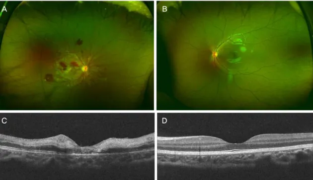
Figure 1. Wide field fundus photography (WFP) and optical coherence tomography (OCT) image on initial presentation. (A) WFP after two
weeks of trauma shows pseudo-cherry red spot and multiple retinal hemorrhages near the optic disc in the right eye. (B) The normal WFP image of the left eye. (C) OCT image of the right eye revealed severe inner retinal edema. (D) The normal OCT image of the left eye.폐쇄는 백혈병,8 해면정맥굴혈전증(cavernous sinus throm- bosis),8 감염성심내막염,8 전신홍반성루프스,9 척스트라우 스증후군,10 전이암에 의한 수막암종증(Meningeal carcino- matosis),11 매독,12 경구피임약의 복용13 등의 경우 국외의 증례보고가 있었지만, 외상에 의해 망막중심동맥폐쇄와 망막중심정맥폐쇄가 동시에 발생한 경우는 보고된 바가 적으며,14 국내에서는 보고된 바가 없다. 더욱이 외상으로 인한 안와첨증후군이 이에 병발한 경우는 더욱 드물다. 저 자들은 외상에 의하여 망막중심동맥폐쇄와 망막중심정맥 폐쇄 및 안와첨증후군이 동시에 발생된 증례를 경험하였 기에 이를 보고하고자 한다.
증례보고
특이병력 없는 4세 여자 환자가 우안 시력 저하를 주소 로 의뢰되었다. 환자는 뛰어가다가 지나가는 차량에 추돌 하면서 의식이 소실되어 내원하였다. 내원 당시 안면부 손 상과 비강출혈이 관찰되었고, 컴퓨터단층촬영에서 급성 경막하출혈(subdural hematoma), 기저두개골절(skull base fracture)을 포함한 오른쪽 안와골, 관골(zygoma), 두정후 두골(parieto-occipital bone) 및 측두골(temporal bone)의 골절과 기뇌증(pneumocephalus), 대뇌부종(cerebral ede- ma)이 확인되어 신경외과로 입원하였다. 기관삽관 및 대 량 출혈에 대한 지혈 및 수혈이 이루어졌다. 다음 날부터
환자의 의식 수준이 서서히 회복되었고, 수상 이틀 후 비 강출혈은 멈추었으나 비인두에서 뇌척수액 누출이 관찰 되어 기관삽관을 제거하고 절대 안정을 유지하였다. 수상 5일 후 우안 동공산대 및 시력 저하 증상으로 안과에 의뢰 되었고, 안과적인 초진이 이루어졌다.
초진 소견 시 나안시력은 우안 안전수동, 좌안 0.8이었 고, 우안 동공은 산대된 상태로 움직이지 않았으며, 좌안 동공 반응은 정상이었다. 우측에 margin reflex distance 1 (MRD1) 0의 눈꺼풀처짐이 있었고, 안근기능검사상 우안 은 전방향에서 -4의 안구운동장애를 보였으며, 좌안은 외 전 기능에 -4의 제한을 보였다. 휴대용 세극등 현미경 검 사상 양안 모두 전안부 소견에 특이 사항은 없었다. 안저 검사에서 우안 망막 전반에 다발성 망막출혈, 정맥 확장 및 구불거림이 관찰되었고, 앵두반점을 동반한 후극부 창 백 소견이 관찰되었다. 좌안은 안저검사에서 정상 소견이 었다. 당시 환자가 절대 안정이 필요한 상태로, 안저촬영 등의 검사는 시행할 수 없었으나, 임상적 소견을 토대로 우 안 망막중심동맥폐쇄 및 망막중심정맥폐쇄로 진단하였다.
일주일 뒤 환자가 보행할 수 있는 상태가 되어 안저촬 영 및 빛간섭단층촬영을 시행하였다. 안저검사에서 우안 에 관찰되던 다발성의 망막출혈은 현저히 감소한 상태였 고, 시신경과 후극부 주위에만 망막출혈이 관찰되었다. 후 극부 소견은 세동맥 주위는 침범하지 않는 망막의 창백 소견(retinal whitening with periarteriolar sparing)과 가성
A
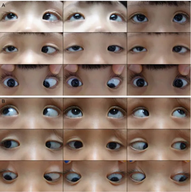
B
Figure 2. Fields of gaze of extraocular movement. (A) Initial examination shows limitation of movement in all directions in the right
eye. Ptosis of the right upper eyelid and abduction limitation of the left eye are also noted. (B) At 6 months after trauma, ptosis of the right upper eyelid was improved. The extraocular muscle movements of both eyes were also improved. However, abduction of the right eye was still limited.앵두반점(pseudo-cherry red spot)이 관찰되어 푸르처 유사 망막병증(Purtsche-like retinopathy)의 양상을 띠었다(Fig.
1A, B). 빛간섭단층촬영에서 우안에 내망막층의 심한 부 종 소견과 시세포층의 손상이 관찰되었다(Fig. 1C, D). 어 린 나이의 환자였기에 침습적 검사인 형광안저혈관조영 술은 시행하지 않았다.
우측 안검하수와 안구운동장애는 초진 당시 소견과 같 은 양상이었다(Fig. 2A). 시신경의 상태를 파악하기 위해 뇌 자기공명영상 촬영을 시행하였고, 시신경결출(optic nerve avulsion) 소견은 없었으나, 기저두개골절과 안와첨단부골 절(orbital apex fracture)로 인해 우측 시신경이 포착되고 (entrapped) 늘어진(stretched) 소견이 관찰되었다(Fig. 3).
시신경 경로를 따라 출혈 소견 또한 관찰되었다. 이에 따 라 안와첨단부골절로 인해 발생한 우측 외상성 시신경(II) 손상을 비롯한 동안신경(III), 도르래신경(IV) 및 가돌림신 경(VI) 마비와 좌안의 외상성 가돌림신경(VI) 마비로 진 단하였다. 수상 이후 급성 단계가 이미 지났고, 뇌척수액 누출 및 발열 증상 등 전신 상태를 고려하여 신경외과와 상의 후 고용량 스테로이드 정맥주입술은 시행하지 않기 로 결정하였다. 이후 발열에 대해 항생제 치료를 시행하였 고, 헤모글로빈수치와 전해질수치 등을 교정 후 전신상태 호전되어 퇴원하였다.
이후 정기적인 안과 외래 경과관찰을 지속하였다. 수상 후 6개월째 우안 나안시력은 안전수동, 좌안 0.8로 초진
A B
Figure 4. Fundus photography (FP) and optical coherence tomography (OCT) images of right eye at 6 months after trauma. (A) FP
shows absorbed retinal hemorrhages and pale disc. (B) OCT image reveals generalized retinal thinning. Long thin white arrows in- dicate OCT scan direction.A
B
C
Figure 3. Computed tomography (CT) image and magnetic res-
onance imaging (MRI) on initial presentation. (A) Axial CT scan demonstrates multiple fractures including orbital apex of the right eye and skull base. (B) Axial MRI (T2 weighted im- age) scan on 7 days after trauma shows bony fragments (dotted arrow) and hematoma (solid arrow) along the optic nerve pathway. (C) Sagittal MRI (T1 weighted image) scan reveals entrapped and stretched optic nerve of the right eye (solid ar- row) because of bony fragments (dotted arrow).당시와 같은 시력으로 측정되었고, 전안부 검진상 우안 홍 채신생혈관 등은 관찰되지 않았다. 안저 검진상 우안에 관 찰되던 다발성의 망막출혈은 모두 흡수되었고, 시신경 유 두는 창백하였다(Fig. 4A). 빛간섭단층촬영상 망막의 전반 적인 얇아짐이 또한 관찰되었다(Fig. 4B). 우측의 MRD1 은 +3으로 눈꺼풀처짐은 호전되었으며, 안근기능검사상 우안의 외전 기능 -1을 제외하고, 양안의 외안근 기능은 모두 개선되었다(Fig. 2B).
고 찰
외상에 의한 망막중심동맥과 망막중심정맥의 동반 폐 쇄 증례는 국내외를 막론하고 매우 드물다. 1987년 Noble and Alvarez15가 타인의 손가락에 찔리는 둔상에 의한 망 막중심동맥과 정맥의 동반 폐쇄를 처음으로 보고한 이후, 2011년 Cumurcu et al16이 축구공에 의한 둔상으로 인해 외상성 시신경병증과 망막중심동맥폐쇄가 병발한 증례를 보고한 바 있다. 2014년 Singh et al14이 교통사고로 안구 주위 둔상을 입고, 본 증례와 비슷한 양상의 외상성 시신 경병증과 망막중심동맥과 정맥이 동반 폐쇄된 1예를 보고 하였다. 국내에서는 1995년 Kim et al4이 부비동 수술 후 안와내벽의 골절과 구후출혈에 의해 망막중심동맥과 정 맥이 폐쇄된 1예를 보고하였고, 2008년 Lim et al5은 백내 장수술 시 시행한 구후마취 직후에 발생한 망막중심동맥 과 정맥의 동반폐쇄 1예를 보고하였다. 또한 최근 Koh and Woo6는 외상이 원인이 된 망막혈관폐쇄 2예를 보고 하였으나 이는 망막중심동맥만 폐쇄된 경우였고, 외상에 의해 망막중심동맥과 정맥이 동반폐쇄된 증례는 아직 국 내에서 보고된 바가 없으며, 외상성 시신경병증을 초래한
안와첨증후군까지 병발된 경우는 국외에도 매우 드물었다.
본 증례에서 교통사고로 인한 기저두개골절과 안와첨 단부골절에 의해 망막중심동맥과 정맥이 폐쇄된 명확한 기전은 확인할 수 없으나 세 가지 가설을 생각해 볼 수 있 다. 첫 번째로 골절에 의한 뼛조각(bony fragments)이 시 신경의 연막혈관을 뚫어 시신경초 내로 출혈이 발생되었 을 가능성이다. 출혈에 의해 시신경초 내의 압력이 상승하 고, 이로 인해 정맥의 유출이 막히면서 정맥내의 혈류정체 에 의해 출혈성 망막병증이 유발된다. 또한 망막중심정맥 폐쇄로 인해 망막혈류순환에 완전한 혈역학적 차단이 일 어나면 망막중심동맥의 폐쇄에까지 이르게 될 수 있다. 두 번째는 외상을 받는 당시 망막의 혈관이 급격히 늘어지거 나 힘이 가해지면서 내피세포의 손상이 일어나게 되고, 이 에 대한 응집반응으로 혈전이 생성되었을 가능성이다. 혈 관의 내피세포에 손상이 발생하면 내막하 조직이 혈류에 노출되고, 손상된 내피세포 주위로 혈소판 응집이 일어나 며, 응고다단계에 의한 혈전이 생성된다. 외상에 의해 손 상받은 혈관의 항상성 반응인 혈관연축(vasospasm)은 이 러한 과정을 더욱 촉진시킨다.6,14,17 마지막으로 외상 그 자체나 골절파편에 의해 혈관이 직접적으로 찢기거나 끊 어지는 손상을 받으며, 혈류의 비관류가 생기고 망막중심 혈관폐쇄의 임상적인 특징이 나타났을 가능성이다. 본 증 례의 경우 뇌자기공명영상촬영에서 시신경 경로를 따라 출혈 소견이 발견된 점, 초진 당시 관찰되던 망막 전반의 다발성 망막출혈, 정맥 확장 및 구불거림 소견이 일주일 뒤 현저히 감소되었던 점으로 미루어보아 첫 번째 기전에 의해 혈관의 폐쇄가 유발되었을 가능성이 가장 높다고 생 각된다. 시신경초 내의 출혈이나 부종이 호전되면서 기계 적인 압박이 줄어들게 되고, 망막중심동맥 및 망막중심정 맥의 재관류가 일어났을 가능성이 있다. 이로 인해 다발성 망막 출혈을 포함한 출혈성 망막병증이 일부 호전되고, 허 혈-재관류 손상(ischemia-reperfusion injury)에 의한 모세혈 관이전세동맥(precapillary arterioles)의 폐쇄가 푸르처 유 사 망막병증의 양상으로 변화된 것으로 추측해 볼 수 있다.
Noble and Alvarez15는 망막중심동맥과 정맥의 동반 폐 쇄는 몇 가지 변수에 의해 다양한 임상적 양상과 안저모 양으로 나타날 수 있다고 설명하였는데, 첫째로 동맥과 정 맥 폐쇄의 상대적 심각도, 둘째로 일부라도 동맥이 재관류 되는 시간, 셋째로 혈관폐쇄를 일으킨 전신질환 등 임상적 상황, 넷째로 환자의 나이를 들었으며, 젊은 환자일수록 혈관폐쇄에 따른 출혈성 망막병증의 정도가 경하기 때문 으로 언급하였다. 1979년 Richards8는 6명의 망막중심동 맥과 정맥의 동반폐쇄 환자들을 보고하면서 출혈성 망막 병증의 양상이 전형적인 망막중심정맥폐쇄의 소견과는
차이가 있었고, 이는 Hayreh18의 원숭이를 이용한 동물 실 험을 통해서도 뒷받침되는 내용이며 아마도 별도의 독자 적인 질환 실체(entity)가 아닐까 하고 제시하고 있었다.
이전의 보고에서 언급했던 증례들이 주로 혈전 성향과 관 련된 전신 질환으로 인해 발생한 것과 달리, 본 증례는 외 상으로 인한 경우이므로 기전이나 임상양상에 대한 가설 을 그대로 적용하기에는 무리가 있다. 외상으로 인한 망막 중심동맥과 정맥의 동반 폐쇄의 보고가 매우 드문 만큼 사례를 더 모아 향후 발생 기전이나 임상양상에 대한 추 가적인 연구가 더 필요할 것으로 생각된다.
외상으로 인한 안와첨증후군의 발생률은 연구에 따라 10-35%까지 보고되고 있다.19,20 외상에 의해 발생한 안와 첨증후군의 경우는 본 증례에서와 같이 대부분 안와첨의 골절에 의해 발생하게 되며,21 시신경을 비롯한 다발성 뇌 신경의 손상은 주로 압박, 좌상, 절단에 의한 기전으로 발 생할 수 있다. 외상 즉시 발생되는 좌상 및 절단에 의한 손상은 비가역적인 변화를 일으키는데 반해, 시신경초 내 의 출혈이나 부종으로 인한 압박에 의한 손상은 출혈과 부종의 감소가 있을 경우 일부 가역적인 회복을 보일 수 있다.22 본 증례에서 수상 후 6개월째 안구운동장애 및 눈 꺼풀처짐 등의 증상이 호전된 것으로 보아, 손상의 기전은 앞서 망막중심동맥과 정맥의 동반 폐쇄를 초래한 기전에 서와 같이 시신경초 내의 출혈과 부종에 의한 압박 때문 에 발생한 것으로 추측해 볼 수 있고, 출혈과 부종이 호전 되면서 일부 가역적인 회복을 보인 것으로 생각된다. 다발 성 뇌신경 손상은 일부 회복을 보였으나 마지막 외래 경 과 관찰 시까지 시력 예후는 좋지 못하였는데, 이것이 시 신경초 내의 출혈이 시신경을 압박하여 축삭 이동(axonal transport)에 비가역적인 장애를 초래하였기 때문인지 아 니면 망막중심동맥폐쇄로 인한 시력 장애인지 명확하게 감별하는 것은 어려워 보인다.
본 증례는 혈전 성향 등과 관련된 전신 질환이 없는 소 아 환자에서 외상에 의해 망막중심동맥과 정맥의 동반 폐 쇄가 발생할 수 있음을 보여주는 드문 증례이며, 안와첨 골절로 인한 안와첨증후군이 이에 병발하였기에 더욱 희 귀한 증례로 생각된다. 안구 주위 외상이 있는 경우, 망막 중심혈관의 폐쇄 및 안와첨증후군이 발생할 수 있음을 염 두에 두고 외상환자에 접근할 필요성이 있겠다.
REFERENCES
1) Park SJ, Choi NK, Seo KH, et al. Nationwide incidence of clin- ically diagnosed central retinal artery occlusion in Korea, 2008 to 2011. Ophthalmology 2014;121:1933-8.
2) Varma DD, Cugati S, Lee AW, Chen CS. A review of central retinal artery occlusion: clinical presentation and management. Eye
= 국문초록 =
외상에 의해 발생한 망막중심동맥과 중심정맥의 폐쇄와 이에 동반된 안와첨증후군 1예
목적: 외상에 의하여 망막중심동맥과 망막중심정맥의 폐쇄 및 안와첨증후군이 동시에 발생된 증례를 경험하였기에 이를 보고하고자 한다.
증례요약: 4세 여자 환자가 차량 추돌로 내원하였다. 내원 당시 두개골의 다발골절과 기뇌증으로 입원하였고, 수상 5일째 우안의 시력 저하로 안과에 의뢰되었다. 초진 시 시력은 우안 안전수동, 좌안 0.8이었고, 우안 동공은 산대된 상태였으며, 우측에 안검하수가 관찰 되었고, 안근기능검사상 우안은 전방향, 좌안은 외전 기능의 제한을 보였다. 안저검사에서 우안 망막 전반에 다발성 망막출혈, 정맥 구불거림, 앵두반점을 동반한 후극부 창백 소견이 관찰되었다. 뇌자기공명영상에서 안와첨단부골절로 인해 우측 시신경이 포착된 소 견이 또한 관찰되었다. 이에 우안 망막중심동맥과 정맥의 동반폐쇄 및 우측의 안와첨증후군, 좌안의 외상성 가돌림신경 마비로 진단 하였다. 수상 후 6개월째 시력은 안전수동이었고, 안저 검진상 우안의 다발성 망막출혈은 모두 흡수되었으나, 시신경 유두 창백 및 망막의 위축이 관찰되었다. 우측의 눈꺼풀 처짐은 호전되었으며, 양안의 외안근 기능은 회복되었다.
결론: 외상에 의한 망막중심동맥과 정맥의 동반폐쇄는 매우 드물며, 안와첨증후군이 이에 병발한 경우는 더욱 드물다. 안구 주위 외상 이 있는 경우 망막중심혈관의 폐쇄 및 안와첨증후군이 발생할 수 있음을 염두에 두어야 하겠다.
<대한안과학회지 2018;59(3):295-300>
(Lond) 2013;27:688-97.
3) Lee WS, Yoon WT, Choi YJ, Park SP. Multiple cerebral infarctions with neurological symptoms and ophthalmic artery occlusion after filler injection. J Korean Ophthalmol Soc 2015;56:285-90.
4) Kim BO, Jin KH, Kwak HW. Retinal vessel obstruction caused by orbital compression after paranasal sinus surgery. J Korean Ophtalmol Soc 1995;36:130-4.
5) Lim HW, Ko BW, Song Y, et al. Combined central retinal vein and artery occlusion after retrobulbar anesthesia: A case report. J Korean Ophtalmol Soc 2008;49:1013-7.
6) Koh JS, Woo SJ. Central retinal artery occlusion after trauma:
Report of two cases. J Korean Ophthalmol Soc 2016;57:324-9.
7) Risk factors for central retinal vein occlusion. The Eye Disease Case-Control Study Group. Arch Ophthalmol 1996;114:545-54.
8) Richards RD. Simultaneous occlusion of the central retinal artery and vein. Trans Am Ophthalmol Soc 1979;77:191.
9) Durukan AH, Akar Y, Bayraktar MZ, et al. Combined retinal artery and vein occlusion in a patient with systemic lupus erythematosus and antiphospholipid syndrome. Can J Ophthalmol 2005;40:87-9.
10) Hamann S, Johansen S. Combined central retinal artery and vein occlusion in Churg-Strauss syndrome: case report. Acta Ophthalmol Scand 2006;84:703-6.
11) Schaible ER, Golnik KC. Combined obstruction of the central reti- nal artery and vein associated with meningeal carcinomatosis.
Arch Ophthalmol 1993;111:1467-8.
12) Smith JL. Acute blindness in early syphilis. Arch Ophthalmol 1973;90:256-8.
13) Stowe GC 3rd, Zakov ZN, Albert DM. Central retinal vascular oc- clusion associated with oral contraceptives. Am J Ophthalmol 1978;86:798-801.
14) Singh NKK, Bhattacharya UK, Kamai GL, Fanai VR. Traumatic optic neuropathy with combined central retinal artery occlusion (CRAO) and central retinal vein occlusion (CRVO) following blunt ocular trauma. J Med Soc 2014;28:128.
15) Noble MJ, Alvarez EV. Combined occlusion of the central retinal artery and central retinal vein following blunt ocular trauma: a case report. Br J Ophtalmol 1987;71:834-6.
16) Cumurcu T, Doganay S, Demirel S, Cankaya C. Traumatic optic neuropathy and central retinal artery occlusion following blunt oc- ular trauma. J Clin Med Res 2011;3:55-7.
17) Scheerlinck TA, Van den Brande P. Post-traumatic intima dis- section and thrombosis of the external iliac artery in sportsman.
Eur J Vasc Surg 1994;8:645-7.
18) Hayreh SS. Occlusion of the central retinal vessels. Br J Ophthalmol 1965;49:626.
19) Keane JR. Cavernous sinus syndrome. Analysis of 151 cases. Arch Neurol 1996;53:967-71.
20) Lenzi GL, Fieschi C. Superior orbital fissure syndrome. Review of 130 cases. Eur Neurol 1977;16:23-30.
21) Garland SD, Maloney PL, Doku HC. Carotid-cavernous sinus fis- tula after trauma to the head. J Oral Surg 1977;35:832-5.
22) Lew H, Lee SY, Jang JW, et al. The effects of high-dose cortico- steroid therapy on optic nerve head blood flow in experimental traumatic optic neuropathy. Ophthalmic Res 1999;31:463-70.