ISSN 0378-6471 (Print)⋅ISSN 2092-9374 (Online)
http://dx.doi.org/10.3341/jkos.2016.57.2.324
Case Report
외상에 의한 망막중심동맥폐쇄 2예
Central Retinal Artery Occlusion after Trauma: Report of Two Cases
고중식1⋅우세준2
Joong Sik Koh, MD1, Se Joon Woo, MD2
서울대학교 의과대학 서울대학교병원 안과학교실1, 서울대학교 의과대학 분당서울대학교병원 안과학교실2 Department of Ophthalmology, Seoul National University Hospital, Seoul National University College of Medicine1, Seoul, Korea Department of Ophthalmology, Seoul National University Bundang Hospital, Seoul National University College of Medicine2, Seongnam, Korea
Purpose: To report two patients who developed central retinal artery occlusion (CRAO) after trauma.
Case summary: A 26-year-old man complained of severe loss of vision in his left eye after falling and bumping his forehead on a staircase. His visual acuity was light perception in the left eye. Fundus examination revealed edematous white retina and a cherry red spot on the macula. Angiography showed severe stenosis in the initial segment of the ophthalmic artery with oph- thalmic arterial embolus. He underwent intra-arterial thrombolysis with a clinical diagnosis of CRAO with ophthalmic artery stenosis. A 57-year-old woman presented with vision loss after falling and striking her face below her right lower eyelid on a wooden stick. Her visual acuity was hand motions in the right eye. Fundus examinations showed white retina with opacity and a cherry red spot on the macula. Fluorescein angiography and optical coherence tomography was performed, and a clinical diag- nosis of CRAO was made.
Conclusions: CRAO must be considered when clinically differentiating visual loss after a trauma.
J Korean Ophthalmol Soc 2016;57(2):324-329
Keywords: Central retinal artery occlusion, Intra-arterial thrombolysis, Trauma
■Received: 2015. 9. 3. ■ Revised: 2015. 10. 22.
■Accepted: 2015. 12. 21.
■Address reprint requests to Se Joon Woo, MD
Department of Ophthalmology, Seoul National University Bundang Hospital, #82 Gumi-ro 173beon-gil, Bundang-gu, Seongnam 13620, Korea
Tel: 82-31-787-7377, Fax: 82-31-787-4057 E-mail: [email protected]
* This study was presented as an e-poster at the 109th Annual Meeting of the Korean Ophthalmological Society 2013.
ⓒ2016 The Korean Ophthalmological Society
This is an Open Access article distributed under the terms of the Creative Commons Attribution Non-Commercial License (http://creativecommons.org/licenses/by-nc/3.0/) which permits unrestricted non-commercial use, distribution, and reproduction in any medium, provided the original work is properly cited.
망막중심동맥폐쇄는 통증이 없는 급격한 시력 저하를 보 이는 망막혈관폐쇄 질환이다.1 한국인을 대상으로 시행되 었던 역학 연구에서 망막중심동맥폐쇄의 연간 발병률은 10 만 명당 1.80명이며 80대까지 나이가 증가할수록 그 발병 률이 서서히 증가하는 것으로 보고되었다.2 망막중심동맥 폐쇄는 고혈압, 당뇨, 혈액학적 이상, 경동맥의 폐쇄 등의
전신질환과 흔히 동반된다.3 그 외에도 의인성 원인으로 내 시경 부비동 수술4, 미용적 필러주입술5, 수술 전 시행한 구 후마취6에 의하여 발생한 국내의 보고가 있다.
외상 직후에 발생한 동맥폐쇄는 두개내동맥7,8, 장골동맥9-11, 쇄골하동맥12 등의 여러 장기에서 보고되었으며 망막중심 동맥폐쇄도 국외의 증례보고13,14가 있었지만 국내에서는 보 고된 바 없다. 이에 저자들은 외상에 의하여 발생한 망막중 심동맥폐쇄 2예를 경험하였기에 이를 보고하고자 한다.
증례보고
증례 1
26세 남자 환자가 내원 11시간 전 갑자기 발생한 좌안의 시력저하를 주소로 내원하였다. 환자는 평소 전신질환이 없이 건강하였으며 안과적 질환의 과거력도 없었다. 환자
Figure 1. Patient case 1. (A) Fundus photography before intra-arterial thrombolysis in case 1. There was edematous retina with typi-
cal cherry-red spot and fragmentation of retinal vessels compatible with central retinal artery occlusion. (B) Computed tomographic images showing left frontal sinus posterior wall fracture (arrow). (C) Arterial phase of left carotid angiogram (arrow). (D) There was severe stenosis of ophthalmic artery (arrow). (E) One day after intra-arterial thrombolysis. (F) Retinal arterial and venous filling was normalized except in the central macular region.는 내원 44시간 전인, 전날 새벽에 계단에서 넘어져 구르면 서 머리를 계단에 여러 번 부딪히면서 좌측 이마를 계단에 부딪혔고 코피가 발생하였다. 환자는 수상 직후에는 시력
저하가 없다가 다음 날 자고 일어나면서 좌안의 급격한 시 력저하를 호소하였다. 내원 시 최대교정시력은 우안 1.0, 좌안 광각무였다. 골드만압평안압계로 측정한 안압은 양안
A B
C D
E F
Figure 2. Patient case 2. Fundus photography (A) and fluorescein angiography (B) in case 2. Retinal arterial filling was markedly
delayed with arterio-venous transit time of 50 secs. (C) Computed tomographic images showing right multifocal retrobulbar hemor- rhage (arrow). Fundus photography (D) and optical coherence tomography (E) 15 months after trauma. Disc pallor and inner retinal atrophy was observed.모두 15 mmHg였으며 안통은 호소하지 않았다. 세극등현 미경 검사에서 전안부 이상소견은 관찰되지 않았다. 안저 검사에서 좌안의 망막은 창백하였으며 부종 및 앵두반점이 관찰되었다(Fig. 1A). 좌안 형광안저혈관조영술에서 팔망 막순환시간이 지연되어 있었고 망막동맥의 형광충만 및 망 막 동정맥 통과시간도 지연되어 있었다. 얼굴 컴퓨터단층 촬영에서 좌측 이마굴 뒷벽의 골절이 관찰되었으나 안와골 절이나 구후출혈은 관찰되지 않았다(Fig. 1B).
내원 당일 혈관조영술을 시행하였고 좌안 안동맥 기시부 근처에서 심한 협착 및 색전이 관찰되어(Fig. 1C, D) 안동
맥폐쇄가 동반된 망막중심동맥폐쇄로 진단하에 동맥내 혈 전용해술을 시행하였다. 동맥내 혈전용해술은 증상 발생 15시간만에 시행되었으며 urokinase 400,000 units와 tir- ofiban 750 mcg을 사용하였다. 시술 도중 시행한 시력 검사 에서 좌안 시력은 광각유로 다소 호전이 있었고 안저검사 에서 망막 관류의 호전을 관찰할 수 있었다. 혈전용해술 직 후 시행한 혈관조영술에서 좌안 안동맥의 순환은 호전된 양상이었고 기시부 협착도 다소 호전되었다. 우안의 안동 맥은 정상소견이었다.
이후 전신질환에 대한 추가적인 검사를 시행하였으며 뇌
A B
C D
E
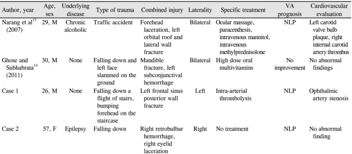
Table 1. Clinical characteristics of patients with CRAO after trauma
Author, year Age,sex
Underlying
disease Type of trauma Combined injury Laterality Specific treatment VA prognosis
Cardiovascular evaluation Narang et al13
(2007) 29, M Chronic
alcoholic Traffic accident Forehead laceration, left orbital roof and lateral wall fracture
Bilateral Ocular massage, paracenthesis, intravenous mannitol, intravenous methylprednisolone
NLP Left carotid valve bulb plaque, right internal carotid artery thrombus Ghose and
Subhabrata14 (2011)
30, M None Falling down and left face slammed on the ground
Mandible fracture, left subconjunctival hemorrhage
Bilateral High dose oral
multivitamins No
improvement No abnormal findings
Case 1 26, M None Falling down a
flight of stairs, bumping forehead on the staircase
Left frontal sinus posterior wall fracture
Left Intra-arterial thrombolysis
NLP Ophthalmic artery stenosis
Case 2 57, F Epilepsy Falling down Right retrobulbar hemorrhage, right eyelid laceration
Right No treatment NLP No abnormal
finding
CRAO = central retinal artery occlusion; VA = visual acuity; M = male; F = female; NLP = no light perception.
자기공명영상 및 자기공명혈관촬영술에서 뇌, 안와 및 혈 관의 이상소견은 관찰되지 않았다. 경식도 심초음파검사, 뇌혈류검사, 24시간 심전도검사에서 정상소견이었고 혈액 응고검사를 포함한 전반적인 혈액 검사에서 중성지방의 상 승(181 mg/dL) 이외에 특이소견은 없었다.
혈전용해술 치료 1일째 좌안 시력은 안전수동이었고 좌 안 형광안저혈관조영술에서 맥락막 충만, 망막동맥의 형광 충만 및 동정맥 통과시간은 정상이었으며(Fig. 1E, F) 빛간 섭단층촬영에서는 심한 황반부종이 관찰되었다. 혈전용해 술 치료 2일째 좌안 시력은 안전수동이었고 환자는 하루 전보다 주관적인 시력호전을 호소하였다. 1달 후 좌안 시력 은 광각유였으며 좌안 형광안저혈관조영술에서 황반부를 제외한 망막 순환은 정상이었다. 빛간섭단층촬영에서 좌안 망막의 얇아짐이 관찰되었으며 망막 전위도 검사에서 전반 적인 진폭의 감소가 관찰되었다. 치료 6개월 후 좌안 시력 은 광각무로 호전이 없었고 안저검사에서 시신경유두는 창 백하였다.
증례 2
57세 여자 환자가 내원 당일 등산하다가 넘어지면서 나 무에 우측 하안검을 찔린 후 갑자기 발생한 우안 시력저하 를 주소로 내원하였다. 환자는 간질약을 복용하고 있었고 8 년 전 좌안 익상편 제거술을 시행한 병력이 있었다. 내원 당시 시행한 최대교정시력은 우안 안전수동, 좌안 1.0이었 다. 골드만압평안압계로 측정한 안압은 우안 28 mmHg, 좌 안 14 mmHg였다. 우측 하안검의 부종이 관찰되었으며 우 측 하안검에 1 cm 길이의 열상이 관찰되었다. 안저검사에
서 우안의 망막은 창백하였으며 앵두반점이 관찰되었다 (Fig. 2A). 우안 형광안저혈관조영술에서 망막동맥의 형광 충만 및 망막 동정맥 통과시간이 지연되어 있었다(Fig. 2B).
우안 망막중심동맥폐쇄 진단하에 내원 당일 뇌 혈관조영술 을 시행하였고 우안 안동맥의 폐색은 관찰되지 않았다. 얼 굴 컴퓨터단층촬영에서 골절은 없었으나 우안의 구후출혈 이 관찰되었다(Fig. 2C). 혈액응고검사를 포함한 전반적인 혈액 검사에서 이상소견은 없었다.
수상 1일 후 우안 안압은 18 mmHg였고 빛간섭단층촬영 에서 우안 망막 내층의 부종이 관찰되었고 우안 형광안저 혈관조영술에서 망막동맥의 형광충만 및 동정맥 통과시간 이 지연되어 있었으나 수상 직후보다는 호전양상이었다.
수상 1달 후 우안 시력은 안전수동이었고 안저 검진에서 시신경유두는 창백하고 망막의 관류는 정상소견이었다. 수 상 6개월 후 우안 시력은 안전수동이었고 빛간섭단층촬영 에서 망막의 전반적인 얇아짐이 관찰되었다. 수상 1년 3개 월 후 우안 시력은 광각무였고 안저 검진에서 시신경 유두 는 창백하였으며(Fig. 2D) 빛간섭단층촬영에서 내층 망막 의 위축이 관찰되었다(Fig. 2E).
고 찰
대부분의 망막중심동맥폐쇄 환자에서는 그 원인을 색전 이나 혈전으로 생각하고 있다.1 드물게 혈관염도 망막중심 동맥폐쇄를 일으킬 수 있으며, 거대세포 동맥염이 망막중 심동맥폐쇄의 잘 알려진 원인이지만1 한국을 비롯한 아시 아 국가에서는 매우 드물다고 보고되었다.15 외상에 의한
망막중심동맥폐쇄는 국내에서는 아직 보고된 바가 없다.
저자들의 증례는 외상 이후 발생한, 통증이 없는 급격한 시력 장애를 호소한 환자들로 모두 응급실로 내원하여 안과 로 의뢰된 환자이다. 증례 1은 기저질환이 없던 환자이며 증 례 2는 간질을 앓았던 병력이 있었다. 두 증례의 수상원인은 추락이었다. 증례 1은 이마굴 뒷벽의 골절이 동반되었고 증 례 2는 동반된 골절은 없었고 구후출혈이 동반되었다. 국외 에서 발표되었던 Narang et al13, Ghose and Subhabrata14의 증례 특징과 저자들의 증례의 특징은 Table 1에 정리하였다.
외상에 의하여 안구가 직접 손상될 경우 혈관 내막 파열 에 의하여 내막하 조직이 혈류에 노출되면 응고다단계에 의한 혈소판의 응집 및 부착으로 혈전이 발생하여 동맥 폐 쇄가 발생할 수 있다.16 반면에 안구외 외상이 발생한 경우, 외상에 의하여 다른 장기에서 비롯된 색전이 원인이 되어 혈관폐쇄가 발생할 수 있다. 또한 외상에 대한 항상성의 반 응으로 촉진된 국소적인 동맥의 수축9도 망막중심동맥폐쇄 에 관여했을 것이다. 내경동맥에서 기인한 플라크나 심장 질환에 의해 발생한 혈전이 망막중심동맥폐쇄의 주요한 위 험인자로 알려져 있지만17 본 증례들에서는 전신검사에서 이런 위험인자는 관찰되지 않았다. 골절에 의하여 발생하 는 지방 색전 증후군도 혈관 폐쇄의 원인이 될 수 있다고 알려져 있다.18 증례 1은 동반되는 골절이 있었지만 지방 색 전 증후군이 대부분 황색 골수를 가지는 장골 골절에서 발 생하므로18 골절 부위는 본 증례와는 부합하지 않는다. 증 례 2의 경우 구후 출혈이 동반되었다. 구후 출혈에 의해 망 막중심동맥폐쇄가 발생하였다는 보고가 있었고6,19,20 구후 출혈에 의한 압박이 동맥 폐쇄에 기여했을 가능성이 있지 만 증례 2는 내원 당시 안압이 28 mmHg로 상승 폭이 크지 않아 이전에 보고되었던 증례들과는 차이가 있다.
망막중심동맥폐쇄의 위험인자로 고혈압, 당뇨, 경동맥질 환, 관상동맥질환, 일과성 허혈 발작, 흡연이 알려져 있다.21 두 증례 모두 망막중심동맥폐쇄와 연관이 있는 심혈관계 위험인자는 없었다. 증례 2는 간질의 병력이 있었으나 간질 과 망막중심동맥폐쇄와의 관계는 알려진 바가 없다.
두 증례에서 일반적인 망막중심동맥폐쇄와의 임상양상 및 진행경과에서 큰 차이점은 관찰할 수 없었다. 망막중심 동맥폐쇄의 빛간섭단층촬영에서 급성기에는 망막의 내층 및 외층의 두꺼워짐이 관찰되고 최종적으로는 망막의 위축 이 관찰된다.22 외상 후 발생한 망막중심동맥폐쇄 환자에서 도 비슷한 변화 양상을 관찰할 수 있었다. 그러나 망막중심 동맥폐쇄는 고령에서 호발한다고 알려져 있지만2 저자들이 겪었던 증례와 기존의 국외 보고 총 네 환자의 평균 나이는 35.5세, 저자들의 두 증례는 각각 26세, 57세로 비교적 젊 은 나이에 발생하였기 때문에 일반적인 망막중심동맥폐쇄
와는 차이가 있음을 추정할 수 있다.
결론적으로 외상 후 망막중심동맥폐쇄가 발생할 수 있음 을 염두에 두고, 외상 후 발생한 시력저하에 대한 감별진단 으로 망막중심동맥폐쇄를 고려해야 한다. 향후 더 많은 사 례를 모아 추가적인 연구가 필요할 것으로 생각한다.
REFERENCES
1) Hayreh SS, Zimmerman MB. Central retinal artery occlusion: vis- ual outcome. Am J Ophthalmol 2005;140:376-91.
2) Park SJ, Choi NK, Seo KH, et al. Nationwide incidence of clin- ically diagnosed central retinal artery occlusion in Korea, 2008 to 2011. Ophthalmology 2014;121:1933-8.
3) Lee JH, Moon HS, Nam DH, Lee DY. Treatment of acute central retinal artery occlusion with ocular ischemic syndrome. J Korean Ophthalmol Soc 2014;55:1242-7.
4) Kim KE, Ahn SJ, Woo SJ, et al. Central retinal artery occlusion caused by fat embolism following endoscopic sinus surgery. J Neuroophthalmol 2013;33:149-50.
5) Park SW, Woo SJ, Park KH, et al. Iatrogenic retinal artery occlu- sion caused by cosmetic facial filler injections. Am J Ophthalmol 2012;154:653-62.e1.
6) Jung EH, Park KH, Woo SJ. Iatrogenic central retinal artery occlu- sion following retrobulbar anesthesia for intraocular surgery.
Korean J Ophthalmol 2015;29:233-40.
7) Nakao Y, Terai H. Embolic brain infarction related to posttraumatic occlusion of vertebral artery resulting from cervical spine injury: a case report. J Med Case Rep 2014;8:344.
8) Tabarki B, el Madani A, Alvarez H, et al. Ischemic cerebral vas- cular accident caused by vertebral artery dissection. Arch Pediatr 1997;4:763-6.
9) Scheerlinck TA, Van den Brande P. Post-traumatic intima dis- section and thrombosis of the external iliac artery in sportsman.
Eur J Vasc Surg 1994;8:645-7.
10) Poon H, Patel A, Vijay S, Downing R. Endovascular repair for left common iliac artery occlusion following blunt trauma without as- sociated bony injury: image in vascular surgery. Vasc Endovascular Surg 2012;46:179-80.
11) Singla AA, McPherson D, Singla AA, et al. External iliac artery occlusion in a paediatric patient following handlebar trauma. J Surg Case Rep 2015;2015. pii: rjv015
12) Sodhi KS, Arora J, Khandelwal N. Post-traumatic occlusion of subclavian artery with clavicle fracture. J Emerg Med 2007;33:
419-20.
13) Narang S, Kochhar S, Gupta S, et al. Bilateral simultaneous central retinal artery occlusion following head injury. Int Ophthalmol 2007;27:387-90.
14) Ghose S, Subhabrata P. Bilateral central retinal arterial obstruction following head trauma: a very rare case report. Indian J Ophthalmol 2011;59:66-8.
15) Cha DM, Lee T, Choe G, et al. Silent giant cell arteritis in an elderly Korean woman. Korean J Ophthalmol 2013;27:224-7.
16) Dalma-Weiszhausz J, Meza-de Regil A, Martinez-Jardón S, Oliver-Fernández K. Retinal vascular occlusion following ocular contusion. Graefes Arch Clin Exp Ophthalmol 2005;243:406-9.
= 국문초록 =
외상에 의한 망막중심동맥폐쇄 2예
목적: 외상에 의하여 발생한 망막중심동맥폐쇄 2예를 경험하였기에 이를 보고하고자 한다.
증례요약: 26세 남자 환자가 계단에서 넘어져 좌측 이마 주위를 계단에 부딪힌 후 발생한 좌안의 급격한 시력저하를 주소로 내원하였 다. 좌안의 시력은 광각유였다. 망막은 창백하였으며 망막 부종 및 앵두반점이 관찰되었고 혈관조영술에서 좌안 안동맥 기시부 근처 에 심한 협착 및 색전이 관찰되어 안동맥폐쇄가 동반된 망막중심동맥폐쇄로 진단하에 동맥내 혈전용해술을 시행하였다. 57세 여자 환자가 넘어지면서 우측 하안검을 나뭇가지에 찔린 후 발생한 급격한 우안의 시력저하를 주소로 내원하였다. 우안의 시력은 안전수동 이었다. 안저검사에서 망막은 창백하고 혼탁하였으며 앵두반점이 관찰되어 형광안저혈관조영술 및 빛간섭단층촬영 시행 후 망막중심 동맥폐쇄로 진단하였다.
결론: 외상 후 발생한 시력저하에 대한 감별진단으로 망막중심동맥폐쇄를 고려해야 한다.
<대한안과학회지 2016;57(2):324-329>
17) Schmidt D, Hetzel A, Geibel-Zehender A, Schulte-Mönting J.
Systemic diseases in non-inflammatory branch and central retinal artery occlusion--an overview of 416 patients. Eur J Med Res 2007;12:595-603.
18) Stein PD, Yaekoub AY, Matta F, Kleerekoper M. Fat embolism syndrome. Am J Med Sci 2008;336:472-7.
19) Goldsmith MO. Occlusion of the central retinal artery following retrobulbar hemorrhage. Ophthalmologica 1967;153:191-6.
20) Moshfeghi DM, Lowder CY, Roth DB, Kaiser PK. Retinal and
choroidal vascular occlusion after posterior sub-tenon tri- amcinolone injection. Am J Ophthalmol 2002;134:132-4.
21) Varma DD, Cugati S, Lee AW, Chen CS. A review of central retinal artery occlusion: clinical presentation and management. Eye (Lond) 2013;27:688-97.
22) Ahn SJ, Woo SJ, Park KH, et al. Retinal and choroidal changes and visual outcome in central retinal artery occlusion: an optical coher- ence tomography study. Am J Ophthalmol 2015;159:667-76.