478
대한안과학회지 2017년 제 58 권 제 4 호 J Korean Ophthalmol Soc 2017;58(4):478-481 ISSN 0378-6471 (Print)⋅ISSN 2092-9374 (Online)
https://doi.org/10.3341/jkos.2017.58.4.478
Case Report
심장 점액종에 의한 중심망막동맥폐쇄 1예
A Case Report of Central Retinal Artery Occlusion Caused by Cardiac Myxoma
김준오1⋅최인호2⋅최경식1
Juno Kim, MD1, In Ho Choi, MD2, Kyung Seek Choi, MD1
순천향대학교 의과대학 서울병원 안과학교실1, 순천향대학교 의과대학 서울병원 병리학교실2
Department of Ophthalmology, Soonchunhyang University Seoul Hospital, Soonchunhyang University College of Medicine1, Seoul, Korea Department of Pathology, Soonchunhyang University Seoul Hospital, Soonchunhyang University College of Medicine2, Seoul, Korea
Purpose: We report the case of a patient diagnosed with central retinal artery occlusion caused by cardiac myxoma who under- went surgery to remove the myxoma.
Case summary: A 47-year-old woman came to our clinic presenting with a sudden decrease of visual acuity in the left eye. At the first visit, left eye visual acuity was hand motion, and intraocular pressure was 15.4 mmHg. A relative afferent pupillary defect was observed in the left eye. On fundus examination, a pale retina and cherry-red spot were observed at the posterior pole. On optical coherence tomography, macular edema was found. On fluorescein angiography and indocyanine green angiography, de- layed blood circulation of the retina and choroid was found at early and late stages. Cerebral angiography was performed in the neurosurgery department and showed no occlusion of the ophthalmic artery. Cardiac ultrasonography and brain magnetic reso- nance imaging were performed. On cardiac ultrasonography, 4.46 × 2.09 cm cardiac myxoma was found. Resection of the car- diac myxoma was conducted in the thoracic and cardiovascular surgery department. Multiple cerebral infarcts were detected by brain imaging, and antithrombotic treatment was administered. After one month, blood circulation in the retina and choroid was observed in fluorescence angiography, but there was no improvement of visual acuity. At the 3-month follow-up visit, macular edema was decreased, but retinal atrophy and epiretinal membrane were observed on optical coherence tomography.
Conclusions: Central retinal artery occlusion is a disease related to one’s general condition. We experienced this case of central retinal artery occlusion caused by cardiac myxoma.
J Korean Ophthalmol Soc 2017;58(4):478-481
Keywords: Cardiac myxoma, Central retinal artery occlusion, Retina
■Received: 2016. 12. 8. ■ Revised: 2017. 1. 20.
■Accepted: 2017. 3. 18.
■Address reprint requests to Kyung Seek Choi, MD Department of Ophthalmology, Soonchunhyang University Seoul Hospital, #59 Daesagwan-ro, Yongsan-gu, Seoul 04401, Korea
Tel: 82-2-709-9114, Fax: 82-2-710-3196 E-mail: [email protected]
ⓒ2017 The Korean Ophthalmological Society
This is an Open Access article distributed under the terms of the Creative Commons Attribution Non-Commercial License (http://creativecommons.org/licenses/by-nc/3.0/) which permits unrestricted non-commercial use, distribution, and reproduction in any medium, provided the original work is properly cited.
중심망막동맥폐쇄는 망막혈관 질환 중 가장 치명적인 질 환이며 뇌졸중, 심근경색, 바이러스 감염 및 심장 종양 등 전신질환과 연관이 있다고 알려져 있다.1 이 중 심장점액종 은 성인에게서 심장에 발생하는 원발성 종양 가운데 가장
흔한 종양이다.2 중심망막동맥 폐쇄를 일으킬 수 있는 다양 한 질병 중 심장점액종에 의한 중심망막동맥폐쇄는 아직까 지 국내에 보고된 바가 없다. 저자들은 갑작스럽게 시력이 저하된 환자에서 중심망막동맥 폐쇄 진단 후, 전신검사에 서 심장점액종을 발견한 증례를 보고하고자 한다.
증례보고
47세 여자 환자가 갑작스럽게 시작된 좌안의 시력저하 주소로 안과에 내원하였다. 현성굴절 검사를 통한 최대교 정시력 우안 1.0, 좌안 안전수동으로 확인되었으며 좌측 편
479 - 김준오 외 : 심장 점액종에 인한 중심망막동맥폐쇄 1예 -
A B
C D
Figure 1. Patient case. (A) Fundus photography at first visit. Ischemic change of the posterior pole and cherry-red spot were observed. (B) Optical coherence tomography showing edematous retina. (C, D) Fluorescein angiography and Indocyanine green an- giography image at first visit. (C) At the early phase of Fluorescein/Indocyanine green angiography (FAG/ICG), we could find de- layed arm to retina time (24 seconds) and delayed choroidal circulation. (D) At the late phase of FAG/ICG (7:40), hypofluorescent lesions are observed in the superior, inferior, and temporal area of the optic disc including fovea.
A B
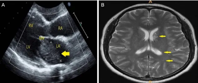
Figure 2. Echocardiographic and brain magnetic resonance imaging findings. (A) Transthoracic echocardiographic finding. Mass with irregular margin in the left atrium (in subcostal view, arrow). (B) Brain magnetic resonance T2 imaging. Multiple micro- infarctions (more than 15) were found (arrows). LA = left atrium; LV = left ventricle; RA = right atrium; RV = right ventricle;
MV = mitral valve.
두통을 호소하였다. 과거력상 전신질환은 없었으며, 안과적 수술 및 과거력도 없었다. 수축기 및 이완기 혈압 110/64 mmHg, 맥박 56회/분으로 생체징후는 안정적이었다. 좌안 에 상대적 구심동공운동장애가 관찰되었고, 안저검사에서 좌안의 창백안저 및 황반부의 앵두반점이 관찰되었으며 (Fig. 1A), 빛간섭단층촬영검사상 황반부의 부종이 관찰되 었다(Fig. 1B). 형광안저혈관조영술상 초기에 팔망막순환시 간의 지연 및 맥락막 및 망막 혈관의 혈류 충만 장애가 관
찰되었으며, 후기에도 망막 및 맥락막의 혈류 충만 장애가 지속되었다(Fig. 1C, D). 좌안의 중심망막동맥 폐쇄로 망막 및 맥락막으로의 혈류유입 장애의 원인을 찾기 위해 신경 외과에서 뇌혈관조영술을 시행하였고, 뇌혈관조영술에서 안동맥은 정상적으로 조영되었으며 혈관폐쇄는 관찰되지 않았다. 치료로 방수생성억제제 점안 및 안구마사지를 시 행하였으며, 혈전용해제의 전신적 투여 및 만니톨 투여, 비강을 통한 2 L 산소투여를 시행하였다. 전신혈액검사와
480
- 대한안과학회지 2017년 제 58 권 제 4 호 -
A B
C D
Figure 3. Gross and pathologic slide pictures of resected myxoma. (A) Grossly, tumor mass showed heterogeneouslymyxoid appear- ance with focal hem orrhage. (B) Tum or cells consisted of hypocellular, fibrom yxoid or loose myxoid strom a (Hem atoxylin and eosin stain [HE] stain, × 40). (C) Tumor cells were spindled or stellated with occasional syncytia and had indistinct cell borders and hyperchromatic nuclei (HE stain, ×400). (D) Tumor cells showed positivity for CD34 (CD34, ×100).
A B
Figure 4. Fluorescein angiography/Indocyanine green angiography (FAG/ICG) image (1 month later from the first visit). Optical co- herence tomography (3 months later from the first visit). (A) At the early phase of FAG/ICG, we could find recovered arm to retina time (18 seconds) and recovered choroidal circulation. (B) Optical coherence tomography shows retinal edema and epiretinal membrane.
전신적 원인을 찾기 위해 심장내과와 신경과에 의뢰하였 고, 심장내과에서 시행한 심장초음파 검사에서 좌심방에 4.46 × 2.09 cm의 심장점액종이 발견되었으며(Fig. 2A), 뇌 자기공명영상검사에서 급성 뇌경색이 진단되었다(Fig. 2B).
신경과에서 급성기 뇌경색 치료를 위해 전신적 혈전용해제 를 사용하였고, 흉부외과에서 좌심방심장점액종 절제술을 시행하였으며, 조직검사에서 심장점액종으로 확진되었다 (Fig. 3). 1개월째 시행한 형광안저혈관조영술상 망막의 혈 액순환이 호전되었으나(Fig. 4A), 좌안의 시력은 안전수동 으로 호전은 없었고, 3개월째 시행한 빛간섭단층촬영상 황 반부망막의 위축과 망막전막이 관찰되었다(Fig. 4B).
고 찰
중심망막동맥폐쇄는 망막의 급성허혈로 인한 망막말단 동맥의 폐쇄이며, 이는 심각한 시력 손상으로 이어질 수 있 다. 중심망막동맥폐쇄는 대부분 고혈압이나, 심질환, 당뇨 및 경동맥 협착 등의 기저질환을 가진 고령자에게서 발생 한다고 알려져있다.3 심장 점액종은 드물지만 가장 흔한 심 장종양이며 이로 인한 세 가지 증상으로는 심장과 직접 관 련된 폐쇄로 인한 경우, 색전에 의한 경우 그리고 발열이나 빈혈 등의 전신 증상이 있다. 주로 좌심방에 위치하며 가장 흔한 증상은 폐쇄성 호흡곤란으로 국내 연구에 따르면 대 개 심장과 관련된 증상이 60%를 차지하며 색전의 경우 22% 정도라고 한다. 심장초음파로 진단할 수 있으며 자기
481
= 국문초록 =
심장 점액종에 의한 중심망막동맥폐쇄 1예
목적: 중심망막동맥폐쇄 진단 후에 심장점액종이 발견되어 흉부외과에서 수술적 제거 후 경과관찰 중인 1예를 보고하고자 한다.
증례요약: 47세 여자 환자가 갑작스럽게 발생한 좌안의 시력저하를 주소로 안과에 내원하였다. 내원 당시 좌안 시력 안전수동, 안압 15.4 mmHg 측정되었으며, 좌안에 상대적 구심동공운동 장애가 관찰되었다. 안저검사에서 앵두반점 및 창백안저가 관찰되었으며, 빛 간섭단층촬영검사상 황반부의 부종이 관찰되었다. 형광안저혈관조영에서 초기 및 후기에 맥락막 및 망막 혈관의 혈류 충만 장애 소견 이 관찰되었고, 이를 바탕으로 좌안의 중심망막동맥폐쇄로 진단하였다. 신경외과에서 시행한 뇌혈관조영술에서 안동맥은 정상적으로 조영되었고, 혈관폐쇄는 관찰되지 않았다. 동맥폐쇄의 원인을 찾기 위해 심장내과와 신경과에 의뢰하였고, 심장초음파검사에서 좌심 방에 4.46 × 2.09 cm의 심장점액종이 발견되었으며, 뇌 자기공명영상검사 후 급성 뇌경색을 진단하였다. 보존적 치료를 시행하며, 급성기 뇌경색 치료로 전신적 혈전 용해제를 사용하였고 흉부외과에서 좌심방점액종 절제술을 시행하였다. 심장점액종 절제술 시행 후 1개월째 시행한 형광안저혈관조영술상 망막의 혈액순환이 호전되었으나 시력은 개선되지 않았고, 3개월째 시행한 빛간섭단층촬영 에서 황반부 부종은 감소했으나 망막층의 위축과 망막전막이 관찰되었다.
결론: 중심망막동맥폐쇄는 눈뿐만 아니라 전신적 질환을 함께 고려해야 하는 질병이다. 고혈압이나 심장질환, 당뇨 및 경동맥협착 등의 다양한 기저질환이 중심망막동맥폐쇄의 원인으로 알려져 있으며, 저자들은 심장점액종에 의해 망막중심동맥이 폐쇄된 증례를 경험하였기에 이를 보고하고자 한다.
<대한안과학회지 2017;58(4):478-481>
- 김준오 외 : 심장 점액종에 인한 중심망막동맥폐쇄 1예 -
공명영상 등도 진단에 유용할 수 있다.4 심장점액종에 의한 안구의 혈관 폐쇄는 흔하지 않으나 심장점액종에 인해 발 생한 색전에 의한 안구 혈관 장애의 보고가 있다.5 1990년 이후 문헌에서 심장점액종에 의한 망막동맥 폐쇄의 증례는 거의 보고되지 않았으며, 국내에서의 증례 보고도 아직까 지 없었다. 심장점액종은 기계적으로 혈류의 장애를 일으 키고 응고 인자를 활성화하여 색전을 형성하기도 하고, 심 장점액종의 일부가 떨어져 나가 색전을 형성하기도 한다.6 심장점액종 환자의 20-45%에서 색전으로 인한 증상이 나 타나며, 색전의 절반이상이 뇌혈관을 막아 뇌경색을 발생 시키는 원인으로 알려져 있으며 초기에 혈전용해제 등의 치료를 적절하게 시행하지 못할 경우 예후가 좋지 않다고 한다. 그 외에도 눈, 폐, 신장 등의 장기와 사지로 가는 혈 관 등을 막을 수 있다고 알려져있다.7 심장점액종의 치료는 흉부외과의 수술적 제거이며 재발이나 수술 후 합병증이 거의 없으며 심방세동이 가장 흔한 수술 후 합병증이다.4 본 증례의 경우 망막동맥폐쇄의 원인으로 심초음파검사에 서 심장점액종이 발견되었고 급성 뇌경색이 동반되었음에 도 두통 이외 특이증상이 없었지만 뇌자기공명영상으로 다 발성의 뇌경색을 발견할 수 있었고 종양을 제거한 후 전신 적 상태는 호전되었으나 시력회복은 없었다. 본 저자들은 중심망막동맥폐쇄에 의한 시력저하의 원인으로 심장점액 종이 동반된 환자를 경험하고 진단하였으며, 국내 첫 증례
로 보고하는 바이다. 결론적으로 심장점액종이 망막동맥폐 쇄를 일으킬 수 있는 원인질환임을 알고, 관련된 문진과 검 사, 심장초음파 및 뇌 자기공명영상 등의 검사들이 필요하 며 심장점액종 진단 후 색전의 원인이 되는 종양의 수술적 절제가 필요하겠다.
REFERENCES
1) Woo SC, Lip GY, Lip PL. Associations of retinal artery occlusion and retinal vein occlusion to mortality, stroke, and myocardial in- farction: a systematic review. Eye (Lond) 2016;30:1031-8.
2) Zheng JJ, Geng XG, Wang HC, et al. Clinical and histopathological analysis of 66 cases with cardiac myxoma. Asian Pac J Cancer Prev 2013;14:1743-6.
3) Varma DD, Cugati S, Lee AW, Chen CS. A review of central retinal artery occlusion: clinical presentation and management. Eye (Lond) 2013;27:688-97.
4) Lee SJ, Kim JH, Na CY, Oh SS. Eleven years’ experience with Korean cardiac myxoma patients: focus on embolic complications.
Cerebrovasc Dis 2012;33:471-9.
5) Schmidt D, Hetzel A, Geibel-Zehender A. Retinal arterial occlu- sion due to embolism of suspected cardiac tumors - report on two patients and review of the topic. Eur J Med Res 2005;10:296-304.
6) Yuan SM, Humuruola G. Stroke of a cardiac myxoma origin. Rev Bras Cir Cardiovasc 2015;30:225-34.
7) Bayir H, Morelli PJ, Smith TH, Biancaniello TA. A left atrial myx- oma presenting as a cerebrovascular accident. Pediatr Neurol 1999;21:569-72.