Neovascular Glaucoma Due to Branch Retinal Vein Occlusion Combined with Branch Retinal Artery Occlusion
4
0
0
전체 글
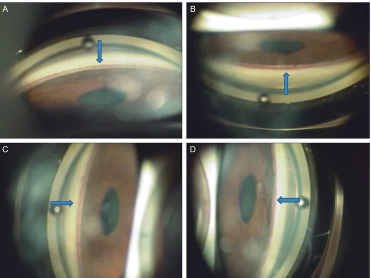
(2) TS An, et al. NVG Due to BRVO Combined with BRAO. A. B. C. D. Fig. 1. Gonioscopic examination at initial examination showed 360 degree angle neovascularization (NVA) of the right eye. Arrows indicate NVA. (A) The gonioscopy revealed inferior NVA. (B) The gonioscopy revealed superior NVA. (C) The gonioscopy revealed nasal NVA. (D) The gonioscopy revealed temporal NVA.. A. B. Fig. 2. (A) In right eye, fundus examination showed scat tered retinal hemorrhage along the inferotemporal vein and ischemic edema in the inferior parafoveal area which was supplied by the small branches of the inferior retinal artery with atheroma at initial examination. (B) In left eye, fundus examination revealed a single peripapillary flame hemor rhage temporally and narrowing of the arterial vessels at initial examination.. 65.
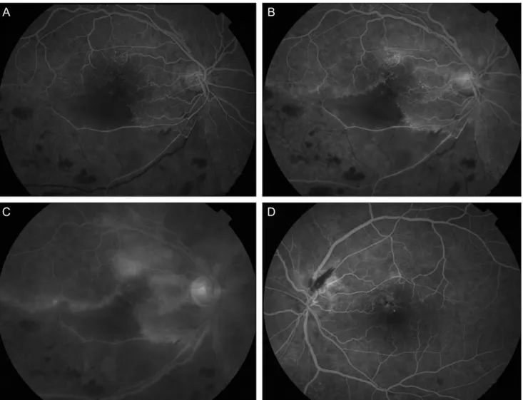
(3) Korean J Ophthalmol Vol.27, No.1, 2013. A. B. C. D. Fig. 3. Fluorescein angiography (FA) at initial visit. (A) In right eye, FA of the right eye showed significant delayed filling of the branches of the inferior retinal artery in the ischemic area (30 seconds). (B) The foveal avascular zone was widened and the superior border was irregular with moderate leakage of dye from the arterioles (70 seconds). (C) A wide area of capillary nonperfusion in the distribution of the inferotemporal vein was also noticed, but choroidal perfu sion was normal in the right eye (10 minutes). (D) In left eye, arteriolar tortuosity and moderate leakage was found near the flame hemorrhage (70 seconds).. and ischemic edema in the inferior parafoveal area which was supplied by the small branches of the inferior retinal artery with atheroma (Fig. 2). Fluorescein angiography (FA) of the right eye showed significant delayed filling of the branches of the inferior retinal artery in the ischemic area. The foveal avascular zone was widened and the superior border was irregular with moderate leakage of dye from the arterioles. A wide area of capillary nonperfusion in the distribution of the inferotemporal vein was also noticed, but choroidal perfusion was normal in the right eye. In her left eye, arteriolar tortuosity and moderate leakage was found near the flame hemorrhage (Fig. 3). FA was consistent with BRAO combined with BRVO in her right eye and the impending state in her left eye. Carotid Doppler sonography and echocardiogram 66. showed no evidence of systemic conditions associated with multiple emboli and thrombosis. Her laboratory data including lipid profile, blood coagulation test, and serum homocystein were normal except for blood glucose. We immediately injected intravitreal and intracameral bevacizumab (0.4 mg/0.05 mL) in her right eye. The next day, we performed scatter photocoagulation in the nonperfusion area. One week after the injection, the NVI and NVA had regressed, and the IOP was 12 mmHg with topical antiglaucoma medication (dorzolamide/timolol fixed combination). One month later, visual acuity returned to 20 / 20 in her right eye and the IOP was 17 mmHg with topical antiglaucoma medication (dorzolamide/timolol fixed combination)..
(4) TS An, et al. NVG Due to BRVO Combined with BRAO. Discussion NVI and the subsequent development of NVG are serious complications seen in patients with ischemic retinal disorders such as diabetic retinopathy, central retinal vein occlusion and central retinal artery occlusion [2,3]. Hayreh et al. [4] have attributed NVI and NVG associated with CRAO to underlying atherosclerotic carotid artery disease and also reported that not one of the 44 eyes in their study with BRAO showed NVI or NVG. In the study by Hayreh et al. [4] on ocular neovascularization associated with retinal vein occlusion, none of the 264 eyes with BRVO developed NVG. The studies have indicated that it usually requires at least half or more of the retina to have ischemic involvement to provide an adequate neovascular stimulus [4,5]. Thus, there is little risk of NVG following BRVO or BRAO. In our case, two rare causes of NVG (BRVO and BRAO) simultaneously developed to cause NVG. We assumed that the angiogenic factors produced by the different lesions interacted synergistically and reached a level sufficient for angiogenic stimuli to develop NVG. In addition to this, an unrevealed underlying disease might have enhanced the angiogenic stimuli. De Salvo et al. [6] reported that when retinal artery and vein occlusion occurred in the same eye, a systemic disorder should be suspected. In our patient, FA revealed multiple arteriolar obstructions in both eyes, which suggest that the embolic and thrombotic event could be systemic or disseminated emboli. Recent multiple brain infarctions could be more evidence of systemic embolism, even though we could not find an underlying disorder.. As observed in our case, ophthalmologists should be aware that NVI and NVG may occur as possible complications of BRAO combined with BRVO, and patients should be followed up carefully with repeated slit-lamp examinations and undilated gonioscopy.. Conflict of Interest No potential conflict of interest relevant to this article was reported.. References 1. Hayreh SS. Neovascular glaucoma. Prog Retin Eye Res 2007;26:470-85. 2. Shazly TA, Latina MA. Neovascular glaucoma: etiology, diagnosis and prognosis. Semin Ophthalmol 2009;24:113-21. 3. Sivak-Callcott JA, O’Day DM, Gass JD, Tsai JC. Evidencebased recommendations for the diagnosis and treatment of neovascular glaucoma. Ophthalmology 2001;108:1767-76. 4. Hayreh SS, Rojas P, Podhajsky P, et al. Ocular neovascularization with retinal vascular occlusion-III. Incidence of ocular neovascularization with retinal vein occlusion. Ophthalmology 1983;90:488-506. 5. Hayreh SS, Podhajsky P. Ocular neovascularization with retinal vascular occlusion. II. Occurrence in central and branch retinal artery occlusion. Arch Ophthalmol 1982;100:1585-96. 6. De Salvo G, Li Calzi C, Anastasi M, Lodato G. Branch retinal vein occlusion followed by central retinal artery occlusion in Churg-Strauss syndrome: unusual ocular manifestations in allergic granulomatous angiitis. Eur J Ophthalmol 2009;19:314-7.. 67.
(5)
수치
관련 문서
Purpose: To compare the intraocular pressure (IOP) in diabetic macular edema (DME) patients and macular edema associated with branch retinal vein occlusion (BRVO) patients
Purpose: To evaluate the factors associated with refractory macular edema (ME) secondary to branch retinal vein occlusion (BRVO) after three times of intravitreal bevacizumab
Predictive Factors for a Favorable Response to Intravitreal Bevacizumab for Macular Edema Associated with Branch Retinal Vein Occlusion.. 강현구 1 , 서유리 2 , 최은영 1 ,
Purpose: To compare the difference of efficacy between intravitreal dexamethasone implant and bevacizumab injection for patients with branch retinal vein occlusion (BRVO) with
