179
Korean J Ophthalmol 2010;24(3):179-181
DOI: 10.3341/kjo.2010.24.3.179
pISSN: 1011-8942 eISSN: 2092-9382
Case Report
Progression of Impending Central Retinal Vein Occlusion to the Ischemic Variant Following Intravitreal Bevacizumab
Na Rae Kim, Hee Seung Chin
Department of Ophthalmology, Inha University School of Medicine, Incheon, Korea
A 60-year-old woman who had experienced two episodes of amaurosis fugax in her right eye presented with vision loss. Two weeks earlier, at a private clinic, she was diagnosed with impending central retinal vein occlusion (CRVO) of the right eye and received an intravitreal injection of bevacizumab. Two weeks after this injection she was diag- nosed with ischemic CRVO. At 11-weeks post-presentation, extremely ischemic features were observed with fluo- rescein angiographic findings of severe vascular attenuation and extensive retinal capillary obliteration. At 22-weeks post-presentation she was diagnosed with neovascular glaucoma; she experienced no visual improve- ment over the following several months.
Key Words: Bevacizumab, Central retinal vein occlusion
ⓒ2010 The Korean Ophthalmological Society
This is an Open Access article distributed under the terms of the Creative Commons Attribution Non-Commercial License (http://creativecommons.org/licenses
/by-nc/3.0/) which permits unrestricted non-commercial use, distribution, and reproduction in any medium, provided the original work is properly cited.
Received: October 11, 2008 Accepted: May 3, 2010
Reprint requests to Hee Seung Chin. Department of Ophthalmology, Inha
University Hospital, #7-206, Shinheung-dong, Jung-gu, Incheon 400-700,
Korea. Tel: 82-32-890-2400, Fax: 82-32-890-2403, E-mail: [email protected]
We report a case of ischemic central retinal vein occlusion (CRVO) in a patient who initially had impending CRVO and who was subsequently treated with intravitreal bevacizumab.
Case Report
A 60-year-old woman with a history of normal tension glaucoma presented because of vision loss in her right eye. Four weeks prior to her first visit to our hospital she had noted a sudden blurring over the entire visual field of her right eye. This lasted for approximately ten minutes and then fully recovered; eight days later, she experienced exactly the same phenomenon. Her previous clinical his- tory was only significant for chronic hepatitis B. She did not have hypertension, diabetes mellitus, or hyperlipidemia.
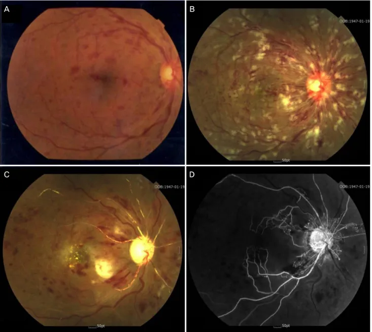
She visited a private clinic on November 1, 2006. On examination, mild dilatation and tortuosity of the retinal veins, some intraretinal hemorrhages at the posterior pole, and a slightly swollen optic disc with small hemorrhages at the disc margin were found in her right eye (Fig. 1A).
Her best corrected visual acuity (BCVA) was 20/20, and her intraocular pressure (IOP) was 12 mmHg. Fluorescein angiography (FAG) revealed normal choroidal filling and
slightly delayed filling of the central retinal vein without capillary nonperfusion. The left eye was normal. She was diagnosed with impending CRVO and underwent a trial of intravitreal bevacizumab (2.5 mg in 0.1 mL) in an at- tempt to improve the vascular stasis.
She was referred to our hospital two weeks after the bevacizumab injection, on November 13. Her BCVA was 20/80, and the IOP was normal in her right eye. Fundus examination revealed numerous retinal hemorrhages in four quadrants, dilated and tortuous retinal veins, and severe disc swelling. Newly developed macular edema and cotton- wool spots were observed (Fig. 1B). FAG revealed normal choroidal circulation, a marked delay in arteriovenous transit time, and extensive areas of capillary nonperfusion.
Optical coherence tomography revealed increased retinal thickness in the macular area. Electroretinography revealed decreased b-wave amplitude, but a normal a-wave. We diagnosed ischemic CRVO and continued to observe the patient without additional treatment. Laboratory data were unremarkable, including hemoglobin, platelet count, white cell count, electrolytes, and lipid profile. Additional ex- aminations were subsequently performed; inflammatory markers and plasma viscosity were unremarkable.
At three-weeks post-presentation, on November 20, the patient noted a significant decrease in visual acuity;
her BCVA had declined to counting fingers. A slit lamp
examination showed a relative afferent pupillary defect. At
six-weeks post-presentation, on December 15, her BCVA
was unchanged and her IOP was 14 mmHg. A fundus ex-