ISSN 0378-6471 (Print)⋅ISSN 2092-9374 (Online)
https://doi.org/10.3341/jkos.2018.59.9.893
Case Report
외상 이후 발생한 안와첨증후군 2예
Two Cases of Orbital Apex Syndrome after Blunt Orbital Trauma
양상철1⋅최희영1,2⋅전혜신1,2
Sang Cheol Yang, MD1, Hee Young Choi, MD, PhD1,2, Hyeshin Jeon, MD1,2
부산대학교 의과대학 안과학교실1, 부산대학교병원 의생명연구원2
Department of Ophthalmology, Pusan National University School of Medicine1, Yangsan, Korea Biomedical Research Institute, Pusan National University Hospital2, Busan, Korea
Purpose: To report two cases of orbital apex syndrome caused by blunt orbital trauma without structural damage of the orbit.
Case summary: (Case 1) A 50-year-old male came to our clinic complaining of visual loss after blunt orbital trauma by a metal bar. The best-corrected visual acuity was no light perception and light reflex was not observed in the affected eye. He also pre- sented with complete ptosis and ophthalmoplegia with relative sparing of adduction and depression. High signal intensity of the orbital soft tissue including the optic nerve sheath was revealed using a T2-weighted image in magnetic resonance imaging.
After starting steroid pulse therapy, his visual acuity improved to counting fingers on the third day. Ocular movement and levator function recovered to the normal range while visual acuity remained counting fingers. (Case 2) A 64-year-old female presented with complete ptosis after trauma to her right eyeball. The best-corrected visual acuity was 20/25 in the right eye. Complete pto- sis and ophthalmoplegia with relative sparing of abduction and depression in the right eye were observed at the initial presentation. Magnetic resonance images showed enhancement of the right periphery optic nerve and distal rectus muscle. Two months after undergoing steroid pulse therapy, levator function and ocular movement recovered completely, and visual acuity improved to 20/20.
Conclusions: The orbital apex syndrome caused by blunt orbital trauma showed good response to steroid pulse therapy. Steroid treatments may therefore be considered for the treatment of traumatic orbital apex syndrome.
J Korean Ophthalmol Soc 2018;59(9):893-898 Keywords: Blunt orbital trauma, Orbital apex syndrome
■Received: 2018. 4. 5. ■ Revised: 2018. 6. 8.
■Accepted: 2018. 8. 26.
■Address reprint requests to Hyeshin Jeon, MD
Department of Ophthalmology, Pusan National University Hospital, #179 Gudeok-ro, Seo-gu, Busan 49241, Korea Tel: 82-51-240-7326, Fax: 82-51-240-7341
E-mail: [email protected]
* This work was supported by clinical research grant from Pusan National University hospital 2017.
*Conflicts of Interest: The authors have no conflicts to disclose.
ⓒ2018 The Korean Ophthalmological Society
This is an Open Access article distributed under the terms of the Creative Commons Attribution Non-Commercial License (http://creativecommons.org/licenses/by-nc/3.0/) which permits unrestricted non-commercial use, distribution, and reproduction in any medium, provided the original work is properly cited.
안와첨증후군(orbital apex syndrome)은 안와첨의 병터로 인해 시신경의 기능소실과 함께 3, 4, 5, 6번 뇌신경의 눈
분지의 손상을 일으킬 수 있는 질환이다. 해부학적 위치에 따라 세 가지 질병군을 포함하고 있는데, 이는 상안와열증 후군(superior orbital fissure syndrome), 안와첨증후군, 그리 고 해면정맥굴증후군(cavernous sinus syndrome)이다.1 상 안와열증후군의 경우 시신경병증 없이 3, 4, 5, 6번 뇌신경 의 눈분지를 침범하는 양상을 보일 수 있고,2 해면정맥굴증 후군은 시신경병증 없이 5번 뇌신경의 위턱분지 및 교감신 경에 영향을 줄 수 있다.3
안와첨증후군은 염증, 감염, 종양, 혈관질환 및 외상이 원 인이 되어서 발생할 수 있다.1,4-6 이 중 외상에 의한 안와첨 증후군의 경우 안와 관통상, 안면부 골절을 동반하는 경우 가 많은데,7,8 이때의 기능 소실은 안와 조직의 직접적인 손
A B
Figure 1. Nine diagnostic position of gaze field in case 1. Complete ptosis and ophthalmoplegia with partial sparing of adduction and
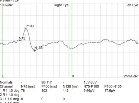
depression in left eye was observed at initial presentation (A). At 5 months after treatment, ptosis and ophthalmoplegia were com- pletely recovered (B).Figure 2. Pattern visual evoked potential (VEP) of Case 1. Pattern VEP showed low amplitude in the affected left eye.
상, 또는 이차적인 염증 혹은 압박으로 인한 간접적 손상에 의해서도 발생할 수 있다.9 국내에서는 안와의 종양 침범에 의한 경우,10 안와 관통상과 연관하여 발생한 경우가 보고 된 바 있다.11,12 그러나 안와 내부 구조물의 직접적인 손상 이 없는 안구의 둔상 이후 발생한 안와첨증후군은 드문 질 환으로 이의 치료 과정을 2예 경험하였기에 이를 보고하고 자 한다.
증례보고
증례 1
50세 남자가 쇠봉에 좌안을 부딪친 후 발생한 좌안의 시 력 저하를 주소로 내원하였다. 좌측 안면부의 감각저하가 동반되어 있었다. 위암 수술을 시행받은 병력 외에는 다른
병력은 없었다. 교정시력은 우안 20/20, 좌안 광각불인지였 으며, 좌안의 대광반사가 소실되어 있었고 상대구심동공반 사 양성이었다. 좌안의 완전 안검하수를 보였으며 약간의 내전 및 하전을 제외한 전방향 안구운동제한을 보였다(Fig.
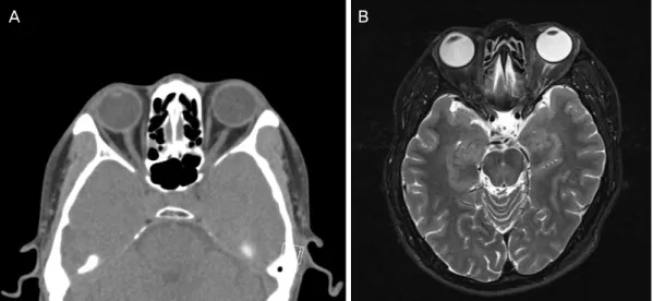
1A). 안구돌출계검사에서 좌안이 4 mm 더 돌출되어 있었 다. 세극등현미경검사에서 좌안의 결막 충혈 및 부종이 발 견되었고, 외상으로 인한 것으로 생각되는 추가적인 이상 소견은 발견되지 않았으며, 안저검사에서도 특이 소견은 관찰되지 않았다. 시유발전위 검사에서 좌안의 진폭이 감 소되어 있었다(Fig. 2). 수상 2일 후 촬영한 안와 전산화단 층촬영 검사에서 안와골절 및 구후출혈 소견은 관찰되지 않았다(Fig. 3A). 또한 안와 자기공명영상의 T2 강조영상에 서 좌측 시신경 및 시신경 주위 조직을 포함한 근원뿔 내 고음영이 관찰되었다(Fig. 3B). 외상으로 인한 안와첨증후
A B
Figure 3. Computed tomography (CT) and magnetic resonance image (MRI) of the case 1 patient. Axial (A) CT scan showed no sign
of orbital wall fracture and retrobulbar hemorrhage. Axial (B) T2-weighted image of magnetic resonance scan presented high signal intensity of the intraconal space including left optic nerve and soft tissue.A B
Figure 4. Nine diagnostic position of gaze field in case 2. Complete ptosis and ophthalmoplegia with partial sparing of abduction and
depression in right eye was observed at initial presentation (A). Ptosis and ophthalmoplegia recovered completely at 2 months after treatment (B).군으로 진단하고 예방적 항생제 및 스테로이드를 정주하였 다. 스테로이드는 methylprednisolone 1 g을 하루 1회 3일간 투여하였으며 이후 경구 스테로이드 치료로 prednisolone을 11일간 60 mg (1 mg/kg/day)을 투여하였고, 이후 2주간 10 mg 감량하여 50 mg, 1주 간격으로 30 mg, 15 mg, 10 mg 씩 복용하였다. 스테로이드 투여 2일째 시력은 안전수동, 3 일째 안전수지로 회복되었다. 5개월 후 안구운동 및 눈꺼풀 기능은 정상범위로 회복되었으나 시력은 안전수지에서 더 이상 회복되지 않았다(Fig. 1B).
증례 2
64세 여자가 10일 전 굵은 철심에 우안을 부딪친 직후 발생한 안검하수 및 안구운동장애를 주소로 내원하였다.
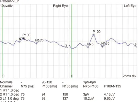
충격 부위 피부의 결손 및 관통상은 없었으나 우측의 상안 검부위의 감각저하를 호소하였다. 교정시력은 우안 20/25,
좌안 20/20이었으며 세극등현미경검사에서 우안의 결막부종 이외 특이 소견은 없었다. 안구돌출도검사에서 우안이 2 mm 더 돌출되어 있었다. 우안의 완전 안검하수를 보였으며 상 전 및 내전이 되지 않았고, 외전 및 하전은 부분적으로 마 비되어 있었다(Fig. 4A). 우안 대광반사 소견은 보였으나 반응속도가 감소되어 있었고 상대구심동공반사는 음성이 었다. 시유발전위검사에서 우안의 진폭이 좌안에 비해 감 소되어 있었으며(Fig. 5), 안저검사에서는 특이 소견이 관찰 되지 않았다. 수상 10일 후 촬영한 안와 자기공명영상에서 시신경주위 및 직근 원위부의 조영 증강이 관찰되었고, 그 외 골절 및 구후출혈 등의 소견은 없었다(Fig. 6). 외상으로 인한 안와첨증후군으로 진단하고 예방적 항생제를 투여하 며 고용량 스테로이드 치료로써 methylprednisolone 1 g을 하루 1회 3일간 정주하였다. 이후 경구 스테로이드 치료로 prednisolone을 11일간 50 mg (1 mg/kg/day)을 투여하였고,
Figure 5. Pattern visual evoked potential (VEP) of Case 2. Pattern VEP showed reduction in the amplitude in the right eye.
A B
Figure 6. Axial (A) and coronal (B) magnetic resonance imaging (MRI) (T2-weighted image) scan at tenth day after injury. MRI
showed enhancement of the right optic nerve sheath, distal rectus muscle and surrounding soft tissue.이후 2주 간격으로 40 mg, 30 mg, 15 mg, 5 mg씩 복용하 였다. 치료 2개월 후 안검하수 및 안구 운동 이상은 모두 호전되었다(Fig. 4B). 우안 시력은 20/20으로 호전되었다.
고 찰
안와첨증후군에서는 병변의 해부학적 위치 및 침범된 뇌 신경의 종류에 따라 다양한 증상이 발생할 수 있으며, 안구 운동 및 동공 마비, 안검하수, 시력 소실이 대표적인 증상 이다.8 감각이 저하되거나 안구 주위 통증이 동반될 수 있
으며, 안구돌출의 정도는 다양하게 나타나는데, 해면정맥굴 증후군에서보다 상안와열증후군이나 안와첨증후군에서 좀 더 두드러지게 나타날 수 있다.13
두 증례의 자기공명영상 촬영 소견에서 이환된 쪽의 T2 강조영상에서 눈 뒤의 고신호음영 및 조영 증강이 관찰된 반면, 외상으로 인한 골절 및 출혈은 관찰되지 않았다. 안 와는 골격 구조에 의해 제한되는 좁은 공간으로 적은 부피 증가에도 안와 내압이 증가할 수 있음을 고려하였을 때, 외 상으로 인해 발생한 염증 및 부종으로 인한 압박으로 안와 첨증후군이 발생하였다고 유추할 수 있었다.
치료는 보존적 치료, 스테로이드 투여 및 수술적 중재까 지 다양하게 보고되었다. 경과관찰만으로 외상으로 인한 안와첨증후군에서의 운동기능이상이 회복되었다는 보고가
있으며,14,15 수술로 인한 추가적인 출혈 및 신경 손상의 위
험성은 수술적 치료를 적극적으로 고려하지 않는 이유가 될 수 있다. 그러나 골절편의 전위, 눈 뒤 혈종, 시신경초의 혈종, 골막하 혈종 등의 이상 소견이 있을 경우, 골절 정복 수술이나 감압술이 효과적일 수 있다.16,17
스테로이드 치료가 외상으로 인한 상안와열증후군에서 부종을 감소시킴으로써 회복에 도움을 줄 수 있다고 알려 져 있지만 그 용량 및 방법에 대해서는 확립된 것이 없는 실정이다. Acartürk et al18은 5명의 환자에서 외상으로 인한 상안와열증후군 혹은 안와첨증후군에서 고용량의 부신피 질호르몬제를 투여하고 좋은 결과를 보고하였다. 이 보고 에서 30 mg/kg의 methylprednisolone에 이어 시간당 5.4 mg/kg의 용량으로 48시간 동안 투여하였으며, 모든 환자에 서 고용량 스테로이드의 합병증 없이 6개월까지 완전 회복 되었다고 하였다. Peter and Pearson9은 골절이 동반되지 않 은 외상성 안와첨증후군 환자에서 경구 덱사메타손 5 mg 을 6주간에 걸쳐 감량하고, 시력과 감각이 호전되었지만 동 공 운동 및 조절기능은 호전되지 않았다고 보고하였다. 국 내에서도 스테로이드 정주 및 경구 복용 치료로 양호한 기 능회복을 보였다는 보고가 있었다.11,12 중추신경계에 손상 이 발생하면 활성산소반응이 일어나는데, 이때 세포성 글 루코코르티코이드 수용체를 포화시키고 남을 정도의 고용 량의 부신피질호르몬제를 투여하면 활성산소반응을 막는 항산화 작용을 하고 허혈, 부종이 있는 부위에 혈중 농도를 높게 유지하여 허혈, 부종의 치료에 도움이 된다고 알려져 있다.19 또한 체내에 높은 용량의 부신피질호르몬이 있으면 혈류 증가, 칼슘 항상성, 에너지대사 등 직접적인 도움을 준다는 보고도 있다.20
두 증례는 안와의 직접적인 손상 없이 둔상에 의해 안와첨 증후군이 발생한 것으로 두 증례 모두에서 스테로이드에 좋 은 반응을 보여 안구 운동은 완전 회복되었지만 시력 저하가 있었던 한 증례에서 시력의 회복은 제한적이었으며, 외상으 로 인하여 시신경의 비가역적인 손상이 병발하였음을 추측 해 볼 수 있다. 이와 같이 외상과 연관한 안와첨증후군이 발 생하였을 때 적극적으로 스테로이드 치료를 고려해 볼 수 있 을 것으로 생각된다. 그러나 현재까지 스테로이드 치료에 대 한 명확한 적응증, 용량 등에 대한 정설은 없는 상태이며 이 에 대한 추가적인 연구가 필요할 것으로 사료된다.
REFERENCES
1) Yeh S, Foroozan R. Orbital apex syndrome. Curr Opin Ophthalmol 2004;15:490-8.
2) Foroozan R, Bhatti MT, Rhoton AL. Transsphenoidal diplopia.
Surv Ophthalmol 2004;49:349-58.
3) Kline LB. The Tolosa-Hunt syndrome. Surv Ophthalmol 1982;27:
79-95.
4) Ing EB, Purvin V. Progressive visual loss and motility deficit. Surv Ophthalmol 1997;41:488-92.
5) Marcet MM, Yang W, Albert DM, et al. Aspergillus infection of the orbital apex masquerading as Tolosa-Hunt syndrome. Arch Ophthalmol 2007;125:563-6.
6) Keane JR. Cavernous sinus syndrome. Analysis of 151 cases. Arch Neurol 1996;53:967-71.
7) Lasky JB, Epley KD, Karesh JW. Household objects as a cause of self-inflicted orbital apex syndrome. J Trauma 1997;42:555-8.
8) Brent BD, May DR. Orbital apex syndrome after penetrating orbi- tal trauma. Ann Ophthalmol 1990;22:267-8.
9) Peter NM, Pearson AR. Orbital apex syndrome from blunt ocular trauma. Orbit 2010;29:42-4.
10) Baik SH, Yeom DJ, Kang YK, et al. Orbital apex syndrome with nasal type natural killer (NK)/t-cell lymphoma of sphenoid and ethmoid sinus. J Korean Ophthalmol Soc 2010;51:286-91.
11) Baek SU, Lee MJ. A case of orbital apex syndrome induced by pen- etrating orbital injury with long-term results. J Korean Ophthalmol Soc 2013;54:1275-81.
12) Han K, Ahn M. A case of superior orbital fissure syndrome in- duced by penetrating orbital injury. J Korean Ophthalmol Soc 2015;56:592-7.
13) Aryasit O, Preechawai P, Aui-Aree N. Clinical presentation, aetiol- ogy and prognosis of orbital apex syndrome. Orbit 2013;32:91-4.
14) Zachariades N. The superior orbital fissure syndrome. Review of the literature and report of a case. Oral Surg Oral Med Oral Pathol 1982;53:237-40.
15) Pogrel MA. The superior orbital fissure syndrome: report of case. J Oral Surg 1980;38:215-7.
16) Imaizumi A, Ishida K, Ishikawa Y, Nakayoshi I. Successful treat- ment of the traumatic orbital apex syndrome due to direct bone compression. Craniomaxillofac Trauma Reconstr 2014;7:318-22.
17) Antonyshyn O, Gruss JS, Kassel EE. Blow-in fractures of the orbit.
Plast Reconstr Surg 1989;84:10-20.
18) Acartürk S, Seküçoğlu T, Kesiktäs E. Mega dose corticosteroid treatment for traumatic superior orbital fissure and orbital apex syndromes. Ann Plast Surg 2004;53:60-4.
19) Hall ED, Braughler JM. Glucocorticoid mechanisms in acute spi- nal cord injury: a review and therapeutic rationale. Surg Neurol 1982;18:320-7.
20) Steinsapir KD, Goldberg RA. Traumatic optic neuropathy. Surv Ophthalmol 1994;38:487-518.
= 국문초록 =
외상 이후 발생한 안와첨증후군 2예
목적: 안와 내부 구조물의 직접적인 손상이 없는 안구의 둔상 이후 발생한 안와첨증후군 및 이의 치료 과정을 2예 경험하였기에 이를 보고하고자 한다.
증례요약: (증례 1) 쇠봉에 부딪친 후 시력 저하를 호소하는 50세 남자가 내원하였다. 좌안 교정시력은 광각불인지였으며 대광반사는 소실되어 있었다. 좌안의 완전 안검하수를 보였으며 부분적인 내전 및 하전을 제외한 전방향 안구운동제한을 보였다. 안와 자기공명 영상에서 시신경 및 시신경 주위 조직을 포함한 근원뿔 내 고음영이 관찰되었다. 스테로이드 정주 치료 3일째 시력은 안전수지로 회복되었고 수상 5개월째 안구운동 및 눈꺼풀 기능은 정상범위로 회복되었다. (증례 2) 굵은 철심에 우안을 부딪친 64세 여자로 수상 안의 교정시력은 20/25, 완전 안검하수, 부분적인 외전 및 하전을 제외한 전방향의 안구운동제한을 보였다. 안와 자기공명영상에서 시신경주위 및 직근 원위부의 조영 증강을 보였다. 스테로이드 정주 치료 후 2개월째 안검하수, 안구운동제한 및 시력은 모두 회복되 었다.
결론: 둔상에 의해 발생한 안와첨증후군이 고용량 스테로이드에 좋은 반응을 보였다. 외상에 의한 안와첨증후군에서 스테로이드 치료 를 적극적으로 고려해 볼 수 있을 것으로 생각된다.
<대한안과학회지 2018;59(9):893-898>
양상철 / Sang Cheol Yang
부산대학교 의과대학 안과학교실 Department of Ophthalmology,
Pusan National University School of Medicine