The Prognostic Value of the Left Ventricular Ejection Fraction Is Dependent upon the Severity of Mitral Regurgitation in Patients with Acute Myocardial Infarction
The prognostic value of the left ventricle ejection fraction (LVEF) after acute myocardial infarction (AMI) has been questioned even though it is an accurate marker of left ventricle (LV) systolic dysfunction. This study aimed to examine the prognostic impact of LVEF in patients with AMI with or without high-grade mitral regurgitation (MR). A total of 15,097 patients with AMI who received echocardiography were registered in the Korean Acute Myocardial Infarction Registry (KAMIR) between January 2005 and July 2011. Patients with low-grade MR (grades 0-2) and high-grade MR (grades 3-4) were divided into the following two sub-groups according to LVEF: LVEF ≤ 40% (n = 2,422 and 197, respectively) and LVEF > 40% (n = 12,252 and 226, respectively). The primary endpoints were major adverse cardiac events (MACE), cardiac death, and all-cause death during the first year after registration. Independent predictors of mortality in the multivariate analysis in AMI patients with low-grade MR were age ≥ 75 yr, Killip class ≥ III, N-terminal pro-B-type natriuretic peptide > 4,000 pg/mL, high-sensitivity C-reactive protein ≥ 2.59 mg/L, LVEF
≤ 40%, estimated glomerular filtration rate (eGFR), and percutaneous coronary
intervention (PCI). However, PCI was an independent predictor in AMI patients with high- grade MR. No differences in primary endpoints between AMI patients with high-grade MR (grades 3-4) and EF ≤ 40% or EF > 40% were noted. MR is a predictor of a poor outcome regardless of ejection fraction. LVEF is an inadequate method to evaluate contractile function of the ischemic heart in the face of significant MR.
Keywords: Mitral Regurgitation; Acute Myocardial Infarction; Left Ventricular Ejection Fraction
Jung Sun Cho,1,2 Ho-JoongYoun,1,2 Sung-Ho Her,1,2 Maen Won Park,1,2 Chan Joon Kim,1,2 Gyung-Min Park,1,2 Myung Ho Jeong,1,3 Jae Yeong Cho,1,3 Youngkeun Ahn,1,3 Kye Hun Kim,1,3 Jong Chun Park,1,3 Ki Bae Seung,1,2 Myeong Chan Cho,1 Chong Jin Kim,1 Young Jo Kim,1 Kyoo Rok Han,1 Hyo Soo Kim,1 and the Korea Acute Myocardial Infarction Registry Investigators
1Korea Acute Myocardial Infarction Registry Investigators of Korean Circulation Society,
2Department of Internal Medicine, The Catholic University of Korea College of Medicine, Seoul;
3The Heart Center of Chonnam National University Hospital, Chonnam National University Research Institute of Medical Sciences, Gwangju, Korea Received: 18 October 2014
Accepted: 1 April 2015 Address for Correspondence:
Myung Ho Jeong, MD
The Heart Research Center of Chonnam National University Hospital, 671 Jaebong-ro, Dong-gu, Gwangju 501-757, Korea Tel: +82.62-220-6243, Fax: +82.62-228-7174
E-mail: [email protected]
Funding: This study was supported by grants from the Korean Society of Cardiology, the Korea Centers for Disease Control and Prevention (2013-E63005-00), and the Korean Health Technology R&D Project (HI13C1527), Ministry of Health &
Welfare, Republic of Korea.
http://dx.doi.org/10.3346/jkms.2015.30.7.903 • J Korean Med Sci 2015; 30: 903-910
INTRODUCTION
Mitral regurgitation (MR) is a frequent complication and a pow- erful predictor of long-term cardiovascular mortality after acute myocardial infarction (AMI) (1-5). Although left ventricular (LV) ejection fraction (EF) is a determinant of poor prognosis and an accurate marker of LV systolic dysfunction, the prognostic value of LVEF after MI has been questioned (6). In patients with chro- nic MR, LVEF underestimates the degree of LV systolic dysfunc- tion because of volume overload. LVEF is also highly influenced not only by LV contractility but also by LV geometry, loading con- dition, and MR severity (7, 8). In patients with AMI, low LVEF can be the result of reduced contractile function due to extensive myocardial damage, LV dilatation, or myocardial stunning (9).
The role of LVEF as a prognostic factor in AMI patients with significant MR has been poorly addressed (10, 11). Furthermore, the prognostic value of LVEF in AMI patients with severe MR has not been previously reported. This study aimed to examine the prognostic significance of LVEF in the long-term outcome of AMI patients with or without severe MR in the clinical setting.
MATERIALS AND METHODS Study population
The Korean Acute Myocardial Infarction Registry (KAMIR) is a Korean, prospective, open, observational, multicenter, on-line registry of AMI data with support from the Korean Society of Cardiology that was initiated in November 2005. The 50 partici- Cardiovascular Disorders
pating hospitals are capable of performing primary percutane- ous coronary intervention (PCI). Details of the KAMIR have been published previously (2, 12, 13). A total of 15,097 patients with AMI who received echocardiography were registered in the KA- MIR between January 2005 and July 2011. Patients with low-grade MR (grade 0-2) were divided into two groups according to LVEF, namely, LVEF ≤ 40% (n = 2,426) or EF > 40% (n = 12,252). Pa- tients with high-grade MR (grade 3-4) were similarly divided into two groups (EF ≤ 40% [n = 197] or EF > 40% [n = 226]).
The endpoints of the study were major adverse cardiac events (MACE), cardiac death, and all-cause death during the year fol- lowing registration. MACEs were defined as the composite of all-cause death, MI, and repeated PCI or coronary artery bypass grafting (CABG) during 12 months of clinical follow-up.
The follow-ups in the outpatient clinic occurred immediately after hospital discharge, one month post-discharge, and at in- tervals of less than 6 months thereafter. Information on events and mortality was obtained from hospital records and phone calls.
The diagnosis of non-ST elevation myocardial infarction (NS- TEMI) and ST elevation myocardial infarction (STEMI) was bas- ed on the definitions from the American College of Cardiology/
American Heart Association (ACC/AHA guidelines) (14, 15).
Coronary angiogram
Coronary angiograms were performed using standard techni- ques. Significant coronary artery disease (CAD) was defined as
≥ 70% stenosis of an epicardial coronary artery. The extent of CAD was characterized by one-, two-, or three-vessel disease or left main disease (14, 15). Percutaneous coronary intervention (PCI) was performed according to the physician’s discretion.
Coronary blood flow in the infarct-related artery before and af- ter stent implantation was graded according to the classifica- tion used in the Thrombolysis in Myocardial Infarction trials.
Echocardiography
All index transthoracic echocardiographs were recorded during routine clinical practice according to the current guidelines (16).
Two-dimensional M-mode echocardiography and Doppler ul- trasound examinations were performed within 3 days of the PCI.
In each patient, LVEF was measured using bidimensional echocardiography from two- and four-chamber apical views by the modified Simpson’s method (17). The wall motion score in- dex was derived according to a 17 segment model (18). For each segment, wall motion was scored from 1 (normal) to 4 (dyski- nesia). The presence and degree of MR were measured using the proximal isovelocity surface area (PISA) method and a vali- dated nomogram for semi-quantitative estimation (19). MR was classified into four degrees of severity (I: mild, II: mild to mod- erate, III: moderate, IV: severe).
Statistical analysis
Data are expressed as the means ± SD for continuous variables and absolute numbers (proportions) for categorical variables.
All comparisons between baseline variables were performed by the Pearson chi-square test for categorical variables and the t- test for continuous variables.
Cox proportional hazards regression was used to estimate the relative mortality risk at 1 yr. We controlled for all available variables considered potentially relevant in all regression analy- sis of low-grade MR (grade 0-2) and high-grade MR (grade 3-4):
age, gender, Killip class, Q wave on electrocardiography (ECG), Ischemic heart disease history (IHD), diabetes mellitus (DM), N-terminal pro-B type natriuretic peptide (NT-proBNP), glu- cose level, high-sensitivity C-reactive protein (Hs-CRP), LVEF, estimated glomerular filtration rate (eGFR), and PCI. Best cut- off values of continuous variables were assessed by the receiver operating curve. All statistical tests were 2-sided, and a P value
< 0.05 was considered statistically significant. Statistical analy- sis was performed using SPSS 18.0 for Windows (SPSS, Inc., Chi- cago, IL, USA). Survival analysis was performed using the Ka- plan-Meier method with log-rank tests to compare survival be- tween groups.
Ethics statement
The institutional review board of all participating institutions approved the study protocol. The approval number of Chon- nam National University Hospital was 05-49. Written informed consent was obtained from all participating patients.
RESULTS
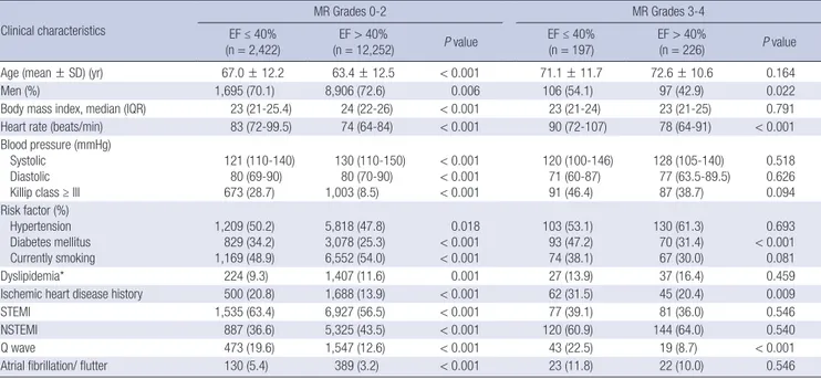
Baseline clinical characteristics and laboratory findings The baseline clinical characteristics are presented in Table 1.
Among the patients with low-grade MR (grade 0-2), the patients in the LVEF ≤ 40% group were older, were more likely to be wo- men, had decreased body mass indexes (BMIs) and blood pres- sures, had higher heart rates and Killip classes and were more likely to have histories of hypertension, DM, and IHD compar- ed with the patients in the LVEF > 40% group. Patients with re- duced LVEF were more likely to present STEMI, Q waves, and atrial fibrillation/flutter on electrocardiography. However, among the patients with high-grade MR (grade 3-4), the patients in the LVEF ≤ 40% group were more likely to be men, have higher heart rates, have histories of DM and IHD, and present Q waves on ECG compared with the patients in the LVEF > 40% group. Re- gardless of MR grade, the LVEF ≤ 40% group had higher glu- cose, Hs-CRP, and NT-proBNP levels compared with the LVEF
> 40% group (Table 2).
Coronary angiogram
In AMI patients with low-grade MR (grade 0-2), the LVEF ≤ 40%
group had more severe coronary angiogram findings than the LVEF > 40% group (Table 3). However, in patients with high-grade MR (grade 3-4), the LVEF ≤ 40% group had no significantly dif- ferent coronary angiographic findings, except for left anterior coronary artery disease, compared with the LVEF > 40% group.
PCI was performed in a significantly lower number of patients in the high grade MR group as compared to the low grade MR group. However, based on LVEF, there was no significant differ- ence between the number of PCI performed on patients in the high grade MR group (Table 3).
Independent predictors of mortality
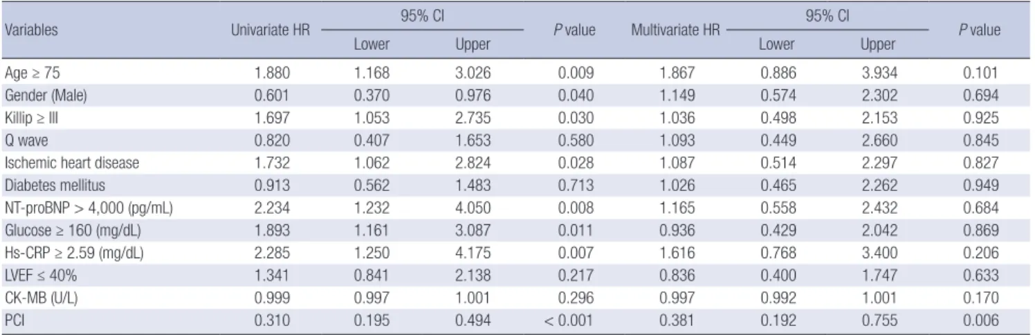
The independent predictors of cardiac death in the multivariate analysis in AMI patients with low-grade MR were age ≥ 75 yr, Killip class ≥ III, IHD history, NT-proBNP > 4,000 pg/mL, Hs- CRP ≥ 2.59 mg/L, eGFR, PCI, and LVEF ≤ 40% (Table 4). How- ever, PCI was an independent predictor in patients with AMI with high-grade MR (Table 5). Primary endpoints in the MR grade 0-2 group based on Kaplan-Meier analysis were signifi- cantly different according to EF (EF ≤ 40% vs. EF > 40%, total death, 330/2,422 vs. 373/12,252, P < 0.001; MACEs, 489/2,422 vs.
1,091/12,252, P < 0.001; cardiac death, 281/2,422 vs. 254/12,252, Table 1. Clinical characteristics of patients
Clinical characteristics
MR Grades 0-2 MR Grades 3-4
EF ≤ 40%
(n = 2,422) EF > 40%
(n = 12,252) P value EF ≤ 40%
(n = 197) EF > 40%
(n = 226) P value
Age (mean ± SD) (yr) 67.0 ± 12.2 63.4 ± 12.5 < 0.001 71.1 ± 11.7 72.6 ± 10.6 0.164
Men (%) 1,695 (70.1) 8,906 (72.6) 0.006 106 (54.1) 97 (42.9) 0.022
Body mass index, median (IQR) 23 (21-25.4) 24 (22-26) < 0.001 23 (21-24) 23 (21-25) 0.791
Heart rate (beats/min) 83 (72-99.5) 74 (64-84) < 0.001 90 (72-107) 78 (64-91) < 0.001
Blood pressure (mmHg) Systolic
Diastolic Killip class ≥ III
121 (110-140) 80 (69-90) 673 (28.7)
130 (110-150) 80 (70-90) 1,003 (8.5)
< 0.001
< 0.001
< 0.001
120 (100-146) 71 (60-87) 91 (46.4)
128 (105-140) 77 (63.5-89.5) 87 (38.7)
0.518 0.626 0.094 Risk factor (%)
Hypertension Diabetes mellitus Currently smoking
1,209 (50.2) 829 (34.2) 1,169 (48.9)
5,818 (47.8) 3,078 (25.3) 6,552 (54.0)
0.018
< 0.001
< 0.001
103 (53.1) 93 (47.2) 74 (38.1)
130 (61.3) 70 (31.4) 67 (30.0)
0.693
< 0.001 0.081
Dyslipidemia* 224 (9.3) 1,407 (11.6) 0.001 27 (13.9) 37 (16.4) 0.459
Ischemic heart disease history 500 (20.8) 1,688 (13.9) < 0.001 62 (31.5) 45 (20.4) 0.009
STEMI 1,535 (63.4) 6,927 (56.5) < 0.001 77 (39.1) 81 (36.0) 0.546
NSTEMI 887 (36.6) 5,325 (43.5) < 0.001 120 (60.9) 144 (64.0) 0.540
Q wave 473 (19.6) 1,547 (12.6) < 0.001 43 (22.5) 19 (8.7) < 0.001
Atrial fibrillation/ flutter 130 (5.4) 389 (3.2) < 0.001 23 (11.8) 22 (10.0) 0.546
Data are expressed as the mean ± SD or number (%), or median (IQR) as appropriate. *Defined as patients who were previously diagnosed by a physician and/or patients re- ceiving lipid-lowering drugs. NSTEMI, non-ST elevation myocardial infarction; STEMI, ST elevation myocardial infarction; MR, mitral regurgitation; EF, ejection fraction.
Table 2. Laboratory findings and echocardiographic parameters Variables
MR Grades 0-2 MR Grades 3-4
EF ≤ 40%
(n = 2,422) EF > 40%
(n = 12,252) P value EF ≤ 40%
(n = 197) EF > 40%
(n = 226) P value
Peak CK-MB (U/L) 162.7 ± 332.3 118.1 ± 223.1 < 0.001 121.8 ± 310.8 98.4 ± 146.8 0.314
Glucose (mg/dL) 188.2 ± 92.3 164.1 ± 75.6 < 0.001 205.6 ± 100.1 179.7 ± 90.2 0.006
Glomerular filtration rate (mL/min/1.73 m2) 59.9 ± 37.3 73.6 ± 40.3 < 0.001 49.9 ± 26.2 53.6 ± 28.1 0.183
Total cholesterol (mg/dL) 178.1 ± 45.7 183.8 ± 43.4 < 0.001 169.6 ± 50.1 174.0 ± 49.8 0.370
Triglycerides (mg/dL) 113.4 ± 73.8 132.9 ± 111.2 < 0.001 101.4 ± 56.9 107.7 ± 57.3 0.273
High-density lipoprotein cholesterol (mg/dL) 44.3 ± 15.3 44.9 ± 21.4 0.221 43.4 ± 14.5 43.7 ± 13.6 0.845 Low-density lipoprotein cholesterol (mg/dL) 114.2 ± 42.8 117.1 ± 39.6 0.003 109.6 ± 46.3 110.2 ± 40.2 0.904 High-sensitivity C-reactive protein (mg/dL) 12.8 ± 41.4 7.28 ± 35.8 < 0.001 19.3 ± 86.4 12.7 ± 45.1 0.035 NT-pro BNP (pg/mL) 5,472.8 ± 9,110.1 1,626.0 ± 443.6 < 0.001 8,984.9 ± 10,105.8 6,116.4 ± 9,101.3 0.009 Echocardiographic parameters
Left ventricular ejection fraction (%) Regional wall motion score
Left ventricular end-diastolic dimension (mm) Left end-systolic dimension (mm)
33.4 ± 6.25 25.3 ± 12.6 52.9 ± 9.30 42.1 ± 9.2
56.0 ± 8.8 18.5 ± 9.3 48.4 ± 8.0 33.3 ± 7.2
< 0.001
< 0.001
< 0.001
< 0.001
31.4 ± 6.7 28.4 ± 13.8 56.1 ± 9.8 43.3 ± 12.3
51.9 ± 7.7 21.3 ± 10.3 50.1 ± 8.3 36.2 ± 8.2
< 0.001
< 0.001
< 0.001
< 0.001 Data are expressed as the mean ± SD. MR, mitral regurgitation; EF, ejection fraction; CK-MB, creatine kinase myocardial band; NT-pro BNP, N-terminal pro-B-type natriuretic peptide.
P < 0.001 by the log-rank test). However, primary endpoints among AMI patients with MR grade 3-4 was not different ac- cording to EF (EF ≤ 40% vs. EF > 40%, total death, 42/197 vs.
37/226, P = 0.216; MACEs, 55/197 vs. 49/226, P = 0.357; cardiac death, 37/197 vs. 31/226, P = 0.216 by the log-rank test) (Fig. 1).
Fig. 1 reveals that patients with grades 3-4 MR and preserved EF exhibit a poor prognosis given the advanced age of this patient
population compared with groups with low-grade MR (Table 1).
DISCUSSION
The present study showed that in the presence of high-grade MR, LVEF is not an independent predictor of mortality. LVEF is the most widely used variable to represent LV systolic function Table 3. Coronary angiography findings
Variables
MR Grades 0-2 MR Grades 3-4
EF ≤ 40%
(n = 2,422) EF > 40%
(n = 12,252) P value EF ≤ 40%
(n = 197) EF > 40%
(n = 226) P value Coronary angiographic findings, n (%) 2,236/2,422 (92.3) 11,942/12,252 (97.4) < 0.001 168/197 (85.2) 199/ 226 (88.0) 0.563 Number of vessels with significant stenotic
0 1 2 3
44/2,236 (2.0) 1,370/2,236 (61.3)
446/2,236 (19.9) 259/2,236 (11.6)
489/11,942 (4.1) 7,439/11,942 (62.3) 2,531/11,942 (21.2) 1,158/11,942 (9.7)
< 0.001
6/168 (3.6) 91/168 (54.0) 37/168 (22.1) 24/168 (14.2)
6/199 (2.6) 114/199 (57.4)
41/199 (21.0) 29/199 (14.6)
0.958
LMA involvement 117/ 2,236 (5.2) 331/ 11,942 (2.8) 10/ 168 (6.1) 9/ 199 (4.4)
Infarct-related artery
Left anterior descending coronary artery Left circumflex coronary artery Right coronary artery LMA
1,737/2,236 (77.6) 499/ 2,236 (22.3) 606/ 2,236 (27.1) 117/ 2,236 (5.2)
6,448/11,942 (54.0) 3,295/11,942 (27.6) 5,087/11,942 (42.6) 331/11,942 (2.8)
< 0.001
< 0.001
< 0.001
< 0.001
94/168 (56.4) 43/168 (26.1) 74/168 (44.2) 10/168 (6.1)
80/199 (40.4) 77/199 (38.9) 112/199 (56.5) 9/199 (4.4)
0.003 0.013 0.026 0.083 ACC/AHA lesion type
A B1 B2 C
84/2,236 (3.8) 330/2,236 (14.8) 595/2,236 (26.6) 1,225/2,236 (54.8)
394/11,942 (3.3) 2,161/11,942 (18.1) 4,119/11,942 (34.5) 5,268/11,942 (44.1)
< 0.001
6/168 (3.4) 31/168 (18.5) 41/168 (24.5) 90/168 (53.6)
5/199 (2.3) 27/199 (13.8) 49/199 (24.9) 118/199 (59.0)
0.616
Initial TIMI flow grade 0 1,113/2,236 (49.8) 5,230/11,942 (43.8) < 0.001 79/168 (47.4) 93/199 (46.9) 1.000 Final TIMI flow grade 3 1,994/2,236 (89.2) 11,321/11,942 (94.8) < 0.001 147/168 (87.5) 187/199 (94.0) 0.072
PCI 2,117/2,422 (87.4) 11,291/12,252 (92.1) < 0.001 136/197 (69.0) 170/226 (75.2) 0.229
Number of stents 1.56 ± 0.87 1.53 ± 0.83 0.180 1.65 ± 0.98 1.61 ± 0.94 0.730
Total stent length (mm) 24.86 ± 7.23 24.01 ± 7.0 < 0.001 24.4 ± 6.8 24.8 ± 6.3 0.657
Thrombolytic therapy 88/2,422 (3.6) 656/12,252 (5.3) 0.007 3/197 (1.5) 5/226 (2.2) 0.347
CABG 17/2,422 (0.7) 34/12,252 (0.2) 0.001 2/197 (1.0) 4/226 (1.8) 0.690
Data are expressed as the number of patients (%). MR, mitral regurgitation; EF, ejection fraction; ACC/AHA, American College of Cardiology/American Heart Association; TIMI, Thrombolysis In Myocardial Infarction; CABG, coronary artery bypass graft; LMA, left main coronary artery; PCI, percutaneous coronary intervention.
lesions
Table 4. Cox regression analysis of cardiac death by 1 yr in the mitral regurgitation grades 0-2 groups of acute myocardial infarction patients
Variables Univariate HR 95% CI
P value Multivariate HR 95% CI
P value
Lower Upper Lower Upper
Age ≥ 75 3.214 2.743 3.765 < 0.001 1.527 1.191 1.957 0.001
Gender (Male) 0.532 0.453 0.625 < 0.001 1.148 0.895 1.473 0.276
Killip ≥ III 5.645 4.797 6.643 < 0.001 1.833 1.405 2.39 < 0.001
Qwave 1.307 1.06 1.611 0.012 1.021 0.76 1.372 0.891
Ischemic heart disease 1.876 1.56 2.256 < 0.001 1.308 0.997 1.716 0.052
Diabetes mellitus 1.877 1.597 2.206 < 0.001 0.954 0.733 1.241 0.725
NT-proBNP > 4,000 (pg/mL) 8.134 6.628 9.982 < 0.001 1.807 1.349 2.42 < 0.001
Glucose ≥ 160 (mg/dL) 2.008 1.71 2.358 < 0.001 1.173 0.915 1.505 0.208
Hs-CRP ≥ 2.59 (mg/dL) 3.063 2.553 3.675 < 0.001 1.668 1.31 2.125 < 0.001
LVEF ≤ 40% 8.223 5.81 11.637 < 0.001 3.802 2.174 6.65 < 0.001
LVEF ≤ 40%*log (time) 1.177 1.07 1.295 0.001 1.204 1.046 1.386 0.010
eGFR (mL/min/1.73 m2) 0.969 0.967 0.972 < 0.001 0.98 0.975 0.985 < 0.001
PCI 0.216 0.182 0.257 < 0.001 0.368 0.283 0.479 < 0.001
CK-MB was excluded because it was not primary variable and had interaction with time. *LVEF ≤ 40% had time dependent hazard ratio (HR = 9.452 on 30 day, 10.504 on 180 day, 10.951 on 365 day). LVEF, Left ventricular ejection fraction; eGFR, estimated glomerular filtration rate; PCI, percutaneous coronary intervention;CK-MB, creatine kinase myo- cardial band; Hs-CRP, high-sensitivity C-reactive protein; NT-proBNP, N-terminal pro-B-type natriuretic peptide.
in patients with AMI. Furthermore, low LVEF was associated with high one-year mortality after AMI in patients without high- grade MR in the present study. Because the majority (97%) of the patients with AMI did not have high-grade MR. However, the prognostic value of LVEF after AMI has been questioned in several studies that could not confirm LVEF as a prognostic fac-
tor in AMI patients. Left ventricular end-diastolic volume (LVE- DV) and left ventricular end-systolic volume (LVESV) were de- scribed as more meaningful prognostic factors than LVEF be- cause a low EF might be attributed to extensive myocardial da- mage, LV dilatation, or a hibernating or stunned myocardium (6, 9). The present study suggests that ischemic high grade MR Table 5. Cox regression analysis of cardiac death by 1 yr in the mitral regurgitation grades 3-4 groups of acute myocardial infarction patients
Variables Univariate HR 95% CI
P value Multivariate HR 95% CI
P value
Lower Upper Lower Upper
Age ≥ 75 1.880 1.168 3.026 0.009 1.867 0.886 3.934 0.101
Gender (Male) 0.601 0.370 0.976 0.040 1.149 0.574 2.302 0.694
Killip ≥ III 1.697 1.053 2.735 0.030 1.036 0.498 2.153 0.925
Q wave 0.820 0.407 1.653 0.580 1.093 0.449 2.660 0.845
Ischemic heart disease 1.732 1.062 2.824 0.028 1.087 0.514 2.297 0.827
Diabetes mellitus 0.913 0.562 1.483 0.713 1.026 0.465 2.262 0.949
NT-proBNP > 4,000 (pg/mL) 2.234 1.232 4.050 0.008 1.165 0.558 2.432 0.684
Glucose ≥ 160 (mg/dL) 1.893 1.161 3.087 0.011 0.936 0.429 2.042 0.869
Hs-CRP ≥ 2.59 (mg/dL) 2.285 1.250 4.175 0.007 1.616 0.768 3.400 0.206
LVEF ≤ 40% 1.341 0.841 2.138 0.217 0.836 0.400 1.747 0.633
CK-MB (U/L) 0.999 0.997 1.001 0.296 0.997 0.992 1.001 0.170
PCI 0.310 0.195 0.494 < 0.001 0.381 0.192 0.755 0.006
eGFR was excluded because it was not primary variable and had interaction with time. LVEF, Left ventricular ejection fraction; eGFR, estimated glomerular filtration rate; PCI, percutaneous coronary intervention; CK-MB, creatine kinase myocardial band; NT-pro BNP, N-terminal pro-B type natriuretic; Hs-CRP, high-sensitivity C-reactive protein.
Fig. 1. Kaplan-Meier analysis of all-cause mortality, major adverse cardiac event (MACE), and cardiac death in the mitral regurgitation grades 0-2 (upper panel) and grades 3-4 (lower panel) groups of acute myocardial infarction patients. Primary endpoints were significant in the groups with mild mitral regurgitation (MR) according to ejection fraction (EF) (≤ 40% and > 40%). However, the groups with severe MR did not exhibit significant differences in all-cause mortality based on EF (≤ 40% and > 40%).
MR Grade 0-2
Follow-up (days)
0 50 100 150 200 250 300 350 Total death
P < 0.001 by log-rank test EF ≤ 40%
EF > 40%
1 0.8 0.6 0.4 0.2 0
Follow-up (days)
0 50 100 150 200 250 300 350 MACE
P < 0.001 by log-rank test EF ≤ 40%
EF > 40%
1 0.8 0.6 0.4 0.2 0
Follow-up (days)
0 50 100 150 200 250 300 350 Cardiac death
P < 0.001 by log-rank test EF ≤ 40%
EF > 40%
1 0.8 0.6 0.4 0.2 0
MR Grade 3-4
Follow-up (days)
0 50 100 150 200 250 300 350 P = 0.216 by log-rank test
EF ≤ 40%
EF > 40%
1 0.8 0.6 0.4 0.2 0
Follow-up (days)
0 50 100 150 200 250 300 350 P = 0.357 by log-rank test
EF ≤ 40%
EF > 40%
1 0.8 0.6 0.4 0.2 0
Follow-up (days)
0 50 100 150 200 250 300 350 P = 0.216 by log-rank test
EF ≤ 40%
EF > 40%
1 0.8 0.6 0.4 0.2 0
could be one reason for this result because LVEDV and LVESV could be increased by volume overload disease, such as ische- mic MR. A previous study reported that the presence of MR was associated with an increased likelihood of adverse outcomes but not LV EF in patients with AMI with MR (11).
Ischemic MR could be caused by global LV remodeling with spherical LV enlargement or local inferior wall remodeling with predominantly posterior leaflet restriction. In case of non-isch- emic MR, an increase in preload and/or a decrease in afterload will result in a falsely higher EF because the EF is load-sensitive (20). However, some have argued that the ‘low impedance leak’
effect might exclusively apply to acute severe MR and that the afterload is likely increased when the LV is dilated. Furthermore, the decline in EF following MVR could result from chordal tran- section. Whitlow et al. (21) have shown that the EF is essentially unchanged at 12 months among patients experiencing signifi- cant reductions in MR and ‘reverse’ remodeling using edge-to- edge clip devices without the confounding effects of CABG, ster- notomy, or chordal transection.
Alternatively, a portion of the LV dysfunction in AMI patient results from afterload excess; this finding could explain why re- duced LVEF has no impact on survival in patients with severe MR. Accordingly, contractile function might be better than the EF suggests because the LV is managing afterload excess.
The LVEF is derived from the LV volume. Although heart rate and fiber shortening both affect LVEF, it is influenced to a far greater extent by LVEDV given that changes in stroke volume tend to be considerably smaller than changes in LVEDV (22). In the AMI setting, LVEDV is not yet fully dilated by acute severe MR. In addition, ischemic MR might have a transiently severe grade.
LVEF is a determinant of the degree of functional ischemic mitral regurgitation in patients with systolic left ventricular dys- function (4). However, there is only a weak correlation between LVEF and MR severity (7, 8). Recently, several small population studies have examined the role of assessing LV systolic function in addition to LVEF in ischemic MR (23, 24).
PCI was performed in a significantly lower number of pati- ents in the group of LVEF < 40% with low grade MR as compar- ed to the group of LVEF > 40% with low grade MR. That could contribute the difference of survival in low grade MR not high grade MR. Furthermore, PCI was an independent predictor of lower cardiac death in high grade MR (Table 4). Reperfusion therapy for patients in AMI with severe MR could be beneficial for survival.
This study was a retrospective study. There are no detailed descriptions of MR volume or effective regurgitant orifice area (ERO) due to a lack of central readings of the echocardiograms in the core laboratory. Furthermore, the etiologies of MR and the presence of non-ischemic MR were not distinguished. Long- term follow-up echocardiography data were limited. The num-
ber of patients with severe MR was considerably lower than that in the other group. Nevertheless, this study demonstrated the prognostic impact of LVEF according to the presence of severe MR in a large, real-world population.
In conclusion, MR is a predictor of poor outcome regardless of ejection infarction. LVEF is an inadequate method to evalu- ate contractile function of the ischemic heart in the face of high grade MR. Other new parameters for assessing LV systolic func- tion beyond LVEF are needed in patients with significant isch- emic MR.
ACKNOWLEDGMENTS
Korean Acute Myocardial Infarction Registry (KAMIR) Investi- gators: Myung Ho Jeong, Young Jo Kim, Chong Jin Kim, Myeong Chan Cho, Youngkeun Ahn, Jong Hyun Kim, Shung Chull Chae, Seung Ho Hur, In Whan Seong, Taek Jong Hong, Dong Hoon Choi, Jei Keon Chae, Jae Young Rhew, Doo Il Kim, In Ho Chae, Junghan Yoon, Bon Kwon Koo, Byung Ok Kim, Myoung Yong Lee, Kee Sik Kim, Jin Yong Hwang, Seok Kyu Oh, Nae Hee Lee, Kyoung Tae Jeong, Seung Jea Tahk, Jang Ho Bae, Seung Woon Rha, Keum Soo Park, Kyoo Rok Han, Tae Hoon Ahn, Moo Hyun Kim, Joo Young Yang, Chong Yun Rhim, Hyeon Cheol Gwon, Seong Wook Park, Young Youp Koh, Seung Jae Joo, Soo Joong Kim, Dong Kyu Jin, Jin Man Cho, Wook Sung Chung, Yang Soo Jang, Jeong Gwan Cho, Ki Bae Seung, Seung Jung Park.
DISCLOSURE
The authors have no conflicts of interest to disclose.
AUTHOR CONTRIBUTION
Conception and coordination of the study: Cho JS, Jeong MH.
Acquisition of data: all authors. Data review: Cho JS, Jeong MH.
Statistical analysis: Cho JS, Her SH. Manuscript preparation: Cho JS, Jeong MH. Manuscript approval: all authors.
ORCID
Jung Sun Cho http://orcid.org/0000-0002-0308-0021 Ho-Joong Youn http://orcid.org/0000-0002-0435-3570 Sung-Ho Her http://orcid.org/0000-0002-1548-4154 Maen Won Park http://orcid.org/0000-0001-5293-8461 Chan Joon Kim http://orcid.org/0000-0003-2929-8325 Gyung-Min Park http://orcid.org/0000-0001-5846-0606 Myung Ho Jeong http://orcid.org/0000-0003-4173-1494 Jae Yeong Cho http://orcid.org/0000-0002-9393-2821 Youngkeun Ahn http://orcid.org/0000-0003-2022-9366 Kye Hun Kim http://orcid.org/0000-0002-6885-1501 Jong Chun Park http://orcid.org/0000-0002-1637-7991
Ki Bae Seung http://orcid.org/0000-0002-7831-8268 Myeong Chan Cho http://orcid.org/0000-0002-0047-0227 Chong Jin Kim http://orcid.org/0000-0002-7460-2835 Young Jo Kim http://orcid.org/0000-0001-8802-8996 Kyoo Rok Han http://orcid.org/0000-0003-2591-4901 Hyo Soo Kim http://orcid.org/0000-0002-7026-6715
REFERENCES
1. Grigioni F, Enriquez-Sarano M, Zehr KJ, Bailey KR, Tajik AJ. Ischemic mitral regurgitation: long-term outcome and prognostic implications with quantitative Doppler assessment. Circulation 2001; 103: 1759-64.
2. Cho JY, Jeong MH, Ahn Y, Jeong HC, Cho SC, Yoo JH, Song JE, Jang SY, Lee KH, Park KH, et al.; Korea Acute Myocardial Infarction Registry In- vestigators. Different impact of mitral regurgitation on clinical outcomes according to timing of percutaneous coronary intervention in patients with non-ST segment elevation myocardial infarction. Int J Cardiol 2013;
168: 4872-4.
3. Lancellotti P, Lebrun F, Piérard LA. Determinants of exercise-induced changes in mitral regurgitation in patients with coronary artery disease and left ventricular dysfunction. J Am Coll Cardiol 2003; 42: 1921-8.
4. Yiu SF, Enriquez-Sarano M, Tribouilloy C, Seward JB, Tajik AJ. Determi- nants of the degree of functional mitral regurgitation in patients with sys- tolic left ventricular dysfunction: A quantitative clinical study. Circula- tion 2000; 102: 1400-6.
5. Perez de Isla L, Zamorano J, Quezada M, Almería C, Rodrigo JL, Serra V, García Rubira JC, Ortiz AF, Macaya C. Prognostic significance of function- al mitral regurgitation after a first non-ST-segment elevation acute coro- nary syndrome. Eur Heart J 2006; 27: 2655-60.
6. Mollema SA, Nucifora G, Bax JJ. Prognostic value of echocardiography after acute myocardial infarction. Heart 2009; 95: 1732-45.
7. Magne J, Pibarot P. Left ventricular systolic function in ischemic mitral regurgitation: time to look beyond ejection fraction. J Am Soc Echocar- diogr 2013; 26: 1130-4.
8. Hetzer R, Dandel M. Early detection of left ventricular dysfunction in patients with mitral regurgitation due to flail leaflet is still a challenge.
Eur Heart J 2011; 32: 665-7.
9. White HD, Norris RM, Brown MA, Brandt PW, Whitlock RM, Wild CJ.
Left ventricular end-systolic volume as the major determinant of surviv- al after recovery from myocardial infarction. Circulation 1987; 76: 44-51.
10. Pecini R, Thune JJ, Torp-Pedersen C, Hassager C, Køber L. The relation- ship between mitral regurgitation and ejection fraction as predictors for the prognosis of patients with heart failure. Eur J Heart Fail 2011; 13: 1121-5.
11. Amigoni M, Meris A, Thune JJ, Mangalat D, Skali H, Bourgoun M, War- nica JW, Barvik S, Arnold JM, Velazquez EJ, et al. Mitral regurgitation in myocardial infarction complicated by heart failure, left ventricular dys- function, or both: prognostic significance and relation to ventricular size and function. Eur Heart J 2007; 28: 326-33.
12. Cho JY, Jeong MH, Ahn Y, Jeong HC, Jang SY, Kim SS, Rhew SH, Jeong YW, Lee KH, Park KH, et al. Impact of high admission blood pressure with- out history of hypertension on clinical outcomes of patients with acute myocardial infarction: From Korea Acute Myocardial Infarction Regis- try. Int J Cardiol 2014; 172: e54-e8.
13. Lee KH, Jeong MH, Ahn Y, Cho MC, Kim CJ, Kim YJ. New horizons of
acute myocardial infarction: from the Korea Acute Myocardial Infarc- tion Registry. J Korean Med Sci 2013; 28: 173-80.
14. American College of Emergency Physicians; Society for Cardiovascular Angiography and Interventions; O’Gara PT, Kushner FG, Ascheim DD, Casey DE Jr, Chung MK, de Lemos JA, Ettinger SM, Fang JC, Fesmire FM, Franklin BA, et al. 2013 ACCF/AHA Guideline for the management of ST-Elevation myocardial infarction: a report of the American College of Cardiology Foundation/American Heart Association Task Force on Practice Guidelines. J Am Coll Cardiol 2013; 61: e78-e140.
15. Anderson JL, Adams CD, Antman EM, Bridges CR, Califf RM, Casey DE Jr, Chavey WE 2nd, Fesmire FM, Hochman JS, Levin TN, et al.; 2011 writ- ing group members; ACCF/AHA task force members. 2011 ACCF/AHA Focused Update Incorporated Into the ACC/AHA 2007 Guidelines for the Management of Patients With Unstable Angina/Non-ST-Elevation Myocardial Infarction: a report of the American College of Cardiology Foundation/American Heart Association Task Force on Practice Guide- lines. Circulation 2011; 123: e426-579.
16. Lang RM, Bierig M, Devereux RB, Flachskampf FA, Foster E, Pellikka PA, Picard MH, Roman MJ, Seward J, Shanewise JS, et al.; Chamber Quan- tification Writing Group; American Society of Echocardiography’s Gui- delines and Standards Committee; European Association of Echocar- diography. Recommendations for chamber quantification: a report from the American Society of Echocardiography’s Guidelines and Standards Committee and the Chamber Quantification Writing Group, developed in conjunction with the European Association of Echocardiography, a branch of the European Society of Cardiology. J Am Soc Echocardiogr 2005; 18: 1440-63.
17. Schiller NB, Shah PM, Crawford M, DeMaria A, Devereux R, Feigenba- um H, Gutgesell H, Reichek N, Sahn D, Schnittger I, et al. Recommen- dations for quantitation of the left ventricle by two-dimensional echocar- diography. American Society of Echocardiography Committee on Stan- dards, Subcommittee on Quantitation of Two-Dimensional Echocardio- grams. J Am Soc Echocardiogr 1989; 2: 358-67.
18. Cerqueira MD, Weissman NJ, Dilsizian V, Jacobs AK, Kaul S, Laskey WK, Pennell DJ, Rumberger JA, Ryan T, Verani MS, et al.; American Heart Association Writing Group on Myocardial Segmentation and Registra- tion for Cardiac Imaging. Standardized myocardial segmentation and nomenclature for tomographic imaging of the heart. A statement for health- care professionals from the Cardiac Imaging Committee of the Council on Clinical Cardiology of the American Heart Association. Circulation 2002; 105: 539-42.
19. Enriquez-Sarano M, Miller FA Jr, Hayes SN, Bailey KR, Tajik AJ, Seward JB. Effective mitral regurgitant orifice area: clinical use and pitfalls of the proximal isovelocity surface area method. J Am Coll Cardiol 1995; 25:
703-9.
20. Enriquez-Sarano M, Tajik AJ, Schaff HV, Orszulak TA, McGoon MD, Bai- ley KR, Frye RL. Echocardiographic prediction of left ventricular func- tion after correction of mitral regurgitation: results and clinical implica- tions. J Am Coll Cardiol 1994; 24: 1536-43.
21. Whitlow PL, Feldman T, Pedersen WR, Lim DS, Kipperman R, Smalling R, Bajwa T, Herrmann HC, Lasala J, Maddux JT, et al.; EVEREST II In- vestigators. Acute and 12-month results with catheter-based mitral valve leaflet repair: the EVEREST II (Endovascular Valve Edge-to-Edge Repair) High Risk Study. J Am Coll Cardiol 2012; 59: 130-9.
22. Rumberger JA, Behrenbeck T, Breen JR, Reed JE, Gersh BJ. Nonparallel
changes in global left ventricular chamber volume and muscle mass dur- ing the first year after transmural myocardial infarction in humans. J Am Coll Cardiol 1993; 21: 673-82.
23. Zito C, Cusmà-Piccione M, Oreto L, Tripepi S, Mohammed M, Di Bella G, Falanga G, Oreto G, Lentini S, Carerj S. In patients with post-infarc- tion left ventricular dysfunction, how does impaired basal rotation affect
chronic ischemic mitral regurgitation? J Am Soc Echocardiogr 2013; 26:
1118-29.
24. Gelsomino S, van Garsse L, Lucà F, Parise O, Cheriex E, Rao CM, Gen- sini GF, Maessen J. Left ventricular strain in chronic ischemic mitral re- gurgitation in relation to mitral tethering pattern. J Am Soc Echocardiogr 2013; 26: 370-80.e11.