490
Clinical and Angiographic Outcome of Sirolimus-Eluting Stent for the Treatment of Very Long Lesions
Jong-Seon Park, MD1, Young-Jo Kim, MD1, Dong-Gu Shin, MD1, Bong-Sup Shim, MD1, Gu-Ru Hong, MD1, Jun-Ho Bae, MD1, Chang-Wook Nam, MD2, Seung-Ho Hur, MD2, Seong-Wook Han, MD2, Kee-Sik Kim, MD2, Yoon-Nyun Kim, MD2,
Kwon-Bae Kim, MD2, Doo-Il Kim, MD3, Dae-Kyung Kim, MD3, Seong-Man Kim, MD3, Tae-Hyun Yang, MD3 and Dong-Soo Kim, MD3
1Division of Cardiology, Yeungnam University College of Medicine, Daegu, 2Division of Cardiology, Keimyung University College of Medicine, Daegu, 3Division of Cardiology, Inje University College of Medicine, Busan, Korea
ABSTRACT
Background and Objectives:Compared to bare metal stent, drug-eluting stent has improved the clinical and angiographic outcomes for de novo, simple lesions. In real world clinical practice, we often encounter more complex, long lesions, which increase the rate of restenosis and cardiovascular events. The aim of this study was to evaluate the clinical and angiographic outcome of sirolimus-eluting stent (SES) for the treatment of very long lesions in real world clinical practice. Subjects and Methods:We implanted multiple SESs (>40 mm in total length) in 113 de novo lesions in 113 patients. The average length of the implanted stents was 58±14 mm (ran- ge: 41-112 mm) and a mean of 2.2 stents were implanted in each lesion and the average stent diameter was 3.0
±0.3 mm. Results:Procedural and angiographic success were achieved in all the patients without death or co- ronary artery bypass surgery. Non-Q wave MI (CK-MB≥ 3 times the normal value) developed in 13 patients (11.5%). Two patients experienced late stent thrombosis after discharge (1.8%). The major adverse cardiac ev- ents (MACE)-free survival was 94% at 12 months. There were two sudden cardiac deaths. Six months follow up angiography was performed on 76 patients (67%) and angiographic binary restenosis developed in 7 patients (9.2%). All of them were the focal type in-stent restenosis and these were found to be located at the distal stents.
Conclusions:In conclusion, long lesion coverage with SESs is feasible with a favorable mid-term outcome in real world clinical practice. (Korean Circulation J 2006;36:490-494)
KEY WORDS:Percutaneous coronary angioplasty;Stents;Outcome.
Introduction
Drug-eluting stent(DES) lowers the amount of in- timal hyperplasia and it significantly improves the cli- nical outcome of percutaneous coronary intervention (PCI) for treating short lesions.1-3) In clinical practice, we encounter more restenosis-prone lesions such as left main lesions, restenotic lesions, small vessels, bifurcat- ion lesions and long lesions than are present in the patients included in clinical trials. The target lesion
length has been shown to be an independent predictor of in-stent restenosis and long lesion stenting had a high risk for revascularization in era of bare metal st- enting.4)5) Yet there is a lack of solid evidence pertain- ing to the safety and effectiveness of DES stents for treating long lesions and there’s a great need to de- monstrate this. Therefore, we evaluated the clinical and angiographic outcomes of long lesion coverage with using serolimus-eluting stents(SES) in the real world clinical practice.
Subjects and Methods
Study subjectsBetween March 2003 and May 2004 at three car- diovascular centers where more than 800 PCIs were performed in a year, 113 long lesions in 113 patients
Received:February 3, 2006 Revision Received:April 10, 2006 Accepted:April 17, 2006
Correspondence:Young-Jo Kim, MD, Division of Cardiology, Yeungnam University College of Medicine, 317-1 Daemyung-dong, Namgu, Daegu 705-717, Korea
Tel: 82-53-620-3835, Fax: 82-53-654-8386 E-mail: [email protected]
were treated with implanting multiple SESs(Cypher, Johnson & Johnson, USA). All the patients had both the clinical indications for PCI and an angiographic diameter stenosis ≥50%. The inclusion criterion was the presence of de novo coronary lesions that were planned to be implanted with multiple SESs(a total stent length ≥40 mm per lesion). Patients with res- tenotic lesions, lesions in saphenous vein grafts and lesions involving the left main coronary artery were excluded from this study. The study protocol conforms to the ethical guidelines of the 1975 Declaration of He- lsinki and all the patients gave us an informed consent.
Stent implantation and medication after stenting All 113 lesions in 113 patients were treated with multiple SESs. The stents were implanted according to the standard interventional techniques. For the sche- duled procedures, all the patients received aspirin 200 mg/day and clopidogrel 75 mg or ticlopidine 500 mg for 3 days before the procedure. The emergency pati- ents received loading dosage of aspirin and clopidogrel.
All lesions were predilated with using an optimal sized balloon according to the decision of the operating do- ctor. All the segments with stenosis ≥20% at the tar- get lesions were completely covered with stents. The length and number of the required stents were decided upon by the operating doctor according to visual esti- mation. All the procedures were performed without intravascular ultrasound guidance. After the procedu- res, the patients received ticlopidine 500 mg/day or clopidogrel 75 mg/day for 6 months and aspirin 200 mg/day indefinitely. Cilostazol 100 mg/twice a day was given to the patients who had very long stents ≥65 mm implanted or if they were were unable to take ti- clopidine or clopidogrel. We did not use abciximab.
Peri-procedural and 12-months clinical follow-up Procedural success was defined as a diameter steno- sis <30% in the treated segment after stent implanta- tion and no major cardiovascular events such as death, Q-wave myocardial infarction or emergency coronary bypass surgery. Myocardial infarction was documented by an increase in the serum creatinine kinase-MB level of more than three times the upper limit, which mea- sured 24 hours after the procedure. Patients were clini- cally followed in the outpatient clinic for 12 months and data was also obtained by serial telephone inter- views. Stent thrombosis was classified as acute(≤1 day), sub-acute(2-30 days) and late(>30 days) thro- mbosis based on the angiographic and clinical findings.
All the patients were monitored for any major cardio- vascular events(MACEs); these were defined as cardi- ac death, myocardial infarction, stent thrombosis or a need for revascularization.
Follow-up angiography
Angiographic follow-up study at 6 months was reco- mmended for all the patients. If any clinical evidence of myocardial ischemia developed at during the follow- up time, then hospital admission and coronary angio- graphy were recommended. Angiographic binary res- tenosis was defined as a narrowing of ≥50% of the vessel diameter at the site of the previous dilatation.
The indication for a new revascularization at a site of previous PCI was the occurrence of restenosis with the correlating symptoms. The restenosis patterns were qu- alitatively assessed using the Mehran classification sys- tem.6)
Angiographic analysis
Quantitative coronary angiographic analysis was pe- rformed using the computer-assisted automated edge detection method(CASS System II, Pie Medical Ima- ging, Netherlands) according to 2 observers. At least 2 orthogonal projections were selected for analysis; these were obtained after intracoronary injection of 200 μg nitrogrycerin. The mean reference vessel diameter, the minimal luminal diameter, the lesion length and the percentage diameter stenosis were analyzed for all the patients. The total length of the implanted stents was measured on the final angiogram with using the length measurement program included in the QCA software.
Statistical analysis
Statistical analysis was performed using SPSS 12 (SPSS Inc., Chicago, Illiois). Qualitative data are pre- sented as frequencies and continuous data are presen- ted as means±SDs.
Results
The baseline clinical data are shown in Table 1. The mean age was 59 years and diabetes mellitus was pre- sent in 34% of the patients. Glycoprotein IIb/IIIa in- hibitors were not given to any patients and all the pa- tients were prescribed more than two antiplatelet age- nts. The coronary angiographic findings are shown in Table 2. The most commonly treated artery was the left anterior descending coronary artery(55%). The mean reference vessel diameter and mean lesion len- gth were 3.0±0.3 mm and 46.8±12.3 mm, respecti- vely. More than two stents were used in all lesions and a mean of 2.2 stents was used in each lesion. The aver- age stent length was 57.6±13.9 mm long(range: 41- 112 mm) and the stent-to-lesion length ratio was 1.2
±0.3. There were 13 peri-procedural non-Q myocar- dial infarctions(11.5%), but any Q-wave myocardial infarction, emergency bypass surgery or death did not occur(Table 3).
During follow-up, late stent thrombosis that was co- nfirmed by coronary angiography occurred in a patient who suffered with dilated cardiomyopathy and a low left ventricular ejection fraction(32%). This develop- ed 50 days after successful implantation of a 56 mm long SES, and it was successfully treated with balloon PCI. The patient was kept on aspirin and clopidogrel until the PCI. Eventually, he suddenly died of ventri- cular fibrillation 273 days after SES implantation. An- other sudden death occurred at home for a patient who suffered with stable angina 512 days after PCI.
The patient had taken the prescribed aspirin and clo- pidogrel only intermittently. This event was perhaps related to late stent thrombosis. Repeat revasculari- zation of the target lesion was required in 6 patients (5.3%). Overall, cardiac events related to the SES occ- urred in 7 patients(7.1%) including non-fatal myo- cardial infarction(n=1), repeat target lesion revascu- larization(n=6) and cardiac death(n=2)(Fig. 1). The
Table 1. Baseline clinical characteristics of the patients
n=113 patients
Age (years) 59±11
Male sex 86 (76.1%)
Diabetes mellitus 34 (30.1%)
Smoking 44 (38.9%)
Hypertension 48 (42.5%)
Previous MI history 16 (14.2%)
Prior PCI 02 (01.7%)
Prior CABG 02 (01.7%)
Hypercholesterolemia 52 (46.0%)
Clinical diagnosis
Stable angina 29 (25.7%) Unstable angina 33 (29.2%) Acute myocardial infarction 48 (42.5%) Silent ischemia 03 (02.7%) Anti-platelet agent medications
Aspirin 113 (100%)
Ticlopidine 11 (09.7%)
Clopidogrel 96 (85.0%)
Cilostazol 41 (36.3%)
LVEF (%) 54.6±12.4%
MI: myocardial infarction, PCI: percutaneous coronary interven- tion, CABG: coronary artery bypass graft, LVEF: left ventricular ejection fraction. Data are expressed as mean±standard deviation
Table 2. Coronary angiographic findings of the patients
n=113 Treated artery
LAD 55 (48.7%)
LCX 17 (15.0%)
RCA 41 (36.3%)
No. of disease vessels
One 39 (34.5%)
Two 55 (48.7%)
Three 19 (16.8%)
Reference vessel diameter (mm) 03.0±0.4 Lesion length (mm) 46.7±12.3 LAD: left anterior descending artery, LCX: left circumflex ar- tery, RCA: right coronary artery
Table 3. Peri-procedure and in-hospital results
n=113 Used stents
Number 2.2±0.4
Diameter (mm) 3.0±0.3
Total length (mm) 57.6±13.9 Primary success 113 (100%)
Complications QMI 0 Non-QMI (CK-MB ≥ x3 normal value) 13 (11.5%)
Acute stent thrombosis 0
Emergency surgery 0
Cardiac death 0
QMI: Q-wave myocardial infarction, MI: myocardial infarction
12 10 8 6 4 2 0
Patient
Cardiac Non-Q Late TLR MACE death MI thrombosis
2 2
1
6 7
Fig. 1. Major cardiovascular events at 12 months follow-up. MI: myo- cardial infarction, PCI: percutaneous coronary intervention, CABG:
coronary artery bypass surgery, TLR: target lesion revascularization, MACE: major adverse cardiac events.
1.0
0.9
0.8
0.7
0.6
MACE-free rate
50 100 150 200 250 300 350 Days after procedure
MACE—free rate at 12 months; 94%
Fig. 2. Freedom from major adverse cardiac events (MACE) defined as cardiac death, myocardial infarction, stent thrombosis, or a need for revascularization was estimated by Kaplan-Meier method.
probability of MACE-free survival at 12 months was 94%(Fig. 2).
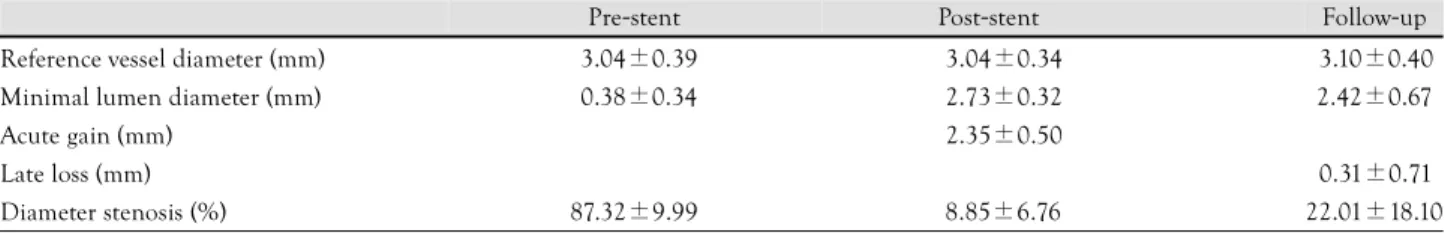
Angiographic follow-up was available for 76 patients (67%). The results of the post-procedural and follow- up angiographic QCAs are listed in Table 4. Angio- graphic binary restenosis occurred in 7 patients(9.2%), and all of them were the focal type without any of the diffuse or proliferative types. Four of the 7 patients had in-stent restenosis in the mid-portion of the far distal stents. Another three restenoses developed in the overlapping segments of two stents.
Discussion
The current study demonstrates that the use of SES for the treatment of very long lesions has improved the clinical and angiographic outcomes over that of the bare metal stent era. Diffuse lesion is a morphological characteristic associated with a poorer immediate and long-term clinical outcome after balloon angioplasty with or without stenting.7)8) In the Additional Value of NIR Stents for Treatment of Long Coronary Lesions (ADVANCE) Study, the MACE-free rate was 76.6%
at 300 days. Several studies that have focused on inter- mediate length lesions have demonstrated favorable re- sults with using SES. Ruiz-Nodar et al9) used 25 mm long SESs in patients who met the exclusion criteria of the RAVEL and SIRIUS studies, and the MACE rate at 6-months was 7%. More recently, obstructed full metal jacket stents that were treated by multiple DESs showed a low 1-year target vessel revascularization rate(7.5%) and MACE rate(18%).10) In that study, the 12 month MACE-free rate was 94% although the average stent length was 58±14 mm long.
Stenting in long segments is associated with more vessel trauma, a smaller vessel size and more exposed metal area, and all of this can lead to more neointimal growth and a higher restenosis rate than short stenting.
In the bare metal stent era, the stented segment length was an independent predictor of restenosis.11) A stent length that exceeded the lesion length increased the risk of restenosis and this was independent of the stented lesion length.12) If the stented segment length was >35 mm, then the restenosis rate was 47.2%.11) So, we have been trying to reduce the stent length, and performing spot stenting has been suggested as an
good strategy.13) But in the DES era, operating doctors are willing to completely cover lesions by stents and they are prone to use more long stents. We completely covered the lesions with >20% stenosis. In the present study, we found 7 angiographic restenosis(9.2%) in patients who were asymptomatic and these were found at the scheduled follow-up angiography at 6 months.
All of those lesions were the focal type, which were found at the stents’ overlapping segments or at the distally implanted stents. Thus, this finding indicates that these overlapping segments and distal small ve- ssels are vulnerable to restenosis and we should try to optimize stent deployment in this type of segments.
Another debatable problem is the subacute and late stent thrombosis. Animal studies have demonstrated that the polymers used in DES are proinflammatory, and this becomes particularly problematic for long stent implantation.14) In the SIRIUS tria,l2) the thro- mbosis rate at 270 days was 0.4% and in more com- plex lesions, the subacute stent thrombosis rate was 1.8%.9) A pooled meta-analysis of randomized clinical trials and registry studies showed the rate of stent thrombosis after DES to be similar to those of BMS.15) The long lesion coverage with long DESs becomes hy- percoagulable and more sensitive to inflammation and thrombosis. DES also delays re-endothelialization, and late stent thrombosis(LST) is a progressive phenom- enon that can occur at more than one year after im- plantation.16) The rate of stent thrombosis is higher in consecutive “real world” patients than in clinical trials, especially after discontinuation of ticlopidine or clo- pidogrel.17)18) In our study, no subacute stent throm- bosis was note although two late stent thrombosis de- veloped at 50 and 512 days after the procedure(1.8%).
The rate of LST was much higher in our study, alth- ough the cumulative incidence of stent thrombosis was similar to previous reports.2) Cilostazol has shown co- mparable antiplatelet activity to ticlopidine after el- ective coronary stenting.19)20) One out of three patients received triple antiplatelet therapy in this study. That may be the major factor for the lower rate of LST, although future randomized clinical trials are need.
However, the high rate of cumulative stent thrombosis underlines the possible need for long term antiplatelet medication for the patients receiving long DES stents.
In conclusion, long lesion coverage with SES is
Table 4. Quantitative coronary angiographic results
Pre-stent Post-stent Follow-up
Reference vessel diameter (mm) 03.04±0.39 3.04±0.34 3.10±0.40 Minimal lumen diameter (mm) 00.38±0.34 2.73±0.32 2.42±0.67
Acute gain (mm) 2.35±0.50
Late loss (mm) 0.31±0.71
Diameter stenosis (%) 87.32±9.99 8.85±6.76 22.01±18.10
feasible, and this has a favorable mid-term outcome in
“real world” clinical practice. We need to optimize the antiplatelet therapy regimen and its duration for the high risk patients.
There are several limitations in this study because of the small size of the study group and there was no bare metal stent control group. Moreover, follow-up angio- graphy was not done for all the patients. If we conduct intravascular ultrasound sonographic study, then the precise cause and mechanism of restenosis in this stu- dy group will be determined and we will be able to fur- ther reduce the rate of in-stent restenosis.
REFERENCES
1) Schofer J, Schluter M, Gershlick AH, et al. Sirolimus-eluting st- ents for treatment of patients with long atherosclerotic lesions in small coronary arteries: double-blind, randomised controlled tr- ial (E-SIRIUS). Lancet 2003;362:1093-9.
2) Holmes DR Jr, Leon MB, Moses JW, et al. Analysis of 1-year clinical outcomes in the SIRIUS trial: a randomized trial of a sirolimus-eluting stent versus a standard stent in patients at high risk for coronary restenosis. Circulation 2004;109:634-40.
3) Schofer J. Lessons from the E-SIRIUS trial. Ital Heart J 2004;5:
1-2.
4) Ajani AE, Waksman R, Cha DH, et al. The impact of lesion len- gth and reference vessel diameter on angiographic restenosis and target vessel revascularization in treating in-stent restenosis with radiation. J Am Coll Cardiol 2002;39:1290-6.
5) Pan M, Suarez de Lezo J, Medina A, et al. Influence of stent tr- eatment strategies in the long-term outcome of patients with long diffuse coronary lesions. Catheter Cardiovasc Interv 2003;58:
293-300.
6) Mehran R, Dangas G, Abizaid AS, et al. Angiographic patterns of in-stent restenosis: classification and implications for long- term outcome. Circulation 1999;100:1872-8.
7) Prieto AR, Przybysz A, Fischell TA. Long balloon angioplasty with focal stenting for the treatment of diffuse coronary artery disease. Catheter Cardiovasc Interv 2002;57:437-43.
8) le Breton H, Bedossa M, Commeau P, et al. Clinical and an-
giographic results of stenting for long coronary arterial ath- erosclerotic lesions. Am J Cardiol 1998;82:1539-43, A8.
9) Ruiz-Nodar JM, Frutos A, Carrillo P, et al. Use of sirolimus-el- uting stents in complex lesions: clinical and angiographic fo- llow-up. Rev Esp Cardiol 2004;57:123-9.
10) Aoki J, Ong AT, Rodriguez Granillo GA, et al. “Full metal ja- cket” (stented length > or =64 mm) using drug-eluting stents for de novo coronary artery lesions. Am Heart J 2005;150:994-9.
11) Kobayashi Y, de Gregorio J, Kobayashi N, et al. Stented segment length as an independent predictor of restenosis. J Am Coll Cardiol 1999;34:651-9.
12) Murphy GJ, Bicknell GR, Nicholson ML. The effect of combined rapamycin/cyclosporine on the changes in pro-fibrotic gene ex- pression that occur during the development of allograft vascu- lopathy in rats, compared with cyclosporine or rapamycin in iso- lation. Transpl Int 2003;16:347-53.
13) Colombo A, de Gregorio J, Moussa I, et al. Intravascular ultra- sound-guided percutaneous transluminal coronary angioplasty with provisional spot stenting for treatment of long coronary le- sions. J Am Coll Cardiol 2001;38:1427-33.
14) Farb A, Heller PF, Shroff S, et al. Pathological analysis of local delivery of paclitaxel via a polymer-coated stent. Circulation 2001;104:473-9.
15) Park DW, Park SW. Stent thrombosis in the era of the drug- eluting stent. Korean Circ J 2005;35:791-4.
16) Decoene C, Vincentelli A, Fabre O, Crepin F, Pol A. Late thro- mbosis of a drug-eluting coronary stent after antiplatelet therapy discontinuation. Ann Fr Anesth Reanim 2005;24:1275-7.
17) Iakovou I, Schmidt T, Bonizzoni E, et al. Incidence, predictors, and outcome of thrombosis after successful implantation of drug- eluting stents. JAMA 2005;293:2126-30.
18) Karvouni E, Korovesis S, Katritsis DG. Very late thrombosis after implantation of sirolimus eluting stent. Heart 2005;91:e45.
19) Ge J, Han Y, Jiang H, et al. RACTS: a prospective randomized antiplatelet trial of cilostazol versus ticlopidine in patients un- dergoing coronary stenting: long-term clinical and angiographic outcome. J Cardiovasc Pharmacol 2005;46:162-6.
20) Byun KH, Shim JK, Min SJ, Jeon SJ, Kim JH. The multiple co- mbined oral platelet aggregation Inhibitors of aspirin, ticlo- pidine and cilostazol after PCI. Korean Circ J 2004;34:443-50.