205
CASE REPORTDOI 10.4070 / kcj.2009.39.5.205
Print ISSN 1738-5520 / On-line ISSN 1738-5555 Copyright ⓒ 2009 The Korean Society of Cardiology
Very Late Thrombosis of a Drug-Eluting Stent After
Discontinuation of Dual Antiplatelet Therapy in a Patient Treated With Both Drug-Eluting and Bare-Metal Stents
Sung Soo Kim, MD
1, Myung Ho Jeong, MD
1,2, Doo Sun Sim, MD
1,2, Young Joon Hong, MD
1,2, Ju Han Kim, MD
1,2, Young Keun Ahn, MD
1,2and Jung Chaee Kang, MD
1,21The Heart Center of Chonnam National University Hospital, Gwangju,
2Cardiovascular Research Institute of Chonnam National University, Gwangju, Korea
ABSTRACT
Drug-eluting stents (DESs) are the treatment of choice for obstructive coronary artery disease when percutaneous intervention is feasible. However, late stent thrombosis seems to occur more frequently with DESs and is closely associated with the discontinuation of dual antiplatelet therapy. We report a case of very late stent thrombosis after discontinuation of dual antiplatelet therapy. The patient suffered from acute myocardial infarction (MI) and underwent bare metal stent (BMS) implantation in the left anterior descending artery (LAD) five years prior to presentation. Three years after BMS implantation, he presented again with acute MI and had a DES implanted in the right coronary artery (RCA). He ran out of his medication, but failed to refill his prescription. Sixteen days after discontinuing medication, he experienced an episode of chest pain and was taken to the cardiac cathe- terization laboratory, where he was found to have thrombosis in the DES, but no thrombosis in the BMS. It is possible that DESs are more vulnerable to late thrombosis than are BMSs, supporting the use of prolonged dual antiplatelet therapy in patients treated with DESs. The patient was successfully treated with balloon angioplasty and thrombus aspiration without complications.
(Korean Circ J 2009;39:205-208)KEY WORDS:
Thrombosis; Stents; Platelets.
Introduction
In recent years, drug-eluting stents (DESs) have been shown to dramatically reduce the rate of restenosis and the need for repeat revascularization.
1-3)Despite these promising results, late stent thrombosis seems to occur more frequently with DESs and seems to be closely as- sociated with the discontinuation of dual aspirin/thieno- pyridine derivative (usually clopidogrel) antiplatelet ther- apy.
1-4)We report a case of late DES thrombosis after discontinuation of dual antiplatelet therapy.
Case
A 49-year-old man was transferred to the emergency
department complaining of squeezing chest pain that had increased in severity over the past four days. Five years prior to presentation, he had undergone percuta- neous coronary intervention (PCI) for acute myocardial infarction (MI), along with implantation of a bare metal stent (BMS) (3.5×28 mm Arthos Inert Stent, AMG, Korea) in the left anterior descending artery (LAD) (Fig.
1). Six months later, he developed angina and underwent balloon angioplasty for in-stent restenosis (Fig. 2). Two years after that, he had a paclitaxel-eluting stent (2.75×
32 mm Taxus Stent, Boston Scientific, USA) placed in the right coronary artery (RCA) after suffering another acute MI (Fig. 3). The patient was treated with dual an- tiplatelet therapy: aspirin (100 mg daily) and clopidogrel (75 mg daily). He ran out of his medication, but failed to refill his prescription. Sixteen days after discontinu- ing medication, he developed chest pain and presented to the emergency department. An electrocardiogram (ECG) performed at that time showed new ST-segment depression and T-wave inversion in leads II, III, and aVF (Fig. 4). Cardiac enzymes were also elevated (creatine kinase-MB 9.9 U/L, Troponin I 7.73 ng/mL, Troponin
Received: May 24, 2008
Revision Received: August 13, 2008 Accepted: September 18, 2008
Correspondence: Myung Ho Jeong, MD,The Heart Center of Chonnam Na- tional University Hospital, 8 Hak-dong, Dong-gu, Gwangju 501-757, Korea Tel: 82-62-220-6243, Fax: 82-62-228-7174
E-mail: [email protected]
206
·Very Late Stent Thrombosis in DES and BMST 2.64 ng/mL). The patient underwent emergency cor- onary angiography, which revealed total occlusion of the DES in the proximal RCA due to very late stent throm- bosis with grade II collateral flow (Fig. 5A and B). The BMS in the LAD was patent (Fig. 5C). After receiving an intravenous glycoprotein IIb/IIIa receptor blocker (abciximab), the patient underwent repeat balloon angio- plasty and thrombus aspiration secondary to recurrent, immediate thrombus formation and coronary occlusion (Fig. 6A). The final angiogram showed good flow without residual stenosis (Fig. 6B).
Discussion
We report this case to draw attention to the phenom-
enon of very late stent thrombosis after DES implanta- tion, which might be associated with serious clinical im- plications after the discontinuation of dual antiplatelet therapy. It is possible that DESs are associated with sub- stantially higher rates of thrombosis when compared with BMSs.
Stent thrombosis (ST) is a generally fatal complication after PCI. It may occur in the acute (<1 day), subacute (<30 days), late (<1 year), or very late (>1 year) periods and may result in serious complications such as MI and death.
5)Very late stent thrombosis (VLST) is defined as a stent thrombosis event that occurs beyond one year.
The risk of VLST was analyzed using trial level data in a meta-analysis of 14 randomized trials in which 6,675 patients had been randomly assigned to PCI with DES
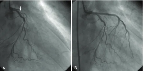
Fig. 2. The first follow-up coronary angiogram and percutaneous coronary intervention for restenosis. A: CAG revealed type III diffuse in-stent restenosis in the proximal left anterior descending artery (LAD) (arrow). B: plain balloon angioplasty was successfully performed for the treatment of in-stent restenosis in the LAD. CAG: coronary angiogram.
B A
Fig. 1. Diagnostic coronary angiogram and primary percutaneous intervention. A: CAG revealed total occlusion of the proximal left anterior descending artery (LAD) (arrow). B: a bare metal stent (3.5×28 mm Arthos Inert Stent) was successfully placed in the occluded LAD. CAG: coronary angiogram.
A B
Sung Soo Kim, et al.·
207
or BMS.
6)The incidences of VLST were 5.0 events per 1,000 patients in the DES group and 0 events per 1,000 patients in the BMS group (relative risk=5.0, 95% con- fidence interval 1.3-19.5). In another report, DESs were reported to have an approximately 0.3 to 0.5 percent
greater risk of VLST compared with BMSs.
7)Several fac- tors were shown to be associated with ST, including older age, black race, diabetes mellitus, bifurcation lesion, in- stent restenosis lesion, post-procedure acute renal fail- ure, and lack of clopidogrel therapy.
8)Discontinuation
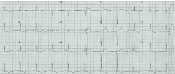
Fig. 4. Follow-up electrocardiographic finding. The ECG showed newly developed ST-segment depression and T-wave inversion in leads II, III, and aVF. ECG: electrocardio-gram.
Fig. 3. The second follow-up coronary angiogram and percutaneous coronary intervention for de novo lesion in right coronary artery. A:
CAG revealed a patent LAD stent (arrow). B: near total occlusion of the proximal RCA was also detected (arrow). C: a TAXUS stent (2.75×
32 mm) was successfully implanted after dilation using a 2.5×20 mm balloon in the proximal RCA. CAG: coronary angiogram, LAD: left anterior descending artery, RCA: right coronary artery.
C B
A
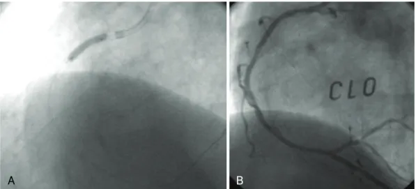
Fig. 5. The third follow-up coronary angiogram. A: total occlusion of the proximal right coronary artery at the level of the stent with evi- dence of thrombosis. B: grade II collateral flow from the left coronary artery (arrow). C: left coronary angiogram revealed a patent bare metal stent in the left anterior descending artery (arrow).
C B
A
208
·Very Late Stent Thrombosis in DES and BMSof antiplatelet therapy, as an independent predictor of stent thrombosis, even several years after DES implan- tation, increases the risk of late stent thrombosis. This finding suggests that clopidogrel compliance may mini- mize the incidence of VLST after DES implantation.
Current recommendations suggest extending dual antiplatelet therapy beyond one year in patients with low bleeding risk. Antiplatelet treatment should be con- tinued even if there is an increased risk of minor bleed- ing complications so that life-threatening complications, such as acute MI, are avoided. Patients with previously implanted DESs who are currently taking dual antiplatelet therapy are at high risk for developing stent thrombosis when a situation arises that requires cessation or inter- ruption of dual platelet inhibition.
9)10)The optimal duration of antiplatelet therapy in pa- tients with coronary artery stents still remains to be de- termined. Further large-scale studies are needed to de- termine the optimal combination and duration of anti- platelet therapy that should be used to prevent these serious thrombotic events.
REFERENCES
1) Serruys PW, Degertekin M, Tanabe K, et al. Intravascular ul- trasound findings in the multicenter, randomized, double-blind RAVEL (Randomized study with the sirolimus-eluting Velocity balloon-expandable stent in the treatment of patients with de novo native coronary artery Lesions) trial. Circulation 2002;106:798- 803.
2) Colombo A, Drzewiecki J, Banning A, et al. Randomized study to assess the effectiveness of slow- and moderate-release polymer-
based paclitaxel-eluting stents for coronary artery lesions. Cir- culation 2003;108:788-94.
3) Zhang F, Ge J, Qian J, et al. Sirolimus-eluting stents in real- world patients with ST-segment elevation acute myocardial in- farction. Int Heart J 2007;48:303-11.
4) Daemen J, Wenaweser P, Tsuchida K, et al. Early and late cor- onary stent thrombosis of sirolimus-eluting and paclitaxel-eluting stents in routine clinical practice: data from a large two-institu- tional cohort study. Lancet 2007;369:667-78.
5) Cutlip DE, Windecker S, Mehran R, et al. Clinical end points in coronary stent trials: a case for standardized definitions. Circul- ation 2007;115:2344-51.
6) Bavry AA, Kumbhani DJ, Helton TJ, et al. Late thrombosis of drug-eluting stents: a meta-analysis of randomized clinical trials.
Am J Med 2006;119:1056-61.
7) Mauri L, Hsieh WH, Massaro JM, et al. Stent thrombosis in randomized clinical trials of drug-eluting stents. N Engl J Med 2007;356:1020-9.
8) Kuchulakanti PK, Chu WW, Torguson R, et al. Correlates and long-term outcomes of angiographically proven stent thrombosis with sirolimus- and paclitaxel-eluting stents. Circulation 2006;
113:1108-13.
9) Park SH, Hong GR, Seo HS, Tahk SJ. Stent thrombosis after successful drug-eluting stent implantation. Korean Circ J 2005;
35:163-71.
10) Iakovou I, Schmidt T, Bonizzouni E, et al. Incidence, predictors, and outcome of thrombosis after successful implantation of drug eluting stents. JAMA 2005;293:2126-30.
11) Flores-Rios X, Marzoa-Rivas R, Abugattás-de Torres JP, et al.
Late thrombosis of paclitaxel-eluting stents: long-term incidence, clinical consequences, and risk factors in a cohort of 604 pa- tients. Am Heart J 2008;155:648-53.
12) Pfisterer M, Brunner-La Rocca HP, Buser PT, et al. Late clinical events after clopidogrel discontinuation may limit the benefit of drug-eluting stents: an observational study of drug-eluting versus bare-metal stents. J Am Coll Cardiol 2006;48:2584-91.
Fig. 6. Percutaneous coronary intervention for the very late stent thrombosis in right coronary artery. A: a balloon angioplasty was per- formed to treat total occlusion of the stented right coronary artery. B: final coronary angiogram showed good distal flow without residual stenosis.
B