205 205
넙치, Paralichthys olivaceus에서
병원성 Photobacterium damselae subsp. damselae의 분리
권문경
†·박상언 ·방종득·박수일
국립수산과학원 동해수산연구소, 국립수산과학원 동해수산연구소 어류연구센터, 부경대학교 수산생명의학과
Isolation of pathogenic Photobacterium damselae subsp. damselae from olive flounder, Paralichthys olivaceus
Mun-Gyeong Kwon†, Saung Un Park , Jong Deuk Bang and Soo-Il Park East Sea Fisheries Research Institute, National Fisheries Research & Development
Finfish research center, East Sea Fisheries Research Institute, National Fisheries Research & Development Department of Aquatic Life Medicine, Pukyong National University
The isolates, which has caused considerable damage to the olive flounder farm located in the eastern coast of Korea showed 99% sequence homology in the comparison of 16s rRNA gene of P. damselae subsp. damselae ATCC 33539. The present P. damselae was identical to the biotype No. 8 in Pedersen et al.
(1997) and the same LPS protein pattern as P. damselae subsp. damselae ATCC 33539. The comparison of infection rates among present P. damselae and Vibrio spp. showed that isolated P. damselae was the high- est, followed by V. anguillarium, V. harveyi, and V. ordalii.
Key words : Photobacterium damselae subsp. damselae, Olive flounder, 16s rRNA, Vibrio, Mortality
우리 나라 동해안 넙치 양식장에서 발생 빈도 가 높은 Vibrio 속 세균을 조사하는 과정에서 thiosulfate citrate bile salts sucrose (TCBS) 배지에 서 청색의 집락을 형성하는 균의 빈도가 특히 높은 점에 주목하여 연구한 결과 Vibrio 과에 속 하는 Photobacterium damselae subsp. damselae로 동정되었다. 이 병원균은 처음 분리될 당시에는
Vibrio 속으로 분류된 세균이었으나 (Love et al.,1981), 이후 분자생물학적 분류법에 따라 검토해 본 결과 다른 Vibrio 속 세균과 DNA homology 가 낮아 V. damselae에서 Photobacterium 속으로 재분류되어 지금은 분류 위치가 다른 속에 속해 있다. Vibrio 속 어병 세균은 대부분 TCBS 배지
상에서 황색의 집락을 형성하지만, 청색의 집락 을 형성하는 종도 다수 존재하므로 질병의 진단 에 어려움이 있으며, P. damselae의 감염증도 비 브리오병에 포함시키고 있는 실정이다 (Wang and Leung, 2000; Villami et al., 2003).
P. damselae는 damselfish, Chromis punctipinnis
에서 처음으로 보고되었으며 (Love et al., 1981), bottlenose dolphins, Tursiops truncatus (Fujioka et
al., 1988), leatherback turtles, Dermochelys cori- acea (Obendorf et al., 1987), 방어, Seriola quin- queradiata (Sakata et al., 1989), 감성돔, Sparus aurata (Vera et al., 1991), baramundi, Lates calcar- ifer (Renault et al., 1994), brown shark, Carcharhi-†Corresponding Author : Mun-Gyeong Kwon, Tel : 033-661-8504, Fax : 033-661-8514, E-mail : shellk@hanmail.net
nus plumbeus (Colwell and Grimes, 1984; Grimes et al., 1984) 및 터봇, Scophthalmus maximus (Fouz et al., 1991; 1992)에 질병을 일으켰다는 보고가
있지만, 아직 양식 넙치, Paralichthys olivaceus에 대한 감염 피해의 발생 예는 알려져 있지 않다.
본 연구에서는 우리 나라 동해안 넙치 양식장 에서 Photobacterium 감염증을 일으키는 P.
damselae와 이 질병과 혼용되어 사용되는 비브
리오병의 원인균을 분리, 동정하고 각 질병의 감 염률을 조사하였다.
재료 및 방법
시료 채취
시료 채취는 2002년 2월부터 2003년 11월까 지 매월 1회씩 동해안에 위치한 13개소의 넙치 양식장을 대상으로 시행하였다 (Fig. 1). 이 양식 장에서 질병 증상을 나타내는 양식 넙치의 체 표, 지느러미 및 아가미 등으로부터 기생충 감염 여부를 조사한 후 세균 배양을 실시하였다. 병원 균의 분리는 병어의 환부, 간, 신장, 비장 및 복수 액을 무균적으로 채취하여 1.5% NaCl 첨가 tryp- tic soy agar (TSA, Difco) 평판배지에 도말 접종 하였다. 분리 균주는 양식장에서 접종한 TSA 배 지를 25℃, 24시간 배양하여 분리한 집락 중
TCBS 배지 상에서 green 색을 나타내는 colony 를 택하여 시험에 사용하였다. 그리고 대조 균주 는 P. damselae ATCC 33539을 사용하였다.
생화학적 성상
분리된 균주 중 TCBS에서 green 색을 나타내 는 colony에 대하여 MacFaddin (1980)에 따라 생화학 검사를 실시한 후 P. damselae에 대하여 Pedersen et al. (1997)의 P. damselae biotype과 비 교하였다.
16s ribosomal RNA (16s rRNA) 유전자 배열 분석
PCR 수
수행 행
시험 균주의 genomic DNA는 DNAzol (Gibco- BRL)을 이용하여 분리한 후 A
260/280nm에서 DNA 농도가 50 ng/㎕가 되도록 준비하였다. PCR은 rTaq (Takara) polymerase와 universal bacterial primer (Bioneer사)로 Sense (5'-agtgtttgatcmtg- gctcag-3')와 antisense (5'-tacggytaccttgttacgactt-3') 를 사용하였다. PCR은 thermal cycler (Perkin- Elmer)로 수행하였으며, 조건은 initial denatura- tion (94℃, 5 min), denaturation (94℃, 30 sec), annealing (55℃, 30 sec), extension (72℃, 45 sec), final extension (72℃, 7 min)의 조건으로 30 cycles 반복하였다.
Gel에
에서 서 PCR 산 산물 물의 의 추 추출 출 및 및 cloning
Prep-A-Gene DNA purification system (BM)을 사용하여 agarose gel 상에서의 PCR 산물을 정제 하였다.
pGEM T-easy vector (Promega)에 T
4ligase로 재조합된 plasmid DNA를 준비하였으며, compe- tent cell (Escherichia coli JM 109)에 heat shock method로 transformation시켰다. 이에 1 ㎖의 LB broth를 첨가하여 37℃에서 1시간 배양한 후 ampicillin (50 ㎍/㎕), IPTG (Isopropylthio-β -D- galactopyranoside, 20 ㎍/㎕)와 X-gal (5-Bromo-4 - chloro-3-indolyl-β -D-galactoside, 20 ㎍/㎕)이 포함
Fig. 1. Sampling sites( ) of diseased olive flounder, Par-alichthys olivaceus in Gyeongbuk and Gangwon province.
된 LB agar (1% Bacto tryptone, 0.5% Bacto yeast extract, 1% NaCl, 1.5% Agar)에 배양하여 재조합 된 white colony를 선별하였다.
DNA sequencing 및
및 염 염기 기 서 서열 열 비 비교 교 분 분석 석 Plasmid DNA를 추출한 후 Sanger et al. (1977) 의 방법에 따라 automatic sequencer (ABI version 3.2, USA)로 sequencing을 실시하였다.
해독한 염기 배열의 조합과 restriction enzyme site를 찾기 위해서 Genetyx program을 사용하였 으며, The National Center for Biotechnology Infor- mation (NCBI)에서 제공되는 BLAST program으 로 이미 밝혀져 있는 균주들의 16S rRNA gene 과 비교 분석하였다.
SDS PAGE
분리 균주 및 참조 균주인 ATCC 33539의 LPS 단백질 profile은 Laemmli (1970)의 방법에 따라 SDS-polyacryl amide gel electrophoresis를 통하여 확인하였다.
Lipopolysaccharide (LPS)는 Schill et al. (1985) 의 rapid method로 분리하였다. 즉, 배양된 균체 는 멸균 PBS로 세척 및 현탁시킨 후 분광광도 계를 이용하여 A
525에서 0.5로 농도를 조정한 다 음 14,000g에서 3분간 원심분리하여 집균된 균 체를 2×SDS-PAGE sample buffer와 동량으로 현탁시켜서 10분간 끓였다. 끓인 sample에 pro- teinase K (Proease type ⅩⅠ; Sigma, St Louis, MO, USA)가 SDS-PAGE sample buffer에 2.5 ㎎/㎕로 현탁된 용액을 10 ㎕ 첨가하여 60℃ water bath 에서 1시간 끓인 후 사용할 때 까지 -20℃에 보 관하였다.
준비된 LPS는 14% (v/v) resolving gel에 10 ㎕ 씩 loading 후 mini-protean Ⅱsystem (BioRad)에 서 40mA, 90분간 전기 영동하였다. Marker는 Prestained Protein Molecular Weight Marker (MBI Fermentas)를 사용하였으며, 전기 영동한 gel은 Coomassie brillant blue R-250 staining solution으 로 염색 및 탈색 과정을 거친 후 확인하였다.
지역별 감염율
P. damselae와 Vibrio 속 어병 세균의 생화학적
성상 결과를 이용하여 월별, 지역별 및 종별 출 현 경향을 조사하였다.
결 과
생화학적 성상
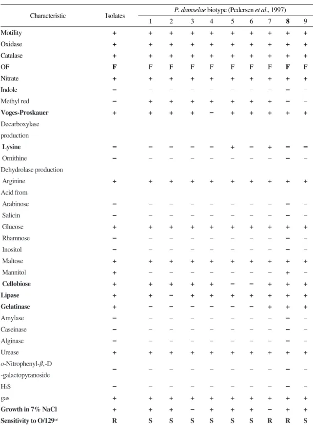
분리된 P. damselae의 생화학적 성상은 Table 1 과 같이 TCBS 배지에서 녹색 집락을 형성하는 그람 음성균이며, 운동성이 있는 nonswarming 간균이다. Catalase, oxidase, methyl red, nitrate에 서는 양성 반응을 나타내었으나, indole과 H
2S는 생성하지 않았다. 또한, arginin dihydrolase는 생 성하였으나, orinithine과 lysin decarboxylase는 생 성하지 못하였다. Urease와 chitinase는 생성하였 으나, amylase, caseinase와 alginase는 생성하지 못하였으나, glucose는 혐기적인 조건에서 가스 를 생성하면서 분해하였다. 그리고, glucose와 maltose에서 산이 생성되었으나 salicin, arabi- nose, rhamnose, inositol과 mannitol에서는 산이 생성되지 않았으며, o-Nitorphenyl-β-D-galactopy- ranoside는 분해하지 못하였다. 균주는 Voges - Proskauer test와 lipase, cellobiose에 대해서 양성 반응을 나타내었으며, 항생제인 O/129에 대하여 내성을 나타내었다. 20℃와 37℃에서 증식할 수 있으며, 7% NaCl에서도 증식하였다. 이와 같은 생리·생화학적 특성을 덴마크의 Pedersen et al.
(1997)이 분리한 P. damselae의 biotype과 특성을 비교한 결과 biotype No. 8에 속하는 것으로 동 정되었다.
16s rRNA 유전자 배열
생화학 성상에서 P. damselae로 동정된 균주의 16s rRNA를 universal primer로 분석하여 Genebank에 등록되어 있는 P. damselae subsp.
damselae ATCC 33539와 비교하여 nucleotide
sequence를 나타내었다 (Fig. 2). 그 결과 99%의
Table 1. Biochemical and physiological characteristics of Photobacterium damselae reference strains and isolates Characteristic Isolates P. damselae biotype (Pedersen et al., 1997)
1 2 3 4 5 6 7 8 9
Motility + + + + + + + + + +
Oxidase + + + + + + + + + +
Catalase + + + + + + + + + +
OF F F F F F F F F F F
Nitrate + + + + + + + + + +
Indole – – – – – – – – – –
Methyl red – + + + + + + + – –
Voges-Proskauer + + + + – + + + + +
Decarboxylase production
Lysine –– – – – – + – + –– ––
Ornithine – – – – – – – – – –
Dehydrolase production
Arginine + + + + + + + + + +
Acid from
Arabinose – – – – – – – – – –
Salicin – – – – – – – – – –
Glucose + + + + + + + + + +
Rhamnose – – – – – – – – – –
Inositol – – – – – – – – – –
Maltose + + + + + + + + + +
Mannitol + – – – – – – – + –
Cellobiose + + + + + – – + + +
Lipase + + – + + + + + + +
Gelatinase + – – – – – – + + +
Amylase – – – – – – – – – –
Caseinase – – – – – – – – – –
Alginase – – – – – – – – – –
Urease + + + + + + + + + +
o-Nitrophenyl-β‚-D
-galactopyranoside – – – – – – – – – –
H2S – – – – – – – – – –
gas + + + + + + + + + +
Growth in 7% NaCl + + + – + + + – + +
Sensitivity to O/129(a) R S S S S S S R R S
S, Sensitive; R, Resistant.
높은 상동성을 나타내었으며, 다른 Vibrio 속 세균 과는 다소 낮은 상동성을 나타내었다 (Table 2).
SDS PAGE
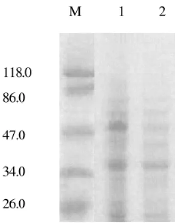
동해안 넙치 양식장에서 분리된 균주와 공시 균주의 LPS 단백질 패턴을 조사한 결과는 Fig. 3 과 같다. 실험에 사용한 P. damselae와 참조 균주 인 P. damselae subsp. damselae ATCC 33539는
47 kDa와 40 kDa 부근에서 두꺼운 밴드를 형성 하여 동일한 패턴을 나타내었다.
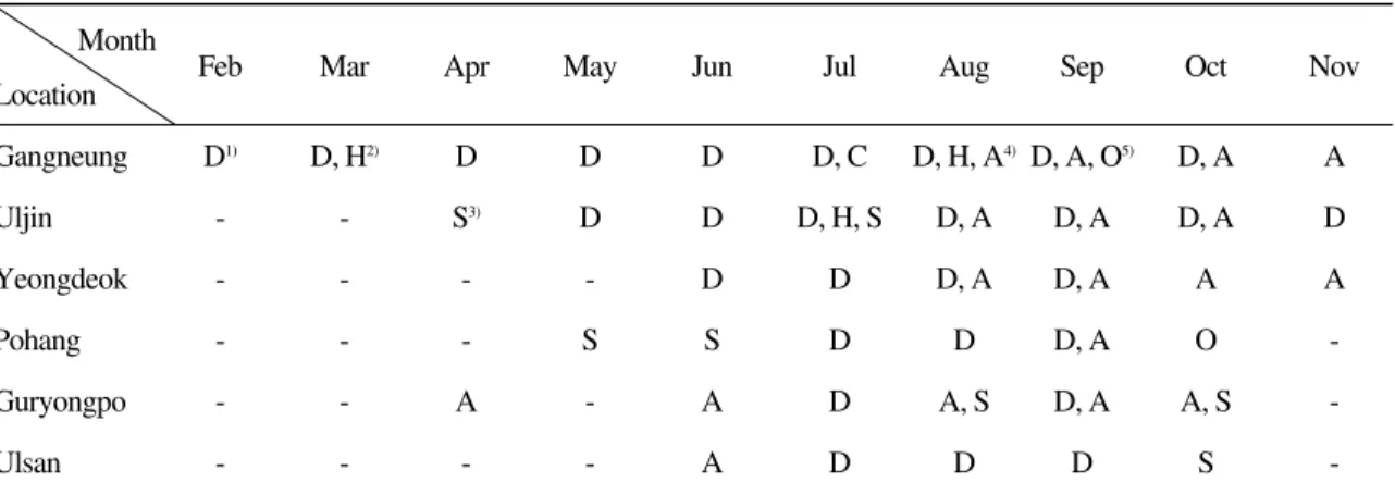
지역별 감염율
생화학적 성상과 16s rRNA 염기 서열 분석에 기초하여 동정된 Photobacterium속 세균과 vib- rio 균의 감염 경향은 Table 3과 같다. Vibrio 균 감염 시에는 균종별에 관계없이 모두 체색 흑 화, 체표 출혈과 궤양, 출혈성 복수증이 나타났 으며 P. damselae에 감염된 어체에서는 에드워드 균 감염증과 유사한 탈장 증상이 나타났다.
발생 시기는 수온과 깊은 관련성이 있는 것으 로 나타났다. 수온이 높은 강릉 지역에서는 P.
damselae가 연중 분리되었으며, 특히 고수온기
에는 V. anguillarum, V. ordaili 및 V. harveyi가 함 께 분리되었다. 강릉을 제외한 다른 지역에서는 주로 고수온기에 P. damselae와 vibrio 균들이 분 리되었으며, 이 중 P. damselae와 V. anguillarum 의 출현율이 높게 나타났다.
2002년 2월에서 2003년 11월까지 분리된 Vib-
rio 속과 Photobacterium 속 균주가 차지하는 비율은 Fig. 4와 같다. 전체 분리 균주 중 P. damse-
lae가 50%로 가장 많이 분리되었으며, V. anguil- Fig. 2. Comparison of 16s rRNA gene sequence of present isolates with P. damselae subsp. damselae ATCC 33539.Fig. 3. Protein profiles of lipopolysaccharide (LPS) from present isolate in SDS-PAGE using 14% acrylamide. M, marker; 1, P. damselae isolated from olive flounder, Par- alichthys olivaceus; 2, P. damselae subsp. damselae ATCC 33539.
M 1 2
118.0 86.0 47.0 34.0 26.0
larum이 29%, V. harveyi가 7%, V. splendidus가
5%, V. ordalii가 4%, 미동정된 Vibrio 속 세균이 5%로 나타났다.
고 찰
본 연구에서는 넙치에 감염되어 질병을 일으 키는 P. damselae를 분리하여 Pedersen et al.
(1997)의 P. damselae biotype과 비교한 결과, 무 지개송어 (Oncorhynchus mykiss)에서 분리된 biotype No. 8과 생화학적 성상이 동일하였다. 본 균주는 94~95년 덴마크에서 평년보다 기온이 5
℃나 높은 이상 기온을 나타낸 7~8월에 많은 세균성 질병과 함께 분리되었으며, 숙주 특이성 이 약한 병원체로 본 연구에서도 고수온기에 분 리율이 높아 같은 경향을 나타내었다. P. damse-
lae의 plasmids는 이전부터 확인되어왔으나(Austin et al., 1997; Fouz et al., 1992), Pedersen et
al. (1997)은 한 양어장 내에서 분리된 plasmid Table 2. Homology comparison of 16s rRNA gene sequence in P. damselae with several vibriosStrains Sequence homology (%)
PD1 VC2 VH3 VS4 VF5 VA6 VI7 VO8
Isolated P. damselae 99 93 92 92 92 91 92 90
1Photobacterium damselae subsp. damselae (ATCC 33539); 2Vibrio cholerae (AY911390); 3Vibrio harveyi (AY928014); 4 Vibrio splendidus; 5Vibrio fischeri; 6Vibrio alginolyticus (AY373027); 7Vibrio ichthyoenteri (AJ437192); 8Vibrio ordalii (AY628643).
Table 3. Photobacterium damselae subsp. damselae and vibrios isolated from the diseased olive flounder, Paralichthys oli- vaceus at culturing farm of east coast in Korea
Month
Feb Mar Apr May Jun Jul Aug Sep Oct Nov
Location
Gangneung D1) D, H2) D D D D, C D, H, A4) D, A, O5) D, A A
Uljin - - S3) D D D, H, S D, A D, A D, A D
Yeongdeok - - - - D D D, A D, A A A
Pohang - - - S S D D D, A O -
Guryongpo - - A - A D A, S D, A A, S -
Ulsan - - - - A D D D S -
* 1), Photobacterium damselae subsp. damselae; 2), Vibrio harveyi; 3), Vibrio splendidus; 4), Vibrio anguillarum; 5), Vibrio ordalii.
Fig. 4. P. damselae and vibrios (% incidence) isolated from the diseased olive flounder, Paralichthys olivaceus at cul- turing farm of east coast in Korea.
Vibrios V. ordalii
V. harveyi
V. anguillarum
P. damlselae V. splendidus
profile이 다양하며 일반적으로 virulence plasmid 는 아니라고 보고하였다.
16s rRNA를 이용하여 분리된 P. damselae의 동정 결과, P. damselae subsp. damselae ATCC 33539와 99%의 상동성을 나타내어 확정 동정 을 할 수 있었으며, 다른 vibrio 속 세균과는 다 소 낮은 상동성을 나타내어 세균 속간의 차이를 확인할 수 있었다.
P. damselae는 8~10월에 높은 감염율로 체표
에 괴사소를 형성하였으며, 탈장과 복강 내벽의 근육부에 점상 출혈이 특징적으로 관찰되었다.
이와 같은 결과는, Love et al. (1981)이 8~10월 에 damselfish 양식장에서 10~70%의 어류에서 괴사를 형성하고, 근육부의 융해를 관찰한 것과 유사한 결과로 생각되었다.
P. damselae와 vibrio 속 세균의 종별 지역별
감염율을 조사한 결과, P. damselae, V. anguil-
larum, V. harveyi, V. splendidus 및 V. ordalii의 순으로 감염율이 나타나, 다른 Vibrio 속 세균에 비 하여 P. damselae의 감염율이 높은 것을 알 수 있었으며, 동해안 넙치 양식장에서 주로 분리된
Vibrio 속 세균의 특징은 아래와 같다.V. anguillarum은 비브리오병 중 가장 최초로
보고된 종으로 1909년에 스웨덴 연안의 감염된 뱀장어 (Anguilla anguilla)에서 수온 상승 시기 에 발생하였다고 처음으로 보고되었으며 본 연 구에서도 수온이 높은 8~10월에 높은 감염율 과 폐사율을 나타내었다. 감염 경로에 대해서는 아직 불확실하지만, Toranzo and Barja (1993)는 세포 표면 성분을 이용하여 숙주에 침입, 부착 후 점액층을 뚫고 들어가 증식하는 것으로 추정 하였으며, Chart and Trust (1984)는 어류에 대한 독성은 multiflagellate를 이용한 화학주성과 점 액층을 침입할 수 있는 것과 관계가 있다고 하 였다. V. anguillarum의 serotype은 O1~O10이 확 인되었으며 전 세계적으로 폐사를 일으키는 것 은 O1, O2와 O3이며, O4~O10은 환경 중에 존 재하는 것으로 알려져 있다 (Toranzo et al., 1987;
Tajima et al., 1985; SФrensen and Larsen, 1986;
Larsen et al., 1988; Wiik et al., 1989; Toranzo and Barja, 1990).
V. harveyi는 새우의 대표적인 병원체로 별개
의 종으로 분류되었던 V. carchariae와 유사하여 현재 한 종으로 분류되고 있으나 (Pedersen et
al., 1998), chitinase 활성은 서로 다르게 나타났다 (Suginta et al., 2000). 어류에서는 Kraxberger- Beatty et al. (1990)에 의해 처음 보고되어 Austin 과 Austin (1993)에 의해 어류의 병원성 vibrio로 확인되어, 최근에는 grouper (Yii et al., 1997), summer flounder (Soffientino et al., 1999)와 우리 나라의 넙치 (원 등, 2003)에서도 폐사가 보고되 고 있는 종으로 본 조사 기간 동안 넙치에서는 감염율이 높지 않았다.
V. splendidus는 어류를 포함한 많은 해양 동물
에서 질병을 일으키는 종으로 넙치류 (Miranda and Rojas, 1996), 터봇과 대구류 (Santos et al., 1997)와 농어류 (Balebona et al., 1998)에서 질병 을 일으켰으며, 새우류 (Bacticados et al., 1990)와 패류 (Nicolas et al., 1996)에서도 감염되어 폐사 를 발생시키는 종으로 본 연구에서는 감염율이 비교적 낮은 것으로 나타났다.
V. ordalii는 V. anguillarum biotypeⅡ (Schiewe et al., 1981)로 분류되었던 종으로 출혈성 패혈
증의 원인균이지만 본 연구에서는 병원성과 분 리율이 높지 않았다.
따라서 우리 나라 동해안 넙치 양식장에서 분 리된 P. damselae는 넙치에서 다른 Vibrio 속 세 균에 비하여 높은 감염율을 나타내었으며, 특히 고수온기에 감염율이 높았다. Pedersen et al.
(1997)이 덴마크에서 분리한 균주와 biotype을
비교한 결과 No. 8에 속하는 균주로 ATCC
33539의 16s rRNA와 99% homology를 나타내
었다. 본 세균에 의한 질병은 일반적으로 비브리
오병으로 분류되고 있으나 (Austin and Austin,
1999; Wang et al., 1998), 유전학적 위치뿐만 아니
라 질병 발생시 세균의 감염 방법, 병원성 발현
인자 및 기작 등에서 비브리오병과는 다를 것으
로 예상되므로 이에 대한 연구가 지속적으로 이
루어져야 할 것으로 생각된다.
요 약
최근 2년간 동해안 지역 넙치 양식장의 양식 넙치에 피해를 일으키는 P. damselae 균을 분리 하였으며, 분리된 P. damselae의 16s rRNA 염기 서열은 P. damselae subsp. damselae ATCC 33539 와 99%의 상동성을 나타내었다. 분리 균주는 Pedersen et al. (1997)의 biotype과 비교한 결과, biotype No. 8과 동일하게 나타났으며, P. damse-
lae subsp. damselae ATCC 33539의 LPS와 동일한 단백질 패턴을 나타내었다. 넙치의 병어로부 터 P. damselae와 Vibrio 속 세균의 감염 상태를 조사한 결과, P. damselae가 가장 높은 감염율을 나타내었고, 그 다음으로 V. anguillarum, V. splen-
didus, V. harveyi와 V. ordalii 순으로 감염율이 나타났다.
감사의 글
본 연구는 국립수산과학원 (해산어 세균성 질 병 특성 연구, RP-2005-AQ-032)의 지원에 의해 운영되었습니다.
참 고 문 헌
Austin, B. and Austin, D. A.: Bacterial fish pathogens disease in farmed and wild fish.
2nd ed. Ellis Horwood, New York, 1993.
Austin, B., Austin, D. A., Blanch, A. R., Cerdá, M., Grimont, F., Grimont, P. A. D., Jofers, J., Koblavi, S., Larsen, J. L., Pedersen, K., Tiainen, T., Verdonck, L. and Swings, J.: A comparison of method for typing of fish- pathogenic Vibrio spp. Syst. Appl. Microbi- ol., 20: 89-101, 1997.
Bacticados, M. C. L., Lavilla-Pitogo, C. R., Cruz- Lacierda, E. R., Pena, L. D. and Sunaz, N.
A.: Studies on the chemical control of lumi- nous bacteria Vibrio harveyi and V. splen-
didus isolated from diseased Penaeus mon- odon larvae and rearing water. Dis. Aquat.Org., 9: 133-139, 1990.
Balebona, M. C., Zorilla, I., Morinigo, M. A. and Borrego, J. J.: Survey of bacterial patholo- gies affecting farmed gilt-head sea bream (Sparus aurata L.) in southwestern Spain from 1990 to 1996. Aquaculture, 166: 19- 35, 1998.
Chart, H. and Trust, T. J.: Characterization of the surface antigens of the marine fish pathogens Vibrio anguillarum and Vibrio
ordalii. Can. J. Microbiol., 3: 703-710, 1984.Colwell, R. R. and Grimes, D. J.: Vibrio diseases of marine fish populations. Helgol, Meeresun- ters, 37: 265-287, 1984.
Fouz, B., Larsen, J. L. and Toranzo, A. E.: Vibrio
damsela as a pathogenic agent causing mor-tality in cultured turbot (Scophthalmus max-
imus). Bull. Eur. Assoc. Fish Pathol., 11: 80-81, 1991.
Fouz, B., Larsen, J. L., Nielsen, B., Barja, J. L. and Toranzo, A. E.: Characterization of Vibrio
damsela strains isolated from turbot Scoph- thalmus maximus in Spain. Dis. Aquat. Org.,12: 155-166, 1992.
Fujioka, R. S., Greco, S. B., Cates, M. B. and Schroeder, J. P.: Vibrio damsela from wounds in bottlenose dolphins Tursiops
truncatus. Dis. Aquat. Org., 4: 1-8, 1988.Grimes, D. J., Colwell, R. R., Stemmler, J., Hada, H., Maneval, D., Hetrick, F. M., May, E. B., Jones, R. T. and Stoskopf, M.: Vibrio species associated with mortality of sharks held in captivity. Microb. Ecol., 10: 271-282, 1984.
Kraxberger-Beatty, T., McGarey, D. J., Grier, H. J.
and Lim, D. V.: Vibrio harveyi and oppor-
tunistic pathogen of common snook, Cen-
tropomus undecimalis (Bloch), held in cap-tivity. J. Fish Dis., 13: 557-560, 1990.
Laemmli, U. K.: Cleavage of structural protein dur- ing assembly of the head of bacteriophage T4. Nature (London), 222: 680-685, 1970.
Larsen, J. L., Rasmussen, H. B. and Dalsgaard, I.:
Study of Vibrio anguillarum strains from different source with emphasis on ecological and pathobiological properties. Appl. Envi- ron. Microbiol., 54: 2264-2267, 1988.
Love, M., Fisher, D. T., Horse, J. E., Farmer, J. J., Hickman, F. W. and Fanning, G. R.: Vibrio
damsela, as a marine bacterium, causes skinulcers on the damselfish, Chromis punctipin-
nis. Science, 214: 1140-1141, 1981.MacFaddin, J. F.: Biochemical tests for identifica- tion of medical bacteria. 2nd ed. Willams &
Wilkms. pp 527, 1980.
Miranda, C. and Rojas, R.: Vibriosis en el lenguado,
Paralichthys adspersus (Steindachner, 1867)en cautiverio. Revista de Biologia Marina, 31: 1-9, 1996.
Nicolas, J. L, Corre, S., Gauthier, G., Rober, R. and Ansquer, D.: Bacterial problems associated with scallop, Pacten maximus larval culture.
Dis. Aquat. Org., 27: 67-76, 1996.
Obendorf, D. L., Carson, J. and McManus, T. J.:
Vibrio damsela infection in a stranded
leatherback turtle (Dermchelys coriacea).
Wildl. Dis., 23: 666-668, 1987.
Pedersen, K., Dalsgaard, I. and Larsen, J. L.: Vibrio
damsela associated with diseased fish inDenmark. Appl. Environ. Microbiol., 63:
3711-3715, 1997.
Pedersen, K., Verdonck, L., Austin, B., Austin, D.
A., Blanch, A. R., Grimont, P. A. D., Jofre, J., Koblavi, S., Larsen, J. L., Tiainen, T., Vignelle, M. and Swings, J.: Taxonomic evi-
dence that Vibrio carchariae is a junior syn- onym of V. harveyi. Int. J. System. Bacteri- ol., 48: 749-758, 1998.
Renault, T., Haffner, P., Malfondet, C. and Weppe, M.: Vibrio damsela as a pathogenic agent causing mortalities in cultured sea bass (Lates calcarifer). Bull. Eur. Assoc. Fish Pathol., 14: 117-119, 1994.
Sanger, F., Nicklen, S. and Coulson, A. R.: DNA sequencing with chain-terminating inhibitors. Proc. Natl. Acad. Sci. USA, 74:
5463-5467, 1977.
Sakata, T., Matsuura, M. and Shimokawa, Y.: Char- acteristics of Vibrio damsela isolated from diseased yellowtail (Seriola quinqueradia-
ta). Nippon Suisan Gakkashi, 55: 135-141,1989.
Santos, Y., Pazos, F., Nunez, S. and Toranzo, A. E.:
Antigenic characterization of Vibrio anguil-
larum-related organisms isolated from turbotand cod. Dis. Aquat. Org., 28: 45-50, 1997.
Schiewe, M. H., Trust, T. and Crosa, J. H.: Vibrio
ordalii sp. nov.: A causative agent of vibrio-sis in fish. Curr. Microbiol., 6: 343-348, 1981.
Schill, W. B., Phelps, S. R., and Pyle, S. W.: Rapid serological analysis of bacterial lipopolysac- charide by electrotransfer to nitrocellulose. J.
Immunol. Methods, 85: 371-382, 1985.
Soffientino, B., Gwaltney, T., Nelson, D. R., Speck- er, J. L., Mauel, M. and Gomez-Chiarri, M.:
Infectious necrotizing enteritis and mortality caused by Vibrio carchariae in summer flounder Paralichthys dentatus during inten- sive culture. Dis. Aquat. Org., 38: 201-210, 1999.
SØrensen, V. B. S. and Larsen, J. L.: Serotyping of
Vibrio anguillarum. Appl. Environ. Microbi-ol., 51: 593-597, 1986.
Suginta, W., Robertson, P. A. W., Austin, B., Fry, S.
C. and Fothergill-Gilmore, L. A.: Chitinases from Vibrio: activity screening and purifica- tion of chiA from Vibrio carchariae. J. Appl.
Microbiol., 89: 76-84, 2000.
Tajima, K., Ezura, Y. and Kimura, T.: Studies on the taxonomy and serology of causative organ- ism of fish vibriosis. Fish Pathol., 20: 131- 142, 1985.
Toranzo, A. E., Baya, A. M., Roberson, B. S., Barja, J. L., Grimes, D. J. and Hetrick, F. M.:
Specificity of slide agglutination test for detecting bacterial fish pathogens. Aquacul- ture, 61: 81-97, 1987.
Toranzo, A. E. and Barja, J. L.: Virulence factors of bacteria pathogenic for coldwater fish.
Annu. Rev. Fish Dis., 3: 5-36, 1993.
Toranzo, A. E. and Barja, J. L.: A review of the tax- onomy and sero-epizootiology of Vibrio
anguillarum, with special reference to aqua-culture in the northwest of Spain. Dis.
Aquat. Org., 9: 73-82, 1990.
Vera, P., Navas, J. L. and Fouz, B.: First isolation of
Vibrio damsela from seabream (Sparus aurata). Bull. Eur. Assoc. Fish Pathol., 11:112-113, 1991.
Villami, V., Figueras, A., Aranguren, R. and Novoa, B.: Non-specific immune response of turbot,
Scophthalmus maximus (L.), experimentallyinfected with a pathogenic Vibrio pelagius. J.
Fish Dis., 26: 321-329, 2003.
Wang, X. H., Oon, H. L., Ho, G. W. P., Wong, W. S.
F., Lim, T. M. and Leung, K. Y.: Internaliza- tion and cytotoxicity are important virulence mechanism in vibrio fish epithelial cell inter- actions. Microbiol., 144: 2987-3002, 1998.
Wang, X. H. and Leung, K. Y.: Biochemical charac- terization of different types of adherence of
Vibrio species to fish epithelial cells. Micro-biol., 146: 989-998, 2000.
Wiik, R., Anderson, K., Daae, F. L. and Hoff, F. A.:
Virulence studies based on plasmid profiles of the fish pathogen Vibrio salmonicida.
Appl. Environ. Microbiol., 55: 819-825, 1989.
Yii, K. C., Yang, T. I. and Lee, K. K.: Isolation and characterization of Vibrio carchariae, a causative agent of gastroentertis in the grouper, Epinephelus coioides. Curr. Micro- biol., 35: 109-115, 1997.
원경미, 홍미주, 김수미, 박수일: 우리 나라 양식 해산어에서 분리된 Vibrio harveyi의 특성.
2003 Joint Meeting of the Korean Societies of Fisheries Science, 320-321, 2003.
Manuscript Received : October 14, 2005 Revision Accepted : December 02, 2005 Responsible Editorial Member : Tae-Sung Jung
(Gyeongsang Univ.)