Original Article
원고 접수일 2011년 12월 19일, 원고 수정일 2012년 1월 16일, 게재 확정일 2012년 3월 20일
책임저자 황대석
(626-770) 양산시 물금읍 범어리 3-3, 부산대학교 치의학전문대학원 구강악안면외 과학교실
Tel: 055-360-5103, Fax: 055-360-5104, E-mail: [email protected]
RECEIVED December 19, 2011, REVISED January 16, 2012, ACCEPTED March 20, 2012
Correspondence to Dae-Seok Hwang
Department of Oral and Maxillofacial Surgery, Pusan National University Hospital
3-3, Beomeo-ri, Mulgeum-eup, Yangsan 626-770, Korea
Tel: 82-55-360-5103, Fax: 82-55-360-5104, E-mail: [email protected]
CC This is an open access article distributed under the terms of the Creative Commons Attribution Non-Commercial License (http://creativecommons.org/licenses/
by-nc/3.0) which permits unrestricted non-commercial use, distribution, and reproduction in any medium, provided the original work is properly cited.
르포트씨 1급 골절단술을 시행 받은 환자들에서 Cone-beam Computed Tomography를
이용한 수술 전, 후의 상악동의 평가
이재열1,3ㆍ김용일2,3ㆍ백영재1ㆍ황대석1,3
부산대학교 치의학전문대학원 1구강악안면외과학교실, 2교정학교실, 3부산대학교병원 의생명연구원
Abstract
Evaluation of Maxillary Sinus Using Cone-beam Computed Tomography in Patients Who Underwent Le Fort I Osteotomy
Jae-Yeol Lee
1,3, Yong-Il Kim
2,3, Young-Jae Baek
1, Dae-Seok Hwang
1,31
Departments of Oral and Maxillofacial Surgery,
2Orthodontics, School of Dentistry, Pusan National University,
3
Biomedical Research Institute, Pusan National University Hospital
Purpose: The aim of this sturdy was to assess the prevalence and change in pathologic findings in the maxillary sinus by using preoperative and postoperative cone-beam computed tomography (CBCT).
Methods: The subjects included 83 patients with maxillary sinus abnormalities who underwent orthognathic surgery between January 2010 to December 2010. The CBCT analyses were classified according to the thickness of maxillary sinus membrane;
Normal (membrane thickness<2 mm), mucosal thickening (membrane thickness≥2 mm and <6 mm), partial opacification (membrane thickness>6 mm but not complete), total opacification, and polypoidal mucosal thickening. The diameters of the maxillary sinus ostium on the coronal cross-sectional view were also calculated.
Results: Out of 166 maxillary sinuses in 83 patients, 42 (25.3%) maxillary sinuses before surgery and 37 (22.3%) maxillary sinuses after surgery showed abnormalities. A decrease in the diameters of maxillary ostium was observed after surgery (
P
<0.05).However, there was no significant difference in mucosal thickness both, preoperatively and postoperatively.
Conclusion: The orthognathic surgery didn't deteriorate the maxillary sinus abnormaility. Despite the low prevalence of sinus complications in orthognathic surgery, all the patients should be informed of the possibility of sinusitis that could require the surgical intervention before surgery.
Key words: Cone-beam CT, Maxillary sinus, Le Fort I osteotomy
서 론
르포트씨 1급 골절단술은 중안모의 골격 부조화를 개선하기 위해 일반적으로 사용되는 술식이다. 이러한 르포트씨 1급 골절단 술은 1859년 Langenbeck에 의해 처음 기술된 이후 합병증이나 부작용을 최소화하는 방향으로 발전되어 왔다. 하지만 여러 연구 에서 르포트씨 1급 골절단술 후 출혈, 신경손상, 골괴사, 상악동 질환의 악화 등이 일어날 수 있음이 보고되고 있다[1,2]. 이러한 합병증 중 르포트씨 1급 골절단술 후 발생하는 상악동염은 연구에 따라 0.24∼20%로 다양하게 보고되고 있다[3,4]. 상악골 절단술 후에 발생하는 상악동염의 발생 가능한 원인으로는 상악동 점막의 섬모운동의 변화, 상악동의 혈병의 생성과 저류, 의원성 손상에 의한 치성 이차 감염, 조직의 허혈, 혈액 공급의 부족, 상악동에 남은 이물질 등을 생각할 수 있다[5,6]. 이 중 상악동 점막의 변화는 점막의 부종과 밀착에 의한 상악동 개구(maxillary os- tium)의 폐쇄에 의해 발생할 수 있으며[7], 이러한 감염이나 자극 에 의해 상악동 골벽을 따라 평행하게 점막의 비후가 나타나며, 더 악화되면 일반적으로 상악동염으로 나타나게 된다[8].
상악동에 대한 평가는 단순 부비동 평가(Water's view), 부비 동 초음파 검사, 전산화 단층촬영(computed tomography, CT), 자기공명영상촬영(magnetic resonance imaging, MRI) 등이 있다[9].
최근에는 악안면 영역에서 많이 사용되고 있는 cone beam CT의 발달로, 악교정 시술 전, 후 평가를 위해 cone beam CT의 촬영이 일반화되고 있다. Cone beam CT는 전통적인 전산화 단층촬영에 비해 우수한 해상도와 적은 방사선 조사, 그리고 촬영 의 편의성 등의 장점을 가진다. 낮은 연조직 해상도가 단점으로 지적되고 있지만, 상악동 점막의 이상소견을 관찰하고 평가하는 데는 큰 어려움이 없다[10,11].
저자는 리포트씨 1급 골절단술을 시행 받은 환자를 대상으로 술 전과 술 후 cone beam CT를 이용하여 상악동 점막의 변화 양상을 파악하고 상악동 개구의 크기 차이가 있는지 조사하기 위해 이 연구를 실시하였다.
연구방법
본 연구의 대상은 2010년 1월부터 2010년 12월까지 부산대학 교병원 구강악안면외과에서 한 명의 외과의에 의해 르포트씨 1급 골절단술을 시행 받은 환자 중 술 전과 술 후 cone beam CT 자료가 모두 있는 환자를 대상으로 하였다. 모든 환자는 술 전, 술 후 6∼8개월에 cone beam CT를 촬영하였으며, 환자 수는 총 83명(166 sinuses)이었으며 평균나이는 22.9세였고, 남자가 32명이었고, 여자가 51명이었다.
Cone beam CT 촬영은 DCT pro (Vatech Co., Seoul,
Korea)를 이용하여 노출시간 24초, 0.3 mm 간격의 영상을 digi- tal imaging and communications in medicine 3.0 file로 전환 하였으며, Ondemand program (Cybermed Inc., Seoul, Korea)을 통해 시상면 및 횡단면 영상으로 변환하여 관찰하였다.
사진의 판독은 구강악안면외과 전문가 1인에 의해 이루어졌다.
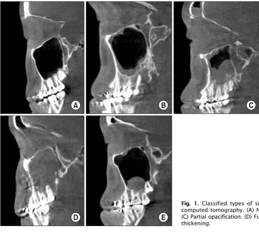
상악동 점막의 변화는 사진상 상악동 내 연조직 음영의 증가가 가장 심한 부위를 기준으로 분류하였으며, 변환된 횡단면 영상에 서 점막 비후가 2 mm 이하는 정상, 2 mm 이상 6 mm 미만은 점막비후(mucosal thickening), 6 mm 이상의 점막 비후는 부분 불투명(partial opacification), 그리고 완전 불투명(full opacifi- caton) 및 폴립형 점막 비후(polypoidal mucosal thickening) 로 분류하였다(Fig. 1). 상악동 개구의 크기는 시상면 및 횡단면 사진에서 상악동 개구부를 국소화시킨 후 횡단면 사진에서 관찰되 는 가장 큰 부위를 측정하였다(Fig. 2).
수술 전, 후의 상악동 점막의 상태에 따른 상악동 개구 크기의 차이를 확인하기 위해 SPSS version 12 (SPSS Inc., Chicago, IL, USA)를 사용하여 ANOVA test를 실시하였고, 수술 전, 후 상악동 점막의 두께 및 상악동 개구의 변화를 알아보기 위해 paired t-test를 사용하였다. 점막의 두께를 관찰할 수 없는 정상 상악동에서는 점막의 두께를 0 mm로, 폴립형 점막 비후인 경우는 폴립 이외의 점막의 두께를 측정하였고 완전 불투명의 경우 상악 동 최대 높이의 1/2을 점막의 두께로 정하였다. 이때 P value가 0.05 이하일 경우 통계적으로 유의하다고 판정하였다.
결 과
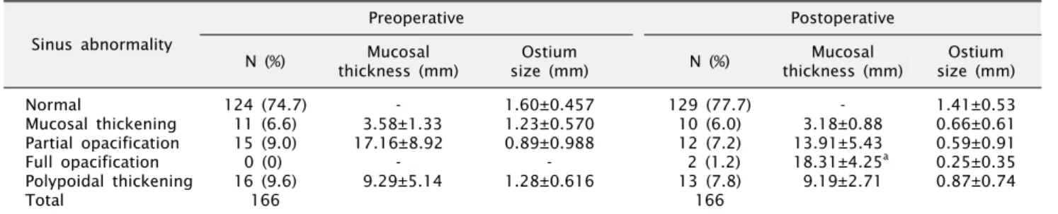
총 83명의 환자의 166개의 상악동을 평가한 결과, 술 전에 점막 비후를 포함하는 비정상적 상악동은 42개(25.3%), 술 후에 는 37개(22.3%)로 나타났다. 술 전과 술 후 모두에서 각각의 상악동 점막의 상태에 따른 상악동 개구의 크기는 정상에 비해 점막 비후, 부분 불투명, 완전 불투명으로 갈수록 작아지는 경향을 보였으나 통계적으로 유의한 차이를 얻지는 못하였다(Table 1).
술 전 상악동 점막의 상태가 정상으로 분류된 경우 술 후 점막의 비후나 폴립형 점막 비후 등 상태가 악화된 경우는 6개(4.8%)였 고, 점막 비후나 부분 불투명을 보이는 상악동의 경우에도 대부분 의 경우 상악동 점막의 상태가 유지 혹은 개선되었다. 하지만 술 전 점막 비후와 폴립형 점막 비후를 보이던 상악동에서 술 후 완전 불투명으로 바뀐 경우도 각각 1개씩 발견되었다(Table 2).
Cone-beam CT 사진상 술 전과 술 후의 상악동 개구의 크기는
통계적으로 유의한 감소를 보였으며( P <0.05), 상악동 점막 두께
변화를 확인한 결과 술 후에서 다소 감소하는 양상을 보였으나
통계적으로 유의성은 없었다(Table 3).
Fig. 2. Measurement of diameter of the maxillary sinus ostium ().
고 찰
이번 연구 결과 술 전 Cone beam CT상 상악동 점막의 이상 소견은 23% (42/166)로 나타났다. 이는 CT를 통한 연구에서 나타나는 63∼83.2%나[12,13] Cone beam CT를 통한 연구에서 나타난 56.3%[10]에 비해 현저하게 낮은 유병률을 보였다. 이는
조사대상의 차이가 그 원인으로 보이며, 특히 나이가 많을수록 상악동 점막 이상 소견이 많이 나타난다는 연구와 같이[14] 이번 연구대상의 나이가 비교적 젊은 것이 영향을 준 것으로 보인다.
방사선학적으로 정상 상악동은 공기로 차 있으므로 방사선 투과상이며 명확한 경계를 가진다. 하지만 질병에 이환된 상악동 의 경우 방사선 사진상에 불투명, 점막 비후 등을 관찰할 수 있다 [15]. 방사선 검사에서 비정상적으로 판정할 만한 연조직 음영 증가의 기준에 대해서는 Rak 등[16]은 MRI (자기공명영상촬영) 에서 4 mm 이상의 점막의 비후가 임상적인 증상과 연관이 있다고 하였으며, 1∼2 mm의 점막 두께는 정상 범주로 정의하였다.
Patel 등[17]은 2 mm 이상을 의미 있다고 하였으며, 현재까지 명확한 기준은 없는 상태이다. 본 연구에서는 2 mm 이상을 비정 상으로 간주하고 분류하였다.
수술 전 상악동의 상태를 관찰하기 위한 검사로 단순 부비동 촬영, 초음파 검사, CT, MRI 등이 있다. 이 중 MRI는 점막의 변화 및 연조직 이상을 관찰하는 데 더 우수하지만 골조직의 변화나 석회화 병변은 관찰하기 힘든 단점이 있어[13], 현재까지 CT가 상악동 평가를 위해 많이 시행되고 있다. 최근에는 촬영시간 이 적고 외래에서 쉽게 사용 가능한 cone beam CT가 구강 악안면 영역에서 많이 이용되고 있다. Cone beam CT는 일반 CT (Multidectector CT)에 비해 높은 해상도와 적은 방사선 선 량, 저렴한 비용의 장점이 있으며, 등방성(isotropic) 복셀(voxel)
Fig. 1. Classified types of sinus abnormalities in the cone-beam
computed tomography. (A) Normal sinus. (B) Mucosal thickening.(C) Partial opacification. (D) Full opacification. (E) Polypoidal mucosal thickening.
Table 1. Prevalence of maxillary sinus abnormalities before and after Le Fort I osteotomy
Sinus abnormality
Preoperative Postoperative
N (%) Mucosal
thickness (mm)
Ostium
size (mm) N (%) Mucosal
thickness (mm)
Ostium size (mm) Normal
Mucosal thickening Partial opacification Full opacification Polypoidal thickening Total
124 (74.7) 11 (6.6) 15 (9.0) 0 (0) 16 (9.6)
166
- 3.58±1.33 17.16±8.92
- 9.29±5.14
1.60±0.457 1.23±0.570 0.89±0.988
- 1.28±0.616
129 (77.7) 10 (6.0) 12 (7.2) 2 (1.2) 13 (7.8) 166
- 3.18±0.88 13.91±5.43 18.31±4.25a
9.19±2.71
1.41±0.53 0.66±0.61 0.59±0.91 0.25±0.35 0.87±0.74
When comparing differences in ostium size by sinus abnormality, there was no significant difference in all groups (ANOVA test).
Values are presented as n (%) or mean±standard deviation.
aHalf of sinus height.
Table 2. Pre- and postoperative changes of maxillary sinus abnormalities
Preoperative
Postoperative
Normal Mucosal
thickening
Partial opacification
Full opacification
Polypoidal
thickening Total Normal
Mucosal thickening Partial opacification Full opacification Polypoidal thickening Total
118 3 3 - 5 129
1 4 2 - 3 10
2 - 9 - 1 12
- 1 - - 1 2
3 3 1 - 6 13
124 11 15 0 16 166
Table 3. Mucosal thickness and ostium size in before and after
Le Fort I osteotomy (n=166)Pre-operative
(mm) Post-operative
(mm)
P-value
Mucosal thickness Ostium size
1.90±5.64 1.48±0.56
1.58±4.40 0.87±1.08
0.373 0.000a Paired t-test.
a
P <0.05.
을 가져 정확도가 높고 금속에 의한 이미지 왜곡이 적다. 하지만 낮은 대조구간(contrast range)과 하운스필드 유닛(Hounsfield units)을 적용하기 어려운 점으로 인해 내부 연조직 평가에 불리한 단점을 가진다[18]. 이러한 단점에도 불구하고 치성기원의 상악동 염의 평가를 비롯하여[10,11,19] 부비동 수술 시 cone beam CT의 사용[20]이 증가하고 있다. 특히 악교정 수술 환자의 경우 술 전 분석과 모의수술, 술 후 평가 등을 위해 cone-beam CT의 촬영이 일반적으로 시행되고 있어 추가적인 검사 없이 상악동을 평가할 수 있는 장점이 있다. 본 연구에서도 상악동 점막의 평가나 상악동 개구의 크기를 확인하는 데 어려움이 없었으며, 소프트웨 어를 사용하면 변환된 영상을 통해 더 정확한 위치 및 크기를 측정할 수 있었다.
상악골 절단술 후에 발생하는 상악동염의 발생 가능한 원인으로는 상악동 점막의 섬모운동의 변화, 상악동의 혈병의 생성과 저류, 의원성 손상에 의한 치성 이차 감염, 조직의 허혈, 혈액 공급의
부족, 상악동에 남은 이물질 등을 생각할 수 있다[5,6]. 이 중
부비동 개구 연합(ostiomeatal unit)의 해부학적 변화는 르포트
씨 1급 골절단술 후 발생하는 상악동염의 가장 흔한 원인으로
지목되고 있다[21,22]. 정상 상악동 개구의 크기는 2 mm 이상으
로 알려져 있으나 본 연구에서는 정상군에서 1.5 mm 정도로
측정되었으며, CT를 통한 다른 연구에서도 비슷한 크기로 관찰되
었다[23]. 이는 정상 상악동 개구의 크기가 0.5∼5 mm까지 변위
가 크며[24], 자세에 따른 변화나[25] 촬영 방법, 측정 위치에
영향을 받는 것으로 보인다. 따라서 절대적인 크기보다는 변화양
상에 더 의미가 있을 것이다. 상악동 개구부가 작은 경우 르포트씨
1급 골절단술 후 점막의 부종으로 인해 상악동 개구의 폐쇄가
더 잘 일어날 수 있고, 이러한 상악동 개구의 폐쇄가 섬모운동의
변화를 야기해 상악동염을 일으킬 수 있다. Moses 등[22]은 이러
한 환자에서 술 중 부가적인 배출 통로를 만들어 주거나 내시경
수술을 통한 상악동 개구부의 확장이 필요하다고 하였다. 하지만
Turvey[26]는 상악동 개구의 크기가 르포트씨 1급 골절단술 후에
증가하여 상악동의 배출능력이 향상된다고 주장하였다. 본 연구
에서는 술 후에는 술 전에 비해 상악동 개구부의 크기가 작아지는
양상을 보였다. 하지만 술 전과 술 후의 상악동 점막의 상태에
따른 차이는 발견되지 않아 상악동 개구의 크기 변화가 상악동
비정상과의 연관성은 없는 것으로 보이며, 상악동 개구의 감소가
크지 않아 측정할 때의 오류를 배제할 수 없어 해석에 주의를
요한다. 그러나, 술 전 검사상 상악동 개구부의 크기가 작은 경우
술 후 상악동 개구부의 폐쇄 가능성에 대한 고려가 필요하리라 생각한다.
르포트씨 1급 골절단술 후 상악동염의 발생빈도는 낮은 것으로 알려져 있다. Bell 등[3]은 술 전과 술 후 증상의 발현에 차이가 없다고 하였으며, Pereira-Filho 등[27]은 통증이나 코막힘 등이 개선되었으며 이는 상악 재위치에 따른 비강 내의 공기 흐름의 개선에 의한 것으로 설명하였다. 하지만 이들 연구들은 대부분 설문지와 일반 부비동 사진을 통해 조사하거나 내시경을 이용해 비강을 관찰한 것으로 상악동 내부의 변화를 확인하기는 어려웠 다. 이번 연구에서는 상악동 내부의 점막 변화를 관찰한 결과 통계적으로 유의한 차이는 없었으나 다소 감소하는 것을 관찰할 수 있었다. 점막의 두께 측정에서 정상 상악동의 수가 너무 많아 통계적 유의성은 없었지만 점막 두께 변화가 있는 상악동만을 대상으로 측정하였을 때는 더 큰 점막 두께의 감소를 확인할 수 있었다(not shown). 이는 상악골 수술이 상악동 점막에 부정 적인 영향을 줄 수도 있으나 동시에 술 중에 상악동 점막의 이상이 나 폴립형 점막 비후 등을 제거하여 일반적인 상악동 수술의 결과와 같은 점막의 치유를 얻을 수 있기 때문으로 생각한다.
일반적인 상악동 수술 후의 점막의 치유는 4단계로 구분할 수 있는데 술 후 7∼12일 동안 수술부위의 피덩이의 생성이 이루어 지며 2∼4주에 걸쳐 육아조직의 형성이 일어난다. 이후 점막의 부종 상태가 지속되다가 약 12∼18주 이후 정상화 단계 및 반흔화 가 진행된다[28]. 하지만, 점막의 치유는 반흔조직으로 치유되어 상악동의 배출능력이 감소할 수 있으므로 술 중 이상 점막의 제거는 최소한으로 줄이는 것이 좋을 것으로 생각한다[29].
이번 연구를 통해 이전 연구들과 마찬가지로 르포트씨 1급 골절단술 후 상악동 점막의 이상이 더 발생하지는 않는 것으로 나타났다. 이번 연구에서는 상악골의 이동 방향이나 악골 부조화 의 종류 등에 따른 분류는 하지 않았지만, 심한 비대칭이나 골 이식 시 상악동염의 발생 가능성이 더 높을 수 있으므로[22] 술 후 상악동 변화에 영향을 주는 요인에 대한 더 많은 연구가 필요하 리라 생각한다.
결 론
본 연구는 르포트씨 1급 골절단술을 시행한 환자에서 술 전과 술 후의 상악동의 변화를 알아보기 위해 시행되었다. Cone beam CT를 이용하여 점막 비후가 2 mm 이하는 정상, 2 mm 이상 6 mm 미만은 점막 비후(mucosal thickening), 6 mm 이상의 점막 비후는 부분 불투명(partial opacification), 그리고 완전 불투명(full opacificaton) 및 폴립형 점막 비후(polypoidal mu- cosal thickening)로 분류하여 술 전과 술 후를 비교하여 다음과 같은 결과를 얻었다.
1. 술 전과 비교하여 술 후에 상악동 점막의 비정상이 더 증가하
지는 않았다.
2. 술 전과 술 후 모두에서 각각의 상악동 점막의 상태에 따른 상악동 개구의 크기는 정상에 비해 점막 비후, 부분 불투명, 완전 불투명으로 갈수록 작아지는 경향을 보였으나 통계적 유의성은 없었다.
3. 술 전과 술 후의 상악동 개구의 크기를 확인한 결과 술 후에서 통계적으로 유의한 감소를 보였다( P <0.05).
3. 술 후의 상악동 점막의 두께 변화는 통계적 유의성이 없었다.
이상의 결과 르포트씨 1급 골절단술을 시행한 환자에서 상악동 점막 이상은 악화되지는 않는 것으로 나타났다. 하지만 술 후 상악동 점막 이상이 악화되는 경우도 있으므로 수술 전 충분한 설명과 술 후 상악동 질환의 발현에 대한 세심한 주의 및 관찰이 필요하리라 생각한다.
Acknowledgements
이 논문은 부산대학교 자유 과제 학술연구비(2년)에 의하여 연구되었다(This work was supported by a 2-Year Research Grant of Pusan National University.).
References