Prevalence of incidental maxillary sinus findings in Italian orthodontic patients: a retrospective cone- beam computed tomography study
Objectives:
To determine the prevalence of incidental maxillary sinus findings in a large sample of orthodontic patients by cone-beam computed tomography (CBCT) with a wide field of view and assess the relationships of such abnor- malities with age and gender.
Methods: Five hundred thirteen CBCT scansobtained for orthodontic diagnosis and treatment planning in a Northern Italian population (N = 513; 292 female and 221 male subjects; 1,026 maxillary sinuses) were studied. The frequencies of pseudocysts and mucosal thickening of the maxillary sinus were recorded. Logistic regression analysis was used to determine the influence of age and gender on these abnormalities. Results: Pseudocysts were detected in 52 patients (10.1%) and 59 sinuses (5.75%). Mucosal thickening was observed in 206 patients (40.1%) and 258 sinuses (25.1%). Gender and age were significantly associated with pseudocysts (p = 0.027) and mucosal thickening (p <
0.001), respectively. Conclusions: Half of the orthodontic patients had incidental maxillary sinus findings. Men were more likely to show pseudocysts, and older patients (aged 41 - 60 years) were more likely to show mucosal thickening.
[Korean J Orthod 2012;42(6):329-334]
Key words: Computed tomography, Three dimensional diagnosis and treatment
planning
Antonio Gracco
aSerena Incerti Parenti
bChristian Ioele
cGiulio Alessandri Bonetti
bEdoardo Stellini
daDepartment of Orthodontics, School of Dentistry, University of Padua, Padua, Italy
bDepartment of Orthodontics, School of Dentistry, University of Bologna, Bologna, Italy
cDepartment of Orthodontics, School of Dentistry,University of Ferrara, Ferrara, Italy
dDepartment of Pedodontics, School of Dentistry, University of Padua, Padua, Italy
Received June 2, 2012; Revised September 8, 2012; Accepted September 10, 2012.
Corresponding author: Serena Incerti Parenti.
Visiting Professor, Department of Orthodontics, School of Dentistry, University of Bologna, Via San Vitale 59, 40125, Bologna, Italy.
Tel +39-0512088133 e-mail [email protected]
©
2012 The Korean Association of Orthodontists.The authors report no commercial, proprietary, or financial interest in the products or companies described in this article.
This is an Open Access article distributed under the terms of the Creative Commons Attribution Non-Commercial License (http://creativecommons.org/licenses/by-nc/3.0) which permits unrestricted non-commercial use, distribution, and reproduction in any medium, provided the original work is properly cited.
pISSN 2234-7518 • eISSN 2005-372X
http://dx.doi.org/10.4041/kjod.2012.42.6.329
INTRODUCTION
The advent of cone-beam computed tomography (CBCT) represents a great improvement in the field of craniofacial imaging.
1Unlike traditional two-dimensional (2D) radio graphy, CBCT avoids structural superimposition and image enlargement and distortion, thus allowing precise three-dimensional (3D) visualization of dental and maxillofacial structures at a lower radiation dose than multislice computed tomography (CT).
1Such imaging frequently presents the dentist or orthodontist with incidental findings and may raise concerns about the need for further diagnostic tests or referral to other specialists.
2,3Incidental findings are common in the maxillary sinus.
4Although earlier studies using CBCT for dental diagnostic purposes have already addressed this issue,
2,4only one study using CBCT focused on incidental maxillary sinus findings in orthodontic patients.
5However, the study was limited by the small sample size and imaging with a narrow field of view (FOV), which does not allow accu- rate assessment of sinus pathology.
The aims of this retrospective study were to determine the prevalence of incidental maxillary sinus findings in a large sample of orthodontic patients by CBCT with a wide FOV and assess the relationships of such abnormalities with age and gender.
MATERIAL AND METHODS
Sample
This study included 680 CBCT images obtained for orthodontic diagnosis and treatment planning at the Depart ment of Orthodontics, University of Ferrara, Italy, between January 2005 and December 2010. The protocol was approved by the local Institutional Review Board.
Images of patients with fixed orthodontic appliances, implants, or metallic prosthesis in the maxillary posterior region that could generate scattering phenomena, cranio- facial syndromes, or cleft palate were excluded. Finally, 513 CBCT scans of Northern Italian patients (N = 513;
292 female and 221 male subjects; 1,026 maxillary sinuses) were selected. For statistical analysis, the subjects were divided into the following age groups
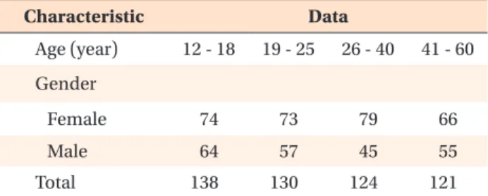
4: 12 - 18, 19 - 25, 26 - 40, and 41 - 60 years. The age and gender distributions are shown in Table 1.
CBCT scanning
CBCT was performed by using a NewTom 3G volume scanner (QR srl, Verona, Italy) under the following conditions: 12-inch FOV, 110 kV (anterior posterior- latero lateral), 2.00 mA (anterior posterior), 1.00 mA (latero lateral), 5.4-s exposure, and 0.50-mm slice thick- ness.
The resulting images were elaborated by using NNT NewTom 3G software to obtain the axial sections in which the maxillary sinus could best be distinguished.
Then, a line was traced through the greatest mesiodistal dimension of the most suitable sections and various 0.2-mm-spaced coronal sections of the maxilla perpen- dicular to this line were obtained.
The CBCT scans were reviewed independently by 2 orthodontists trained by a radiologist with experience of over 1,000 NewTom scans at the time of the study. The diagnostic criteria were developed on the basis of the published literature, and in the case of a disagreement, a consensus was reached after a discussion between the observers. Each observer separately performed all the measurements by using the same computer (50-inch Table 1. Age and gender distributions of the sample
Characteristic Data
Age (year) 12 - 18 19 - 25 26 - 40 41 - 60 Gender
Female 74 73 79 66
Male 64 57 45 55
Total 138 130 124 121
Figure 1. CBCT scans with
incidental findings of pseudocysts associated with mu
cosal thickening in the right maxillary sinus.
liquid crystal display monitors, ASUS VW246H; ASUS, Taipei, Taiwan) and screen resolution (bright ness, 300 cd/
m
2; resolution, 1,920 × 1,080).
Assessment of pseudocysts
Pseudocysts were diagnosed as homogeneous, dome- shaped, noncorticated soft tissue opacities with a smooth and well-defined outline in the maxillary sinus (Figure 1).
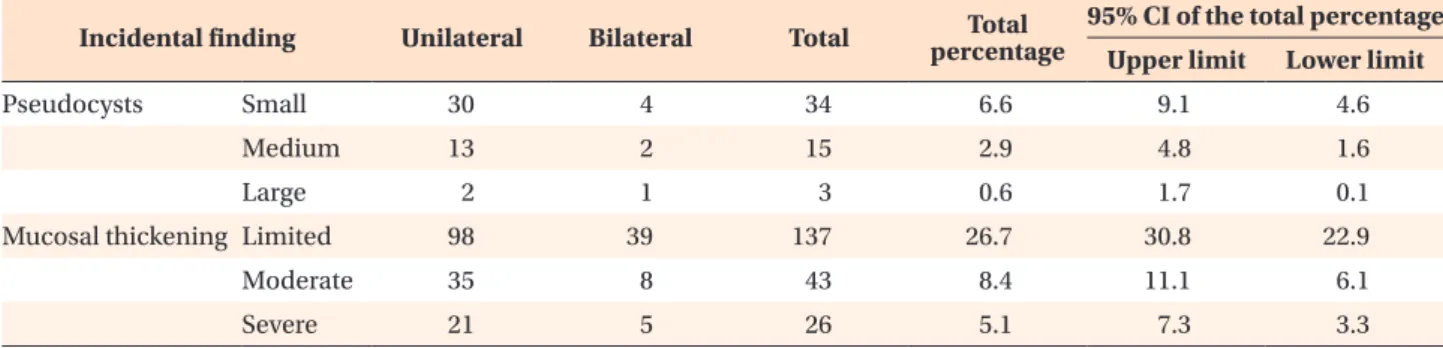
6,7For statistical analysis, the pseudocysts were cate- gorized according to their greatest diameter (small, < 1 cm; medium, 1 - 2 cm; large, > 2 cm) and location (right, left, or bilateral).
Assessment of mucosal thickening
Mucosal thickening was considered present when the thickness of the sinus mucosa was ≥ 1 mm as measured from the floor of the sinus to the highest border of the mucosa (Figure 2).
8For statistical analysis, this patho logy was classified according to the degree of sinus opaci- fication (limited, < 1/3; moderate, 1/3 - 2/3; severe, > 2/3) and location (right, left, or bilateral).
Calibration and reliability
The 2 observers were calibrated for evaluation by toge- ther reviewing and discussing the findings of 20 sam- ple CBCT images (10 per pathology) that had been
Figure 2. CBCT scans with in
cidental findings of mucosal thickening in the right maxil
lary sinus.
Table 2. Unilateral incidental findings in the maxillary sinuses
Pseudocysts Mucosal thickening
Small Medium Large Limited Moderate Severe
Left sinus 20 7 2 89 21 16
Right sinus 18 10 2 87 30 15
Total 38 17 4 176 51 31
Table 3. Data of the unilateral and bilateral incidental findings in the maxillary sinuses
Incidental finding Unilateral Bilateral Total Totalpercentage
95% CI of the total percentage Upper limit Lower limit
Pseudocysts Small 30 4 34 6.6 9.1 4.6
Medium 13 2 15 2.9 4.8 1.6
Large 2 1 3 0.6 1.7 0.1
Mucosal thickening Limited 98 39 137 26.7 30.8 22.9
Moderate 35 8 43 8.4 11.1 6.1
Severe 21 5 26 5.1 7.3 3.3
CI, Confidence interval.