©Copyights 2012. The Korean Academy of Conservative Dentistry.
50
This is an Open Access article distributed under the terms of the Creative Commons Attribution Non-Commercial License (http://creativecommons.org/licenses/
by-nc/3.0) which permits unrestricted non-commercial use, distribution, and reproduction in any medium, provided the original work is properly cited.
Endodontic management of a maxillary lateral incisor with dens invaginatus and external root irregularity using cone-beam computed tomography
Young-Jun Lim
1, Sook- Hyun Nam
1, Sung-Ho Jung
1, Dong-Ryul Shin
2, Su-Jung Shin
3, Kyung-San Min
1*
1Department of Conservative Dentistry, Wonkwang University School of Dentistry, Iksan, Korea
2Gangnam Luden Dental Clinic, Seoul, Korea
3Department of Conservative Dentistry, Gangnam Severance Dental Hospital, Yonsei University College of Dentistry, Seoul, Korea
Cone-beam computed tomography (CBCT) is a useful diagnostic tool for identification of both internal and external root configurations. This case report describes the endodontic management of a lateral incisor with both dens invaginatus and external root irregularity by using CBCT. Nonsurgical endodontic retreatment was performed on the lateral incisor with dens invaginatus. A perforation through the dens invaginatus and external concavity was repaired using mineral trioxide aggregate. After 18 mon of follow-up, there were no clinical symptoms. Recall radiographs appeared normal and showed healing of the periapical pathosis. The understanding of both internal root canal configuration and external root irregularity using CBCT can ensure predictable and successful results. (Restor Dent Endod 2012;37(1):50-53)
Key words: Cone-beam computed tomography; Dens invaginatus; External root irregularity; Perforation
Received October 10, 2011;
Last Revision December 23, 2011;
Accepted December 26, 2011.
1Lim YJ, DDS, Resident; Nam SH, DDS, MSD; Jung SH, DDS, MSD, Graduate student; Min KS, DDS, PhD, Associate Professor, Department of Conservative Dentistry, Wonkwang University School of Dentistry, Iksan, Korea
2Shin DR, DDS, PhD, Gangnam Luden Dental Clinic, Seoul, Korea
3Shin SJ, DDS, MSD, Assistant Professor, Department of Conservative Dentistry, Gangnam Severance Dental Hospital, Yonsei University College of Dentistry, Seoul, Korea
*Correspondence to Kyung-San Min, DDS, PhD
Associate Professor, Department of Conservative Dentistry, Wonkwang University School of Dentistry, 344-2 Shinyong-dong, Iksan, Korea 570-749 TEL, +82-63-850-6930; FAX, +82-63- 859-2932; E-mail, [email protected]
Case report
ISSN 2234-7658 (print) / ISSN 2234-7666 (online) http://dx.doi.org/10.5395/rde.2012.37.1.50
Introduction
Dens invaginatus is a dental malformation caused by an infolding of the enamel organ into the adjacent dental papilla during tooth development.1 According to many studies, dens invaginatus is a rare dental malformation with an incidence of 0.04 - 10%.2 The permanent maxillary lateral incisor appears to be the most frequently affected tooth, and the posterior teeth are less likely to be affected.1,3,4 However, the cause of dens invaginatus is unclear.
The complex anatomy of dens invaginatus makes conservative endodontic treatment of such teeth difficult and unpredictable. Although conventional intraoral periapical radiography is an important diagnostic tool in endodontics for assessing the root canal configuration, it is usually insufficient for understanding the complicated morphology of the root canal system in cases of dens invaginatus. These problems might be overcome by the use of newer diagnostic methods such as cone-beam computed tomography (CBCT).5,6 CBCT is known to be a useful diagnostic tool for assessing the root configuration.7,8
The use of CBCT aids in evaluation of not only internal root canal anatomy but also external root irregularities, especially those in the buccolingual plane. External root irregularities can be formed naturally or iatrogenically by clinicians (e.g., root perforation). Root perforation is closely related to external root irregularities. It is well known that the presence of concavities on root surfaces increases the risk of perforation. In this respect, appropriate use of CBCT might help in avoiding tragic results during access cavity preparation and canal preparation.
Several case reports have described the assessment of dens invaginatus using CBCT.9,10
※This paper was supported by Wonkwang University in 2010.
51 www.rde.ac
http://dx.doi.org/10.5395/rde.2012.37.1.50
However, to our knowledge, no case report has described the management of dens invaginatus combined with external root irregularity. This case report describes the endodontic management of a lateral incisor with both dens invaginatus and external root irregularity by using CBCT.
Report
A 46-year-old woman was referred from a private clinic for management of dens invaginatus of the maxillary left lateral incisor. The patient’s medical history was noncontributory. The previous dentist created a perforation during access cavity preparation. Clinical examination revealed a non-carious cervical defect incompletely filled with glass ionomer cement (Figure 1a).
The maxillary left incisor responded negatively to thermal and electric pulp testing (Digitest Pulptester, Parkell Inc, Edgewood, NY, USA). Periodontal probing revealed a normal and intact periodontium. No mobility was noted, and the tooth was not tender to percussion. The periapical radiograph showed a dens invaginatus with a periapical area of radiolucency (Figure 1b). However, the morphology of the invagination was not clear from the conventional intraoral periapical radiograph. To confirm this unusual morphology, CBCT imaging (Alphard VEGA, Asahi Roentgen Ind. Co., Kyoto, Japan) of the tooth was performed. Informed consent was obtained from the patient. Morphology of the dens invaginatus was evaluated from transverse, axial, and sagittal sections of 0.1 mm
thickness.
From the clinical and radiographic findings, we diagnosed chronic apical periodontitis of the maxillary left lateral incisor. The CBCT images revealed perforation and external root irregularity on the labial aspect (Figure 2). A rubber dam was placed, and the tooth was prepared to provide adequate endodontic access. With the use of a microscope (OPMI pico Dental Microscope, Carl Zeiss, Oberkochen, Germany), 1 root canal and invagination were found.
Working length was determined using an apex locator (Root ZX, Morita, Tokyo, Japan) followed by confirmation with a radiograph. The root canal was cleaned and shaped with ProTaper Universal rotary instruments (Dentsply- Maillefer, Ballaigues, Switzerland) under copious irrigation with 2.5% sodium hypochlorite. The finishing of canal preparation was performed until an F2 file reached the full working length. A plug of mineral trioxide aggregate (MTA) (ProRoot, Dentsply, Tulsa, OK, USA) was then condensed into the perforation site. After a cotton pellet was applied, the access cavity was restored with temporary sealing material (Caviton, GC, Tokyo, Japan). At the next appointment, the patient was asymptomatic, and the main root canal was dried and filled with thermoplasticized gutta-percha and sealer (AH Plus, Dentsply DeTrey, Konstanz, Germany) (Figure 3a). The access and cervical defect were then permanently restored with a universal composite resin restorative material (Z350, 3M ESPE, St.
Paul, MN, USA).
After 18 months of follow-up, there were no clinical
Figure 1. (a) Clinical view of maxillary lateral incisor showing cervical non-carious defect; (b) Radiographic view of dens invaginatus. Periapical radiolucency can be seen; (c) Sagittal CBCT section of the tooth. White arrow indicates the perforation site.
(a)
(c)
(b)
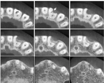
Figure 2. A set of transverse CBCT scan showing external concavity of the maxillary left lateral incisor. Note perforation communicates with periodontium through this concavity (white arrow). CBCT, cone-beam computed tomography.
Maxillary lateral incisor with complex anatomy
52 www.rde.ac
symptoms. Recall radiographs appeared normal and showed healing of the periapical pathosis (Figure 3b).
Discussion
Endodontic treatment of dens invaginatus can be challenging because of the bizarre morphology often associated with the condition and insufficient information provided by conventional periapical radiographs.11 The use of CBCT has provided new capabilities for visualization and treatment of these anomalies. Patel suggested that the true nature of dens invaginatus cannot always be estimated from conventional radiographs and that CBCT is a useful diagnostic tool in the management of this anomaly.9 Moreover, CBCT is helpful for identification of external root irregularities. Kamburoğlu et al. showed that CBCT images are more useful in detection and localization of external cervical root resorption than conventional film images.12 In the present case, the tooth showed not only dens invaginatus but also external concavity on the facial root surface, and these anomalies were obvious on CBCT images. With the use of this information, we were able to set up proper treatment strategies, including prevention of additional complications and repair of the present perforation.
We found that the perforation had been made by the previous dentist through the dens invaginatus and that it communicated with the external root concavity. Shemesh et al. demonstrated that CBCT scans showed a significantly higher sensitivity than periapical radiographs for detection of strip root perforations, which are also related to external
root concavity.13 Furthermore, CBCT is more useful for detection of perforations in the faciolingual plane than is conventional radiography. In this respect, we can assume that the risk of perforation increases during access cavity preparation when internal malformations such as dens invaginatus and external irregularity coexist and that this iatrogenic complication may be prevented or identified with the aid of CBCT.
MTA has profound advantages when used as perforation repair material because of its superior biological properties and excellent sealing ability. The reports of Main et al.
confirm the superiority of MTA as a perforation repair material.14-17 In our case, we used white MTA to repair the perforation, because of the potential discoloration effect of gray MTA. According to Asgary et al., the major difference between white MTA and gray MTA appears to be in the concentrations of carborundum, periclase, and especially ferrous oxide.18 The observed values for each of these oxides is considerably lower in white MTA than in gray MTA.18 Differences in the observed ferrous oxide concentration are thought to be primarily responsible for the variation in color of the white MTA compared with gray MTA. In this case, we did not observe any tooth discoloration until the 18 months recall.
Conclusions
In the present case, successful nonsurgical endodontic retreatment of a lateral incisor that had dens invaginatus, external root concavity, and iatrogenic perforation was performed with the aid of CBCT. The complete understanding of both internal and external anatomy using CBCT can ensure predictable and successful results.
References
1. Hülsmann M. Dens invaginatus: aetiology, classification, prevalence, diagnosis, and treatment considerations.
Int Endod J 1997;30:79-90.
2. Hovland EJ, Block RM. Nonrecognition and subsequent endodontic treatment of dens invaginatus. J Endod 1977;3:360-362.
3. Conklin WW. Bilateral dens invaginatus in the mandibular incisor region. Oral Surg Oral Med Oral Pathol 1978;45:905-908.
4. Lee AM, Bedi R, O’Donnell D. Bilateral double dens invaginatus of maxillary incisors in a young Chinese girl. Aust Dent J 1988;33:310-312.
5. Arai Y, Tammisalo E, Iwai K, Hashimoto K, Shinoda K. Development of a compact computed tomographic apparatus for dental use. Dentomaxillofac Radiol 1999;
28:245-248.
6. Mozzo P, Procacci C, Taccoci A, Martini PT, Andreis IA.
A new volumetric CT machine for dental imaging based Figure 3. (a) Radiographic view showing root fillings.
White arrow indicates the MTA to repair perforation;
(b) Radiographic view after 18 months showing apical healing. MTA, mineral trioxide aggregate.
(a) (b)
http://dx.doi.org/10.5395/rde.2012.37.1.50 Lim YJ et al.
53 www.rde.ac
http://dx.doi.org/10.5395/rde.2012.37.1.50
Maxillary lateral incisor with complex anatomy
on the cone-beam technique: preliminary results. Eur Radiol 1998;8:1558-1564.
7. La SH, Jung DH, Kim EC, Min KS. Identification of independent middle mesial canal in mandibular first molar using cone-beam computed tomography imaging.
J Endod 2010;36:542-545.
8. Song CK, Chang HS, Min KS. Endodontic management of supernumerary tooth fused with maxillary first molar by using cone-beam computed tomography. J Endod 2010;36:1901-1904.
9. Patel S. The use of cone beam computed tomography in the conservative management of dens invaginatus: a case report. Int Endod J 2010;43:707-713.
10. Durack C, Patel S. The use of cone beam computed tomography in the management of dens invaginatus affecting a strategic tooth in a patient affected by hypodontia: a case report. Int Endod J 2011;44:474- 483.
11. Pai SF, Yang SF, Lin LM. Nonsurgical endodontic treatment of dens invaginatus with large periradicular lesion: a case report. J Endod 2004;30:597-600.
12. Kamburoğlu K, Kurşun S, Yüksel S, Oztaş B. Observer ability to detect ex vivo simulated internal or external cervical root resorption. J Endod 2011;37:168-175.
13. Shemesh H, Cristescu RC, Wesselink PR, Wu MK. The use of cone-beam computed tomography and digital periapical radiographs to diagnose root perforation. J Endod 2011;37:513-516.
14. Main C, Mirzayan N, Shabahang S, Torabinejad M. Repair of root perforations using mineral trioxide aggregate: a long-term study. J Endod 2004;30:80-83.
15. Oh MJ, Jeong YN, Bae IH, Yang SY, Park BJ, Koh JT, Hwang YC, Hwang IN, Oh WM. Biocompatibility of experimental mixture of mineral trioxide aggregate and glass ionomer cement. J Korean Acad Cons Dent 2010;
35:359-367.
16. Cho YB. Mineral trioxide aggregate and its substitutes.
J Korean Acad Cons Dent 2010;35:149-151.
17. Jeong YN, Yang SY, Park BJ, Park YJ, Hwang YC, Hwang IN, Oh WM. Physical and chemical properties of experimental mixture of mineral trixoxide aggregate and glass ionomer cement. J Korean Acad Cons Dent 2010;35:344-352.
18. Asgary S, Parirokh M, Eghbal MJ, Brink F. Chemical differences between white and gray mineral trioxide aggregate. J Endod 2005;31:101-103.