https://doi.org/10.5624/isd.2019.49.1.45
Introduction
The anatomic abnormalities and pathological conditions of the sino-nasal complex can be easily identified using computed tomography(CT).1,2 As cone-beam CT(CBCT) has become more widespread, features of the nose and paranasal sinuses have become more easily identified by oral radiologists, with higher accuracy.3 In the osteo-me- atal complex region, concha bullosa(CB) is considered the most common anatomic variation.4 It can be simply de- scribed as a pneumatized middle turbinate. The causes of pneumatization are unclear, although trauma, nasal septal
deviation(NSD), and mouth breathing have been estab- lished as predisposing factors of CB.5 Earwaker reported a prevalence of CB of 55% of his sample,6 whereas Zinreich et al.7 reported that CB was present in 34% of their sample.
CB can compromise the middle meatus, particularly the osteo-meatal complex, which in turn may make maxillary sinus disease more likely due to the compromised mucocil- iary drainage.3,7 Although the majority of cases of CB are asymptomatic, ongoing research is being conducted into its association with sinus disease, when the osteo-meatal com- plex is blocked.8,9
NSD is a very common problem among individuals from the Middle East.10 The most common reported cause of septal deformity is trauma during infancy and childhood.5 However, there is little evidence regarding the possibility that NSD or pneumatization of the middle turbinate may act as a potential contributor to MSV changes and sub-
Concha bullosa, nasal septal deviation, and their impacts on maxillary sinus volume among Emirati people: A cone-beam computed tomography study
Natheer H Al-Rawi 1,*, Asmaa T Uthman 2, Elaf Abdulhameed 1, Ahmed S Al Nuaimi 3, Zahra Seraj 1
1Department of Oral and Craniofacial Health Sciences, College of Dental Medicine, University of Sharjah, Sharjah, United Arab Emirates
2Department of Dental Surgical Sciences, College of Dentistry, Gulf Medical University, Ajman, United Arab Emirates
3Department of Clinical Research-Clinical Affairs Directorate, Primary Health Care Corporation (PHCC), Doha, Qatar
ABSTRACT
Purpose: To determine the prevalence of concha bullosa(CB) and nasal septal deviation(NSD) and their impact on maxillary sinus volume(MSV).
Materials and Methods: Cone-beam computed tomographic(CBCT) images of 106 Emirati people were used in this study. The direction and angle of septal deviation were calculated. The presence of CB, which could be unilateral, contralateral, or bilateral in relation to the direction of NSD, was also recorded. MSV was measured using reconstructed Digital Imaging and Communication in Medicine images on Dolphin 3D imaging software version 11.8 premium(Dolphin Imaging, Chatsworth, CA, USA). P values<0.05 were considered to indicate statistical significance.
Results: CB was detected in 37.7% of the sample; 20.7% of the sample showed single unilateral CB and 16.6% had single bilateral CB. NSD was seen in 74.5% of the sample. In the participants with CB, 45.5% showed mild deviation, 34.4% showed moderate deviation, and only 12.5% showed severe septal deviation. CB, but not NSD, was associated with significantly higher MSV on the affected side(P=0.001).
Conclusion: Although NSD was observed in more than two-thirds of the sample and CB was present in more than one-third of the sample, only CB had a significant impact on MSV.(Imaging Sci Dent 2019; 49: 45-51)
KEY WORDS: Turbinate; Nasal Septum; Maxillary Sinus; Cone-Beam Computed Tomography
Copyright ⓒ 2019 by Korean Academy of Oral and Maxillofacial Radiology
This is an Open Access article distributed under the terms of the Creative Commons Attribution Non-Commercial License(http://creativecommons.org/licenses/by-nc/3.0) which permits unrestricted non-commercial use, distribution, and reproduction in any medium, provided the original work is properly cited.
Imaging Science in Dentistry·pISSN 2233-7822 eISSN 2233-7830 Received October 14, 2018; Revised December 13, 2018; Accepted December 18, 2018
*Correspondence to: Prof. Natheer H Al-Rawi
Department of Oral and Craniofacial Health Sciences, College of Dental Medicine, University of Sharjah, PO Box: 27272, Sharjah, United Arab Emirates
Tel) 971551100169, E-mail) [email protected]
sequent sinus pathology. In the literature, NSD and CB have been found to interfere with proper air flow, which in turn may increase an individual’s predisposition to si- nus disease, but the relationship of this phenomenon with volumetric changes of the maxillary sinuses remains to be clarified.11 The objective of this study was to determine the prevalence of CB, NSD, and sinus pathology in a selected sample of Emirati individuals and to explore the possible association of these findings with MSV changes.
Materials and Methods
The data collection for this retrospective study was ap- proved by the Medical Ethics Committee of the University of Sharjah, United Arab Emirates(UAE). A total of 106 maxillofacial CBCT scans were taken over the course of 2 years(from October 2015 to October 2017) at the College of Dental Medicine, University of Sharjah(Sharjah, UAE).
The study sample was limited to Emirati adults over the age of 18 years. Patients’ medical records were reviewed to exclude individuals with a positive history of any prior sino-nasal surgery and/or craniofacial trauma.
All scans were taken using the Galileo 3D X-Ray system (Sirona Dental Systems, Long Island City, NY, USA) with a voxel size of 150μm. The images were reproduced and observed on the axial, coronal, and sagittal planes, and all scans were reviewed for any abnormalities in the sino-nasal complex, especially CB, NSD, and maxillary sinusitis. The direction of NSD and the angle of deviation were measured on coronal CBCT images. The angle of deviation was de- fined as “the angle between the crista galli and the most prominent point of deviation”.12 The convex part of the nasal septum was identified as the direction of deviation.
Patients were subsequently split into 3 groups according to the severity of NSD, defined by the angle of deviation:
mild(less than 9°), moderate(9°-15°), and severe(greater than 15°)12 (Fig. 1). CB were classified according to the di- rection of NSD as unilateral, contralateral, or bilateral.
In accordance with Rak et al.,13 maxillary sinusitis was defined as a radiographic mucosal thickening greater than 3mm. The likelihood of asymmetries arising as a result of incorrect patient positioning and lack of position standard- ization was considered, and reference lines were used to correct the measurement plane in all patients. Multiplanar reconstructed coronal and sagittal images were generated and reviewed, and the greatest dimensions of the maxil- lary sinus were recorded. Sinus height was measured on coronal reconstructed images, from the lowest point of the sinus floor to the highest point of the sinus roof. Maxillary
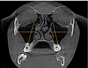
sinus width was measured on coronal reconstructed images as the longest perpendicular distance from the medial wall of the sinus to the outermost point of the lateral wall of the maxillary sinus(Fig. 2). The anteroposterior dimension of the sinus was measured as the longest distance antero-pos- teriorly from the most anterior point to the most posterior point on sagittal reconstructed images(Fig. 3). MSV was measured using the airway tool of Dolphin 3D imaging software version 11.8 premium(Dolphin Imaging, Chats- worth, CA, USA) from reconstructed Digital Imaging and Communication in Medicine images(Fig. 4). To determine the reproducibility and reliability of the measured variables, intra-examiner calibration was performed by comparing 20
Fig. 1. Measurement of the angle of septal deviation(the angle be- tween the crista galli and the most prominent point of deviation).
Fig. 2. Measurement of maxillary sinus height(from the lowest point of the sinus floor to the highest point of the sinus roof) and width(from the longest perpendicular distance from the medial wall of the sinus to the outermost point of the lateral wall of the maxillary sinus).
randomly selected CBCT measurements made by the same radiologist(AU) at a 2-week interval using the paired-sam- ple t-test. No statistically significant difference was noted between the first and second measurements(P>0.05).
SPSS for Windows(version 23.0; IBM Corp., Armonk, NY, USA) was used to perform the statistical analysis.
Categorical variables were presented as percentage and fre- quency, and continuous variables were indicated by mean, standard deviation, range, and their minimum/maximum values. The independent and paired t-tests were also used to compare mean values. A multiple linear regression mod- el was constructed using a backward selection logarithm.
For the statistical comparisons that were made, statistical significance was considered to be present at P<0.05.
Results
The average age of the patient population examined was 39.25±15.61years, and the age range was 18-71years.
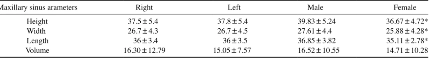
The mean right and left MSV was 16.30±12.79cm3 and 15.05±7.57cm3, respectively. The difference between sides was not statistically significant(P>0.05)(Table 1), although males tended to have significantly greater sinus dimensions than females. Nonetheless, the MSV did not show a significant difference according to sex(Table 1).
CB was found in 37% of females and in 38.8% of males, with no statistically significant sex difference(Fig. 5).
NSD was found in 68.5%(n=77) of females and 80.8%
of males, with no statistically significant difference by sex.
The prevalence of sinus pathology was significantly higher
Fig. 5. Bilateral concha bullosa from a coronal slice of cone-beam computed tomography.
Fig. 3. Measurement of the anteroposterior dimension of the maxil- lary sinus from the most anterior point to the most posterior point.
Fig. 4. Reconstructed 3-dimensional image of the maxillary sinuses for volume measurements.
Table 1. Differences in maxillary sinus volume according to sex and side
Maxillary sinus arameters Right Left Male Female
Height 37.5±5.4 37.8±5.4 39.83±5.24 36.67±4.72*
Width 26.7±4.3 26.7±4.5 27.61±4.4 25.88±4.28*
Length 36±3.4 36±3.5 36.85±3.82 35.11±2.78*
Volume 16.30±12.79 15.05±7.57 16.52±10.55 14.71±10.28
*P<0.05
in females(64.8%) than in males(88.5%)(Table 2). Over- all, the prevalence of bilateral CB was higher in males than in females(23.1% and 11.1%, respectively), while a higher frequency of unilateral CB was detected in females than in males(25.9% and 15.4%, respectively); however, this dif- ference was not significant. Bilateral sinus pathology was more common in males(65.4%) than in females(37%), while an almost equal proportion(~25%) of unilateral sinus pathology was observed in both sexes.
Although the relative frequency of CB was clearly high- er among individuals with NSD(40.5%) than among those without NSD(29.6%), the association between CB and NSD was not statistically significant(P>0.05)(Table 3).
Similarly, there was no significant relationship between NSD severity and the prevalence of CB, although it was noted that individuals with mild NSD had a higher rate of CB(45.5%) than those with severe NSD(12.5%).
The relative frequency of detected sinus pathology was higher among individuals with NSD(79.7%) than among those without NSD(66.7%), but the association was not statistically significant. Likewise, although this tendency was not significant, those with mild NSD had a higher fre- quency of sinus pathology(84.8%) than those with severe NSD(62.5%)(Table 3). The presence of middle CB had no statistically significant association with the presence of si- nus pathology.
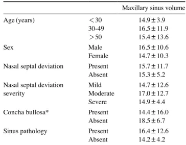
Table 4 shows the impact of several studied parameters on MSV. Only the presence of middle CB was associated with a significantly higher MSV.
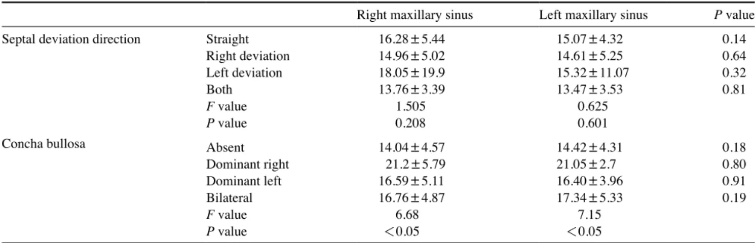
The presence or absence of NSD did not affect MSV, al- though the sinus size appeared smaller on the deviated side than on the non-deviated side. Bilateral CB was linked to a larger MSV in comparison to cases without CB or with only unilateral CB, and the MSV was significantly higher in the presence of CB(Table 5).
Discussion
CBCT is a cost-effective method that is being used with increasing frequency for the diagnosis of maxillofacial structures.14 Maxillofacial and oral radiologists should be familiar with the sino-nasal complex region. The sino-na- sal cavity is conventionally imaged using CBCT exams of the jaws. However, some anatomic variations may lead to chronic sinusitis or complications during sinus surgery, including procedures such as sinus lift surgery, which is performed prior to dental implant placement. Thus, a me- ticulous understanding of the anatomy of the sino-nasal
Table 2. Sex differences in the prevalence of concha bullosa, na- sal septal deviation, and sinus pathology(number and percentage)
Male Female P value
Concha bullosa 20(38.5) 20(37.0) 0.88
Nasal septal deviation 42(80.8) 37(68.5) 0.148 Sinus pathology 46(88.5) 35(64.8) <0.05 Concha bullosa
Absent 32(61.5) 34(63.0)
0.16
Unilateral 8(15.4) 14(25.9)
Bilateral 12(23.1) 6(11.1)
Sinus pathology
Absent 6(11.5) 19(32.5)
<0.05
Unilateral 12(23.1) 15 (27.8)
Bilateral 34(65.4) 20(37.0)
Table 3. The relative frequency of concha bullosa, nasal septal deviation, and sinus pathology(number and percentage)
Present Absent Concha bullosa
Nasal septal deviation Present 32(40.5) 47(59.5) Absent 8(29.6) 19(70.4) Septal deviation severity Mild 15(45.5) 18(54.5) Moderate 11(34.4) 21(65.6) Severe 1(12.5) 7(87.5) Sinus pathology Present 32(80.0) 49(74.2) Absent 8(20.0) 17(25.8) Sinus pathology
Nasal septal deviation Present 63(79.7) 16(20.3) Absent 18(66.7) 9(33.3) Septal deviation severity Mild 28(84.8) 5(15.2) Moderate 26(81.3) 6(18.8) Severe 5(62.5) 3(37.5)
Table 4. Maxillary sinus volume in relation to the study parameters (unit: mm3)
Maxillary sinus volume
Age(years) <30 14.9±3.9
30-49 16.5±11.9
>50 15.4±13.6
Sex Male 16.5±10.6
Female 14.7±10.3
Nasal septal deviation Present 15.7±11.7
Absent 15.3±5.2
Nasal septal deviation
severity Mild 14.7±12.6
Moderate 17.0±12.7
Severe 14.9±4.4
Concha bullosa* Present 14.4±16.0
Absent 18.5±6.7
Sinus pathology Present 16.4±12.6
Absent 14.2±4.2
*P<0.05
complex is of great importance for minimizing and/or elim- inating surgical complications.15 NSD is the most common anatomical variation of the nasal cavity, and it was reported by Earwaker that “NSD occurs in 44% of individuals with a slight right-side predominance and equal gender distribu- tion.”6 In the present study, more than two-thirds(79.9%) of the studied sample had NSD, primarily mild or moder- ate NSD(45.5% and 34.4%, respectively). Deviation of the septum may exacerbate snoring or obstruct breathing if it is severe enough that the septum contacts the lateral nasal wall. In the present investigation, severe NSD was only seen in 7.5% of the studied sample. Shin suggested that the development of unilateral sinusitis might be influenced by NSD.16 According to a meta-analysis conducted by Col- let et al., there was “no relationship between the degree of deviation and the presence of sinusitis.”17 Kapusuz et al.
reported that the MSV was influenced by severe septum deviation and suggested that the MSV was usually small- er on the deviated side than on the opposing side.18 This tendency was also suggested by Lee et al., and is in agree- ment with the present findings of this study.19 Moorthy et al. demonstrated that an S-shaped NSD was significantly correlated with sinus diseases, including cases where na- sal symptoms were absent.20 Erkan et al.21 suggested that both NSD and CB might affect each other physically. In the literature, few studies were found that sought to assess the correlation between NSD and the presence of middle turbinate pneumatization; however, a study demonstrated a highly significant correlation between the presence of mid- dle CB and a deviated septum on the contralateral side.18 It has been suggested that the abnormal space created in the nasal septum by the concave part may provide conditions suitable for pneumatization of the middle turbinate.22 NSD was usually accompanied with a dominant large CB.22,23 In
contrast, in this study, only 40.5% of the individuals with NSD had CB, suggesting there may not be an association.
Some investigators have suggested that there may be a rela- tionship between having CB and sinusitis, but other reports found no such relationship.16,24,25 In this study, positivity for both NSD and CB had no significant impact on the pres- ence or absence of sinus pathology(P>0.05). The exact mechanism of CB formation is not yet clear, but the prev- alence of middle CB has been reported to range from 13%
to 72.2%.3,5,26 CB may originate from unilateral or bilateral
“variation in the pneumatization of the bone plate caused by an extension of ethmoid cells.”27 Miranda et al.27 sug- gested that “the degree of pneumatization varies and may cause obstruction of the middle meatus or infundibulum.”
In the present study, middle CB was observed in 37.7% of the sample; 20.7% of the sample had single unilateral CB, and 16.6% had single bilateral CB. This differs from the findings described in previous studies,24,28 and according to the study conducted by Subramanian et al.,29 the prevalence of CB among females was higher than among males. How- ever, in the present study, CB was detected in both sexes equally, with single unilateral CB being the most common variant among females. Abnormalities of the concha may predispose individuals to osteo-meatal obstruction, which may lead to chronic sinusitis.29,30 There also seems to be a connection between increased MSV and CB. Addition- ally, the sinus volume in the presence of bilateral and/or dominant CB was slightly larger than when CB was not present. This could be attributed to the intensified growth of the maxillary sinus as a result of inadequate ventilation secondary to the presence of CB. Smith et al. reviewed 883 CT scans, in order to determine the prevalence of CB and NSD and their possible association with maxillary sinus morphology, and failed to find a significant relationship
Table 5. Maxillary sinus volume according to the direction of septal deviation and presence or absence of concha bullosa (unit: cm3) Right maxillary sinus Left maxillary sinus P value
Septal deviation direction Straight 16.28±5.44 15.07±4.32 0.14
Right deviation 14.96±5.02 14.61±5.25 0.64
Left deviation 18.05±19.9 15.32±11.07 0.32
Both 13.76±3.39 13.47±3.53 0.81
F value 1.505 0.625
P value 0.208 0.601
Concha bullosa Absent 14.04±4.57 14.42±4.31 0.18
Dominant right 21.2±5.79 21.05±2.7 0.80
Dominant left 16.59±5.11 16.40±3.96 0.91
Bilateral 16.76±4.87 17.34±5.33 0.19
F value 6.68 7.15
P value <0.05 <0.05
between them.31 Similarly, Göçmen et al.32 reviewed 300 CBCT scans, were not able to demonstrate “a definite role for NSD, CB and Haller’s cells on sinus pneumatization,”
and suggested the need for further studies in order to clari- fy the relationship among these phenomena. In the present study, maxillary sinus asymmetry was not affected by the presence of CB and/or NSD, which is in agreement with the study conducted by Demir et al.33 Uygur et al.28 sug- gested that “the NSD angle plays an important role on the pneumatization of the concha on the opposite side.” In the present study, a non-statistically significant difference was observed between the angle of deviation and the direction of CB(P<0.05). Kucybata et al.34 retrospectively ana- lyzed 214 paranasal sinus CT scans in order to investigate whether positivity for NSD and CB was associated with the occurrence of maxillary sinusitis, and concluded that only NSD was related to maxillary sinusitis. Additionally, they demonstrated that bilateral CB had an effect on MSV, which agrees with the present study.
The prevalence and distribution of CB in this study are similar to the findings of studies conducted in Poland and Iran,34,35 whereas the prevalence of NSD in this study is similar to the findings of studies conducted in Poland and Portugal.23,34
In conclusion, NSD and CB were found to be common radiographic findings in asymptomatic Emirati patients. Al- though NSD was observed in more than two-thirds of the sample and CB was observed in more than one-third of the sample, only CB had a significant impact on MSV.
References
1. Uthman AT, Al-Rawi NH, AL-Naaimi AS, Al-Timimi JF. Eval- uation of maxillary sinus dimensions in gender determination using helical CT scanning. J Forensic Sci 2011; 56: 403-8.
2. Uthman AT, AL-Rawi NH, Al-Naaimi AS, Tawfeeq AS, Suhail EH. Evaluation of frontal sinus and skull measurements using spiral CT scanning: an aid in unknown person identification.
Forensic Sci Int 2010; 197: 124.e1-7.
3. Bolger WE, Butzin CA, Parsons DS. Paranasal sinus bony an- atomic variations and mucosal abnormalities: CT analysis for endoscopic sinus surgery. Laryngoscope 1991; 101: 56-64.
4. Tonai A, Baba S. Anatomic variations of the bone in sinonasal CT. Acta Otolaryngol Suppl 1996; 525: 9-13.
5. Aktas D, Kalcioglu MT, Kutlu R, Ozturan O, Oncel S. The rela- tionship between the concha bullosa, nasal septal deviation and sinusitis. Rhinology 2003; 41: 103-6.
6. Earwaker J. Anatomic variants in sinonasal CT. Radiographics 1993; 13: 381-415.
7. Zinreich SJ, Mattox DE, Kennedy DW, Chisholm HL, Diffley DM, Rosenbaum AE. Concha bullosa: CT evaluation. J Comput Assist Tomogr 1988; 12: 778-84.
8. Yiğit O, Acioğlu E, Cakir ZA, Sişman AS, Barut AY. Concha bullosa and septal deviation. Eur Arch Otorhinolaryngol 2010;
267: 1397-401.
9. Hatipoğlu H, Çetin M, Yüksel E. Concha bullosa types: their relationship with sinusitis, ostiomeatal and frontal recess dis- ease. Diagn Interv Radiol 2005; 11: 145-9.
10. Alharethy S, Aldrees T, Aljrid R, Alanazi A, Algaryan SK, Jang YJ. Common nasal deformities among rhinoplasty patients in a university hospital in Saudi Arabia. Ann Saudi Med 2017; 37:
2017-211.
11. Wang RG, Jiang SC, Gu R. The cartilaginous nasal capsule and embryonic development of human paranasal sinuses. J Otolar- yngol 1994; 23: 239-43.
12. Serifoglu I, OZ İİ, Damar M, Buyukuysal M, Tosun A, Tok- göz O. Relationship between the degree and direction of nasal septum deviation and nasal bone morphology. Head Face Med 2017; 13: 3.
13. Rak KM, Newell JD 2nd, Yakes WF, Damiano MA, Luethke JM. Paranasal sinuses on MR images of the brain: significance of mucosal thickening. AJR Am J Roentgenol 1991; 156: 381- 14. Al-Rawi NH, Uthman AT, Sodeify SM. Spatial analysis of man-4.
dibular condyles in patients with temporomandibular disorders and normal controls using cone beam computed tomography.
Eur J Dent 2017; 11: 99-105.
15. Kantarci M, Karasen RM, Alper F, Onbas O, Okur A, Karaman A. Remarkable anatomic variations in paranasal sinus region and their clinical importance. Eur J Radiol 2004; 50: 296-302.
16. Shin HS. Clinical significance of unilateral sinusitis. J Korean Med Sci 1986; 1: 69-74.
17. Collet S, Bertrand B, Cornu S, Eloy P, Rombaux P. Is septal de- viation a risk factor for chronic sinusitis? Review of literature.
Acta Otorhinolaryngol Belg 2001; 55: 299-304.
18. Kapusuz Gencer Z, Ozkırış M, Okur A, Karaçavuş S, Saydam L.
The effect of nasal septal deviation on maxillary sinus volumes and development of maxillary sinusitis. Eur Arch Otorhinolar- yngol 2013; 270: 3069-73.
19. Lee DH, Shin JH, Lee DC. Three-dimensional morphometric analysis of paranasal sinuses and mastoid air cell system using computed tomography in pediatric population. Int J Pediatr Otorhinolaryngol 2012; 76: 1642-6.
20. Moorthy PN, Kolloju S, Madhira S, Jowkar AB. Clinical study on deviated nasal septum and its associated pathology. Int J Otolaryngol Head Neck Surg 2014; 3: 75-81.
21. Erkan SO, Erkan ZA, Tuhanioğlu B, Haytoğlu S, Güney Z. The relationship between septal deviation and concha bullosa. Ku- lak Burun Bogaz Ihtis Derg 2017; 27: 74-8.
22. Sazgar AA, Massah J, Sadeghi M, Bagheri A, Rasool E. The in- cidence of concha bullosa and the correlation with nasal septal deviation. B-ENT 2008; 4: 87-91.
23. Stallman JS, Lobo JN, Som PM. The incidence of concha bullo- sa and its relationship to nasal septal deviation and paranasal sinus disease. AJNR Am J Neuroradiol 2004; 25: 1613-8.
24. Calhoun KH, Waggenspack GA, Simpson CB, Hokanson JA, Bailey BJ. CT evaluation of the paranasal sinuses in symptom- atic and asymptomatic populations. Otolaryngol Head Neck Surg 1991; 104: 480-3.
25. Danese M, Duvoisin B, Agrifoglio A, Cherpillod J, Krayenbuhl
M. Influence of naso-sinusal anatomic variants on recurrent, persistent or chronic sinusitis. X-ray computed tomographic evaluation in 112 patients. J Radiol 1997; 78: 651-7.
26. Arslan H, Aydinlioglu A, Bozkurt M, Egeli E. Anatomic varia- tions of the paranasal sinuses: CT examination for endoscopic sinus surgery. Auris Nasus Larynx 1999; 26: 39-48.
27. Miranda CM, Maranhão CP, Arraes FM, Padilha IG, Farias LP, Jatobá MS, et al. Anatomic variations of paranasal sinuses at multi-slice computed tomography: what to look for. Radiol Bras 2011; 44: 256-62.
28. Uygur K, Tüz M, Doğru H. The correlation between septal de- viation and concha bullosa. Otolaryngol Head Neck Surg 2003;
129: 33-6.
29. Subramanian S, Lekhraj Rampal GR, Wong EF, Mastura S, Razi A. Concha bullosa in chronic sinusitis. Med J Malaysia 2005; 60: 535-9.
30. Lee JS, Ko IJ, Kang HD, Lee HS. Massive concha bullosa with secondary maxillary sinusitis. Clin Exp Otorhinolaryngol 2008;
1: 221-3.
31. Smith KD, Edwards PC, Saini TS, Norton NS. The prevalence of concha bullosa and nasal septal deviation and their relation- ship to maxillary sinusitis by volumetric tomography. Int J Dent 2010; 2010. pii: 404982.
32. Göçmen G, Borahan MO, Aktop S, Dumlu A, Pekiner FN, Göker K. Effect of septal deviation, concha bullosa and Haller’s cell on maxillary sinus’s inferior pneumatization; a retrospec- tive study. Open Dent J 2015; 9: 282-6.
33. Demir UL, Akca ME, Ozpar R, Albayrak C, Hakyemez B. An- atomical correlation between existence of concha bullosa and maxillary sinus volume. Surg Radiol Anat 2015; 37: 1093-8.
34. Kucybala I, Janik KA, Ciuk S, Storman D, Urbanik A. Nasal septal deviation and concha bullosa - do they have an impact on maxillary sinus volumes and prevalence of maxillary sinusitis?
Pol J Radiol 2017; 82: 126-33.
35. Bahemmat N, Hadian H. The frequency of nasal septal devia- tion and concha bullosa and their relationship with maxillary si- nusitis based on CBCT finding. Int J Med Res Health Sci 2016;
5: 152-6.