Synergistic anti-inflammatory effects of Laminaria japonica fucoidan and Cistanche tubulosa extract
Jangbeen Kyung
1,#, Dajeong Kim
1,#, Dongsun Park
1, Yun-Hui Yang
1, Ehn-Kyoung Choi
1, Sung-Pyo Lee
2, Tae-Su Kim
2, Yoon-Bok Lee
3, Yun-Bae Kim
1*
1
College of Veterinary Medicine, Chungbuk National University, Cheongju, Korea
2
Misuba RTech Co., Ltd., Asan, Korea
3
Central Research Institute, Dr. Chung’s Food Co., Ltd., Cheongju, Korea
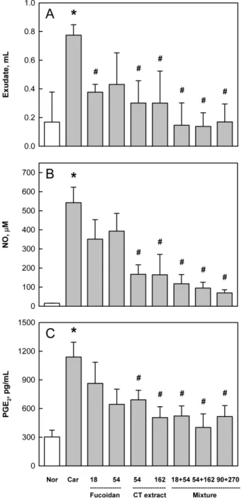
The anti-inflammatory effects of fuciodan and Cistanche tubulosa (CT) extract were investigated in vitro macrophage culture system and in vivo carrageenan-induced air pouch inflammation model. CT extract inhibited nitric oxide production from activated RAW 264.7 macrophage cells, while fucoidan was inactive. In vivo air pouch inflammation model, carrageenan-induced vascular exudation and increased nitric oxide and prostaglandin E
2concentrations in the exudates were synergistically suppressed by co- administration of fucoidan or CT extract. Moreover, tissue inflammation was substantially attenuated by the combinational therapy. However, there was no synergistic effect against the inflammatory cell infiltration, although fucoidan and CT extract each markedly reduced the cell numbers. Therefore, it is suggested that fucoidan blocks infiltration of inflammatory cells, while CT extract inhibits activation of the cells, and that their combinational treatment could be a promising candidate for the relief of various types of inflammation.
Key words: Carrageenan, inflammation, fucoidan, Cistanche tubulosa extract
Received 12 May 2012; Revised version received 21 May 2012; Accepted 24 May 2012
As an immune response against foreign antigens, macrophages release pro-inflammatory cytokines such as tumor-necrosis factor- α (TNF-α), interleukin-1β (IL- 1 β), IL-6 and others [1,2]. Such cytokines induces chemotactic influx of granulocytes, monocytes, lymphocytes, and mast cells to damaged tissue that promotes antigen removal and tissue recovery [3,4].
However, excessive infiltration and activation of the cells aggravates tissue injuries, leading to edema (vascular exudation) and pain which are well-known phenomena of inflammation.
TNF- α is an important factor for inducing nitric oxide synthase (iNOS) gene expression in several cell lines.
iNOS activation leads to nitric oxide (NO) production
[5-7] that not only modulates several physiological functions such as bactericidal and vasodilatation, but also causes inflammation [8,9]. After tissue injury, degradation by phospholipases of cell membrane phospholipids generates arachidonic acid. Arachidonic acid is further decomposed by cyclooxygenase (COX) to prostaglandins (PGs) [10]. Excessive amount of PGE
2formed by COX-II induces several cytokines for inflammation. Since both NO and PGE
2act as major factors for inflammation and pain induction, TNF- α–NO and COX-II–PGE
2pathways are main streams of inflammatory process, which are inhibited by corticosteroids and non-steroidal anti-inflammatory drugs (NSAIDs), respectively [10,11].
http://dx.doi.org/10.5625/lar.2012.28.2.91
#