https://doi.org/10.9721/KJFST.2020.52.5.482
482
©The Korean Society of Food Science and Technology
Anti-inflammatory effects of puffed turmeric extract with
oriental herb extract in acute colitis mice
Hyunsung Kim1, Yohan Choi1, Seungmin Yu1, Gwang-Woong Go2, Mooyeol Baik1, and Wooki Kim1,*
1Department of Food Science and Biotechnology, Graduate School of Biotechnology, Kyung Hee University 2Department of Food and Nutrition, Hanyang University
Abstract Cases of inflammatory bowel diseases including ulcerative colitis are increasing in Korea and development of non-steroidal anti-inflammatory drugs (NSAIDs) are widely investigated. Natural products with anti-inflammatory properties are rising as safe candidates for NSAIDs. The extract of turmeric or puffed turmeric mixed with herbal extract (goji berry, liquorice, lycium root, and dong quai) was treated to acute colitis mice by oral gavage. The symptoms of colitis, i.e., body weight loss, fecal score, and shortened colon length, were significantly attenuated by puffed turmeric extract with the herbal extract. Non-puffed turmeric extract with herbal extract, however, exhibited a very marginal recovery. Tissue culture supernatant of colons further revealed that both puffed turmeric and non-puffed turmeric extracts with herbal extract suppressed pro-inflammatory cytokine production at a comparable level. These results indicate that puffing is a simple and promising process of turmeric for enhancement of anti-inflammatory properties.
Keywords: puffing, turmeric, colitis, oriental herbs, inflammation
Introduction
The domestic prevalence of inflammatory bowel diseases (IBD) in Korea was reported to drastically increase from 57.4 cases per 100,000 persons in 2009 to 95.6 cases per 100,000 persons in 2016 (Kwak et al., 2019). Steroidal drugs are potent for the amelioration of the IBD symptoms (Ardite et al., 1998), but multi-organ-targeting side effects restrict their usages. In an effort to develop safe alternatives for steroids, termed non-steroidal anti-inflammatory drugs (NSAIDs), natural extracts mainly composed of polyphenolic compounds are highlighted for their long history of consumption as food or herbal medicine, especially in oriental countries. However, the lack of detailed working mechanisms and lower efficacy are also hurdling their wider applications.
Among various herbal products, goji berry (fruit of Lycium chinense), Korean name gugija, was previously shown to suppress the development of colitis in interleukin (IL)-10-deficient mice (Kang et al., 2018). Liquorice (Glycyrrhiza uralensis), Korean name gamcho, also has been historically widely used as herbal medicine and previously was shown to attenuate murine colitis for which glabridin was identified as a functional compound (Kwon et al., 2007). In addition, lycium root bark (Lycii radices cortex), Korean name jigolpi, was reported to suppress murine macrophages
resulting in the suppressed osteroclasts (Kim et al., 2016) and inflammation (Yang et al., 2017). It was further identified that lyciumlignan D and lyciumphenyl propanoid A are the major active components of lycium root bark (Yang et al., 2017). Similarly, it was demonstrated that dong quai (Angelica gigas), Korean name danggui, was shown to suppress murine colitis and the polysaccharides were identified as the active compounds (Hay et al., 2019). On the other hand, turmeric (Curcuma long L), a widely used spice, has multiple health beneficial effects including antioxidant, anti-cancer, antimicrobial, and anti-inflammatory effects (Hay et al., 2019). In an effort to enhance its biological functions, the increased antioxidant and anti-inflammatory properties of the puffed turmeric were reported by using murine RAW 264.7 cells as an inflammatory model system (Choi et al., 2019). However, it still remains to be clarified if puffing enhances anti-inflammatory properties of dietary turmeric in animal models.
The oral administration of dextran sodium sulfate (DSS) in free access of tap water is a well-established mouse model for the study of IBD, especially with a focus on acute ulcerative colitis (Whittem et al., 2010). In fact, it was previously reported that DSS-induced colitis model shares many of human IBD etiology, including intestinal microbial imbalance, metabolomic changes, mucin barrier integrity, and immune responses (Whittem et al., 2010). Therefore, in a process of health-beneficial beverage development, the current study sought the effect of puffed turmeric extracts vs non-puffed control turmeric extract at the presence of extracts of aforementioned anti-inflammatory oriental herbs in a DSS-treated colitis status. Following the assessment of several key symptoms of acute colitis in mice, the results reveals an easy way for enhancement of anti-inflammatory properties of turmeric in the presence of herbal extracts, providing a basal formula for the production of functional foods.
*Corresponding author: Wooki Kim, Ph.D., Associate Professor, Department of Food Science and Biotechnology, Graduate School of Biotechnology, Kyung Hee University, Yongin, Gyeonggi 17104, Korea
Tel: +82-31-201-3482 Fax: +82-31-204-8116 E-mail: [email protected]
Received September 10, 2020; revised September 23, 2020; accepted September 25, 2020
Materials and Methods
Preparation of extracts
Sliced and dried turmeric (cultivated in Jindo-gun, Republic of Korea and harvested at October-December, 2019), goji berry, liquorice, lycium root bark, and dong quai were purchased from Bibong Herb Co. (Yangju-si, Korea). Turmeric was puffed at a pressure of 980 kPa using a customized gun puffing machine in the presence of 4x weighed dried rice for the prevention of carbonization as previously reported (Kwon et al., 2019; Choi et al., 2019). Food grade 70% ethanol purchased from Ethanol Supplies World Co. (Jeonju-si, Korea) was used for extraction of either puffed turmeric (PTE), non-puffed control turmeric (TE) or herb combination of goji berry, liquiorice, lyceum root bark, and dong quai (1:1:1:1, HE). Briefly, 200 mL of 70% ethanol was added to 5 g of the ground herbs (20:1, v/w), followed by stirring with a magnet at room temperature for 30 min. The extracts were vacuum filtered and the extract mixtures of TE+HE (7:3) or PTE+HE (7:3) were lyophilized for further experiments.
Animal oral gavage and induction of colitis
All the animal studies followed the guidelines approved by Institutional Animal Care and Use Committee, Kyung Hee University (approval number KHGASP-20-256). To investigate the modulation of acute colitis by the extract mixtures, 40 male BALB/c mice at 4 weeks of age were purchased from Raon Bio (Yongin-si, Korea) and housed under 12:12-h light:dark cycle. The mice were allocated to 4 groups (n=10) and fed AIN-76A diet for 1 week acclimation. Mice in each experimental group were fed 200 ìg of specific extract powder dissolved in 100 ìL distilled water or control vehicle by oral gavage for 3 days prior to the onset of colitis. DSS (MP Biomedicals, Santa Ana, CA, USA) was added in the tap water at 5% (m/v) and mice were freely accessed to the water consumption for the development of acute colitis for 8 days. Daily oral gavage of the extracts or vehicle was continued until day 7 of the experiment.
Assessment of body weight, fecal score and colon length During 7 days of colitis development, body weight of individual mouse was daily recorded. The feces of each mouse were collected and the fecal disease score was determined as following criteria; normal stool for 0 point, loose stool for 1 point, diarrhea for 2 points, and apparent bloody stool for 3 points. At the day of 8 following the DSS treatment, the mice were euthanized by CO2 inhalation and the colon was collected from cecum to rectum. The length of the colon was measured to determine the severity of the colitis.
Cytokine production by colon tissue
The distal part of the colon was dissected and weighed. The dissected colon tissue was further incubated in 1 mL DMEM media supplemented with 5% fetal bovine serum (Hyclone, Logan, UT, USA) and streptomycin/penicillin antibiotics (Hyclone) for 24 h. The supernatant was collected and the pro-inflammatory cytokines, i.e., IL-6 and TNF-α, were quantified by ELISA
following the manufacturer’s protocol (Invitrogen, Carlsbad, CA, USA). Briefly, 100μL of diluted capture antibody (Ab) was added to each well and incubated overnight at 4oC. Supernatant was removed and washed 3 times, repeating the addition and removal of buffers. Next, 200μL of assay diluent was added to each well and incubated for 1 h. Then, 100μL of purified standard included in the ELISA kits and culture supernatant were added to each well and incubated for 2 h. Next, 100μL of mixed detection Ab and streptavidin conjugated with horse radish peroxidase (SAv-HRP) was added to each well. After washing, 100μL of substrate solution was added and incubated for 30 min in the dark. Next, 50 ìL of stop solution was added to each well. Finally, the cytokine concentrations were quantified by using a standard curve on the basis of absorbance measured by a microplate reader (Bio-Rad, Hercules, CA, USA).
Results and Discussion
Attenuated body weight loss during colitis by the extract mixtures
Body weight loss is a hallmark symptom of DSS-induced colitis (Taghipour et al., 2016). In the current study, acute colitis induced by 5% DSS in drinking water (Fig. 1, closed circle) exhibited a significant loss of body weight (98.65±0.02%, p<0.05) at day 3 after onset of colitis as compared to tap water treated mice (open circle, 99.63±0.01%). DSS treated mice further lost their body weight to 88.60±0.07% of the initial body weight at day 7 of colitis. The previous studies also reported the comparable loss of body weight in murine DSS colitis models (Taghipour et al., 2016; Chao et al., 2017) indicating that the current study design worked well. Following daily oral gavage of extract mixtures in DSS-induced colitic mice, DSS+TE+HE intervention (Fig. 1, closed square) demonstrated no difference to the DSS disease model, only except for a slight retardation of the weight loss at day 5 (DSS 94.94±0.03% vs DSS+TE+HE 97.56±0.04%, p<0.05). In contrast, DSS+PTE+HE treated mice (Fig. 1, closed triangle) showed a significant retardation of weight loss. Specifically, DSS+PTE+HE treatment maintained the body weight identical to water drinking healthy mice until day 5 after DSS colitis. At the end-point of the experiment at day 7, DSS+PTE+HE mice weighed 94.54±0.0% of initial body weight resulting in a significant delay (p<0.05) of weight loss as compared to both DSS disease control and DSS+TE+HE treated groups. As for anti-inflammatory effects of turmeric in DSS colitis mice, curcumin (Deguchi et al., 2007) and essential turmeric oil (Toden et al., 2017) were demonstrated to ameliorate the diseases. Similarly, it was previously reported that dietary intervention of goji berry powder slightly but significantly prevented DSS-induced body weight loss in mice (Kang et al., 2017). However, there is no report regarding the enhancement of anti-inflammatory properties by puffing process or a mixture of aforementioned herbs, to date.
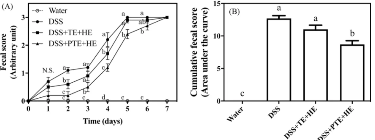
Improvement of fecal score by the extract mixtures The feces of the individual mouse were daily collected for scoring as described in the “Materials and Methods”. At day 0 of
colitis, all mice scored 0, indicating that the feces were in blood-free solid status. However, until day 2 after the onset of colitis, DSS disease mice (Fig. 2A, closed circle) increasingly scored to 1.10±0.01 demonstrating the development of loose stools. At the same time point, DSS+TE+HE (closed square) significantly down-regulated the fecal disease status at the score of 0.60±0.01 as compared to the DSS control mice (p<0.05). Of interest, DSS+PTE+HE (closed triangle, 0.20±0.01) further suppressed the development of loose stools resulting in no difference to the water-treated control mice (open circle, p>0.05) at day 2. DSS treatment further caused diarrhea at day 4 (score 2.20±0.01) and apparent fecal bleeding (score 3.00) at day 5. The gavage of TE+HE or PTE+HE significantly suppressed DSS-induced development of diarrhea scoring (1.70±0.01 or 1.20±0.01, respectively) at the time point of day 4. DSS+TE+HE treatment exhibited the identical scoring to DSS control at day 5 and day 6, where the feces of DSS+PTE+HE treated mice showed significantly lower disease scoring of 2.40±0.01 and 2.70±0.01, respectively. All DSS treated mice, regardless of gavage, scored 3.00 at the end-point of the
experiment at day 7.
The dynamics of disease development during 7 days of DSS treatment was quantitatively analyzed by integrating the area under the curve (Fig. 2B) as previously suggested (AlSharari et al., 2013). In the current study, the cumulative score of DSS treated control mice was determined as 12.70±0.42 (arbitrary unit), which was significantly decreased to 8.70±0.57 by the oral gavage of PTE+HE. However, DSS+TE+HE treatment (11.00±0.65) exhibited no difference to the DSS control, indicating the enhanced suppression of colitis by the puffing of turmeric but not non-puffed control.
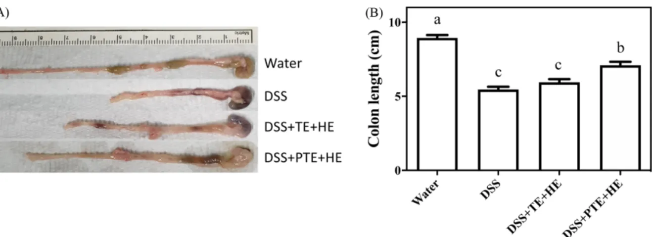
Suppressed shortening of colon length by extract mix-tures in DSS-induced colitis
It is previously well described that colon length is shortened in the process of acute colitis (Chassaing et al., 2014; Taghipour et al., 2016). As shown in Fig. 3A, the representative images of colons in each treatment clearly demonstrates that DSS treatment reduced the colon length as compared to water drinking normal colon in the current study. The statistical analysis, as shown in Fig. 2B, further revealed that colons of DSS treated mice (5.46±0.19 cm) were significantly shorter than colons of water drinking mice (8.94±0.20 cm, p<0.05). The gavage of TE+HE (5.94±0.21 cm) to DSS colitis did not show any difference to DSS control mice, but DSS+PTE+HE treatment significantly (p<0.05) reversed the DSS-induced shrink of colons to 7.09±0.24 cm.
Down-regulated cytokine production of colon tissues by extract mixtures
The local inflammatory status of colons was further quantified by the assessment of pro-inflammatory cytokine production following 24 h incubation of colon tissues ex vivo. The secretion of IL-6 (Fig. 4A) into the culture medium by the colon tissues of water drinking mice (112.6±17.88 pg/mL/mg colon) was significantly increased by DSS addition (549.5±83.79 pg/mL/mg colon, p<0.05). Interestingly, the colon tissues from DSS+TE+HE or DSS+PTE+HE treated mice exhibited significantly reduced IL-6 production (156.3±22.10 and 224.2±37.62 pg/mL/mg colon, respectively) to
Fig. 2. The quantitative assessment of colitis development following DSS treatment with or without extract mixtures. (A) The kinetic changes of fecal scores as daily assessed for the individual mouse. (B) The cumulative evaluation of fecal disease scores for the 7 days of DSS-induced colitis. The mean values with different letters significantly differ within the specific day (p<0.05, n=10). DSS, dextran sulfate sodium; TE, turmeric extract; PTE, puffed turmeric extract; HE, herbal extract.
Fig. 1. The relative body weight changes to the initial value during DSS-induced colitis. The mean values with different letters significantly differ within the specific day (p<0.05, n=10). DSS, dextran sulfate sodium; TE, turmeric extract; PTE, puffed turmeric extract; HE, herbal extract.
the comparable level of water drinking mice (p>0.05). In contrast, DSS treatment exhibited a tendency toward increased TNF-α secretion (296.7±102.7 pg/mL/mg colon) by colon tissues as compared to the water control group (64.41±10.67 pg/mL/mg colon), even no significance was observed (Fig. 4B, p>0.05). TNF-α secretion by colons of DSS+TE+HE (434.60±66.78 pg/mL/mg colon) or DSS+ PTE+HE (214.3±60.29 pg/mL/mg colon) treated group exhibited no significant difference to DSS control group (p>0.05). Among various pro-inflammatory cytokines, IL-6 (Mudter & Neurath, 2007) and TNF-α (Egger et al., 2000) were shown to play major roles in the exacerbation of local to systemic inflammation. The current results indicate that the impaired local inflammation by DSS in colons were partially blocked for systemic propagation by both TE+HE and PTE+HE.
Conclusion
Overall, the current study sought the anti-inflammatory effects of mixtures of herbal extracts. The oral gavage of the mixture of turmeric extract with herbal extracts, consisted of goji berry,
liquorice, lycium root bark, and dong quai, exhibited very similar disease status to DSS only treated control mice. Mixture of puffed turmeric extract with the same herbal extract, however, showed significant recovery from DSS-induced acute colitis as assessed by body weight change, fecal score, colon length, and colon tissue cytokine production. It was previously reported that puffing of turmeric increases the extract yield of total phenolic compounds and Maillard reaction products, resulting in the increment of antioxidant and anti-inflammatory properties (Choi et al., 2019). Together, these results demonstrate that the current extract mixture can be used for the development of anti-inflammatory beverages, with emphasis on puffing as a simple and powerful processing method for the enhancement of biofunctions of turmeric.
Acknowledgment
This work was supported by the Rural Development Administration, Republic of Korea (PJ01314401). None of the authors of this study has any financial interest or conflict with industries or parties.
Fig. 3. (A) The representative images of colons at the end-point of the DSS-induced colitis. (B) The statistical analysis of the colon length following DSS treatment with or without extract mixtures. The mean values with different letters significantly differ (p<0.05, n=10). DSS, dextran sulfate sodium; TE, turmeric extract; PTE, puffed turmeric extract; HE, herbal extract.
Fig. 4. The production of pro-inflammatory cytokine IL-6 (A) and TNF-α (B) by cultured colon tissues following DSS treatment with or without extract mixtures. The mean values with different letters significantly differ (p<0.05, n=10). DSS, dextran sulfate sodium; TE, turmeric extract; PTE, puffed turmeric extract; HE, herbal extract.
References
AlSharari SD, Akbarali HI, Abdullah RA, Shahab O, Auttachoat W, Ferreira GA, White KL, Lichtman AH, Cabral GA, Damaj MI. Novel Insights on the Effect of Nicotine in a Murine Colitis Model. J. Pharmacol. Exp. Ther. 344: 207-217 (2013)
Ardite E, Panés J, Miranda M, Salas A, Elizalde JI, Sans M, Arce Y, Bordas JM, Fernández-Checa JC, Piqué JM. Effects of steroid treatment on activation of nuclear factor êB in patients with inflammatory bowel disease. Br. J. Pharmacol. 124: 431-433 (1998)
Chao L, Zheng P, Xia L, Yong Y, Lu G, Tang F, Zhao Z. Calycosin attenuates dextran sulfate sodium (DSS)-induced experimental colitis. Iran. J. Basic Med. Sci. 20: 1056-1062 (2017)
Chassaing B, Aitken JD, Malleshappa M, VijayKumar M. Dextran Sulfate Sodium (DSS)Induced Colitis in Mice. Curr. Protoc. Immunol. 104: 1-14 (2014)
Choi Y, Ban I, Lee H, Baik M-Y, Kim W. Puffing as a Novel Pro-cess to Enhance the Antioxidant and Anti-Inflammatory Proper-ties of Curcuma longa L. (Turmeric). Antioxidants 8: 506 (2019) Deguchi Y, Andoh A, Inatomi O, Yagi Y, Bamba S, Araki Y, Hata
K, Tsujikawa T, Fujiyama Y. Curcumin Prevents the Develop-ment of Dextran Sulfate Sodium (DSS)-Induced ExperiDevelop-mental Colitis. Dig. Dis. Sci. 52: 2993-2998 (2007)
Egger B, Bajaj-Elliott M, MacDonald TT, Inglin R, Eysselein VE, Büchler MW. Characterisation of Acute Murine Dextran Sodium Sulphate Colitis: Cytokine Profile and Dose Dependency. Diges-tion 62: 240-248 (2000)
Hay E, Lucariello A, Contieri M, Esposito T, De Luca A, Guerra G, Perna A. Therapeutic effects of turmeric in several diseases: An overview. Chem. Biol. Interact. 310: 108729 (2019)
Kang Y, Xue Y, Du M, Zhu M-J. Preventive effects of Goji berry on dextran-sulfate-sodium-induced colitis in mice. J. Nutr. Biochem. 40: 70-76 (2017)
Kang Y, Yang G, Zhang S, Ross CF, Zhu M-J. Goji Berry Modulates Gut Microbiota and Alleviates Colitis in IL-10-Deficient Mice.
Mol. Nutr. Food Res. 62: 1800535 (2018)
Kim J-H, Kim E-Y, Lee B, Min J-H, Song D-U, Lim J-M, Eom JW, Yeom M, Jung H-S, Sohn Y. The effects of Lycii Radicis Cortex on RANKL-induced osteoclast differentiation and activation in RAW 264.7 cells. Int. J. Mol. Med. 37: 649-658 (2016)
Kwak MS, Cha JM, Lee HH, Choi YS, Seo SI, Ko KJ, Park D Il, Kim SH, Kim TJ. Emerging trends of inflammatory bowel dis-ease in South Korea: A nationwide populationbased study. J. Gas-troenterol. Hepatol. 34: 1018-1026 (2019)
Kwon H-S, Oh S-M, Kim J-K. Glabridin, a functional compound of liquorice, attenuates colonic inflammation in mice with dextran sulphate sodium-induced colitis. Clin. Exp. Immunol. 151: 165-173 (2007)
Kwon Y, Yu S, Choi GS, Kim JH, Baik M, Su ST, Kim W. Puffing of Rehmannia glutinosa enhances anti-oxidant capacity and down-regulates IL-6 production in RAW 264.7 cells. Food Sci. Biotech-nol. 28: 1235-1240 (2019)
Mudter J, Neurath MF. Il-6 signaling in inflammatory bowel disease: Pathophysiological role and clinical relevance. Inflamm. Bowel Dis. 13: 1016-1023 (2007)
Taghipour N, Molaei M, Mosaffa N, Rostami-Nejad M, Asadzadeh Aghdaei H, Anissian A, Azimzadeh P, Zali MR. An experimental model of colitis induced by dextran sulfate sodium from acute progresses to chronicity in C57BL/6: correlation between condi-tions of mice and the environment. Gastroenterol. Hepatol. from Bed to Bench 9: 45-52 (2016)
Toden S, Theiss AL, Wang X, Goel A. Essential turmeric oils enhance anti-inflammatory efficacy of curcumin in dextran sulfate sodium-induced colitis. Sci. Rep. 7: 814 (2017)
Whittem CG, Williams AD, Williams CS. Murine Colitis modeling using Dextran Sulfate Sodium (DSS). J. Vis. Exp. 23: 6016-6029 (2010)
Yang Y, An Y, Wang W, Du N, Zhang J, Feng Z, Jiang J, Zhang P. Nine compounds from the Root Bark of Lycium chinense and their anti-inflammatory activities. Acta Pharm. Sin. B 7: 491-495 (2017)