• 검색 결과가 없습니다.

Anti-oxidant and Anti-inflammatory Effects of Acanthopanacia Cortex Hot Aqueous Extract on Lipopolysaccharide(LPS) Simulated Macrophages

N/A
N/A
Protected

Academic year: 2021

Share "Anti-oxidant and Anti-inflammatory Effects of Acanthopanacia Cortex Hot Aqueous Extract on Lipopolysaccharide(LPS) Simulated Macrophages"

Copied!
7
0
0

로드 중.... (전체 텍스트 보기)

전체 글

(1)

Anti-oxidant and Anti-inflammatory Effects of Acanthopanacia Cortex Hot Aqueous Extract on Lipopolysaccharide(LPS) Simulated Macrophages

Na Young Jo and Jeong Du Roh*

Department of Acupuncture & Moxibustion Medicine, Je-Cheon Hospital of Traditional Korean Medicine, Semyung University

[Abstract]

Objectives : This study is to investigate the effects of Acanthopanacis Cortex hot aqueous extract on nitric oxide(NO), prostaglandin E2(PGE2) production and DPPH(1,1-diphenyl-2-picryl hydrazyl) radical scavenging activity in macrophages.

Methods : Acanthopanacis Cortex (200 g) was heated at 100 ℃ with distilled water(2 L) for 4hrs.

The extract was filtered and concentrated to 100 ㎖ using a rotary evaporator and was frozen at -80 ℃, then was freeze-dried. The RAW 264.7 macrophages were subcultured. In order to evaluate cytotoxicity, MTT assay was performed. Experimental groups were divided into five(control, AC 25, 50, 100 and 200 ㎍/㎖) and we measured cytotoxicity. The concentrations of NO were preprocessed by Griess assay. The RAW 264.7 macrophages was pretreated by 10 ㎍/㎖ LPS and experimental groups were divided into five and we measured NO production.

The concentrations of PGE

2

were measured by enzyme immunoassay. The RAW 264.7 macrophages was pretreated by 10 ㎍/㎖ LPS. Experimental groups were divided into five and we measured PGE

2

production. Antioxidant activity was measured by the DPPH method.

experimental groups were divided into four(AC 25, 50, 100 and 200 ㎍/㎖) and we measured DPPH radical scavenging activity.

Results :

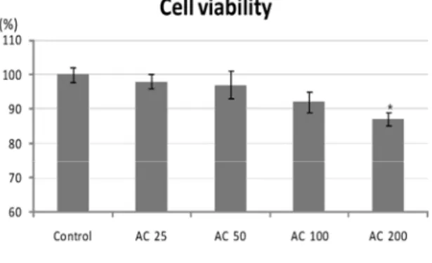
1. Viability of RAW 264.7 macrophages did not significantly decrease in 25, 50 and 100 ㎍/㎖

Acanthopanacis Cortex hot aqueous extract compared to control group.

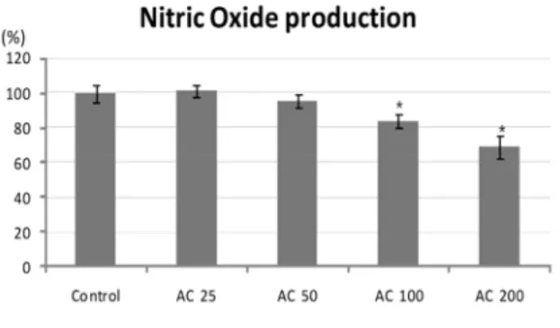
2. NO production in LPS-stimulated RAW 264.7 macrophages significantly inhibited in 100, 200 ㎍/㎖ Acanthopanacis Cortex hot aqueous extract compared to control group.

3. PGE

2

production in LPS-stimulated RAW 264.7 macrophages significantly inhibited in 100, 200 ㎍/㎖ Acanthopanacis Cortex hot aqueous extract compared to control group.

4. DPPH radical scavenging capability of Acanthopanacis Cortex hot aqueous extract in RAW 264.7 macrophages had the high level in 100, 200 ㎍/㎖.

Conclusion : According to the results, Acanthopanacis Cortexx hot aqueous extract has ability to suppress NO, PGE

2

production and improve DPPH free radical scavenging activity. So Acanthopanacis Cortex hot aqueous extract may have an anti-inflammation effect and antioxidant activity.

Key words :

Acanthopanacis Cortex;

Anti-inflammation;

Antioxidant activity;

Hot aqueous extract;

Korean medicine

Received : 2014. 02. 18.

Revised : 2014. 02. 27.

Accepted : 2014. 02. 27.

On-line : 2014. 03. 20.

✱ Corresponding author : Department of Acupuncture & Moxibustion Medicine, Je-Cheon Hospital of Traditional Korean Medicine, Semyung University, 65, Semyung-ro, Jecheon-si,

Chungcheongbuk-do, 390-711, Republic of Korea Tel : +82-43-649-1816 E-mail : [email protected]

This is an Open-Access article distributed under the terms of the Creative Commons Attribution Non-Commercial License (http://creativecommons.org/licenses/by-nc/3.0) which permits unrestricted non-commercial use, distribution, and reproduction in any medium, provided the original work is properly cited.

The Acupuncture is the Journal of Korean Acupuncture & Moxibustion Medicine Society. (http://www.TheAcupuncture.or.kr)

Copyright © 2014 KAMMS. Korean Acupuncture & Moxibustion Medicine Society. All rights reserved.

(2)

Ⅰ. Introduction

Acanthopanacis Cortex(AC) is the bark of the roots and stem of acanthopanax tree. In Korean medicine, Acanthopanacis Cortex has been widely used for treatment of orthopedic disease

1)

. When the tissue have been damaged, inflammatory reaction is shown to combat the pathogen

2)

.The inflammatory response is a cause of pain in sprains, tendinitis, aponeurositis, gout and osteoarthritis

3)

.

Previous studies have been reported in the antioxidant

4-6)

, fat reduction

7,8)

, antidiabetic

9)

, anticancer

10,11)

, increasing cerebral blood flow

12)

, IL-8 product inhibition

13)

, blood making, immunodulatory

14)

and alcoholysis

15)

effect of Acanthopanacis Cortex.

Likewise, many studies have suggested that the Acanthopanacis Cortex might be anti-inflammation effective. But Acanthopanacis Cortex was not enough in researching of the anti-inflammation.

Therefore, this study is to investigate the effects of Acanthopanacis Cortex hot aqueous extract on Nitric Oxide(NO), Prostaglandin E

2

(PGE

2

) production and DPPH(1,1-diphenyl-2-picryl hydrazyl) radical scavenging activity in macrophages.

II. Experimental materials and methods

A. Materials

Acanthopanacis Cortex used in the experiment were purchased from the Omniherb(Kyongbuk, Korea).

Extracting method is as follows. Acanthopanacis Cortex 200 g mixed with 2 L of distilled water, and boiled for 4 hours at 100 ℃ . The extract was filtered with filter paper, and centrifuged at 3,000 xg. And then secondary filtered with 0.03 mm filter paper (Nalgene, New York, USA). The filtrate was concen- trated to 100 ml, and frozen at -80 ℃ . Freezing solution to be freeze-dried for 7 days through the freezing

Cortex extract was obtained. The yield was 7 %.

B. Cell culture

RAW 264.7 macrophages purchased in the ATCC (Manassas, USA) were used in this experiment. RAW 264.7 macrophages were placed in the Dulbecco's modified eagle's medium(DMEM) including 10% Fetal bovine serum(FBS) and were cultivated inside the incubator at 37 ℃ under 5 % CO

2

.

C. Measuring cytotoxicity

In order to evaluate cytotoxicity, MTT assay was performed. RAW 264.7 macrophages were cultivated to concentration of 1 × 10

5

/well in a 96 well plate and were cultured for 16 hrs at 37 ℃ under 5 % CO

2

. Experimental groups were divided into five(Control, AC 25, 50, 100 and 200 ㎍ / ㎖ ). Stable cells were treated in the MTT reagent and absorbance was measured at 570 nm.

D. Measuring NO production

RAW 264.7 macrophages were cultivated to concentration of 1 × 10

5

/well in a 96 well plate and were cultured for 16 hrs at at 37 ℃ under 5 % CO

2

. Experimental groups were divided into five(control, AC 25, 50, 100 and 200 ㎍ / ㎖ ) and were treated with the 10 ㎍ / ㎖ Lipopolysaccharide(LPS) reagents. After mixed supernatant 100 ㎕ and Griess reagent 100 ㎕ , absorbance was measured at 540 nm. Griess reagent were prepared with 0.1 % naphthylethyleneiamine dihydrochloride 50 ㎕ and 1% sulfanilamide 50 ㎕ dissolved in 5 % H

3

PO

4

.

E. Measuring PGE

2

production

PGE

2

was measured using the commercial com-

(3)

Fig. 1. This graph describes that concen- trations of Acanthopanacis Cortex hot aqueous extract make a difference in the viability of RAW 264.7 macrophage

Control : non-treated group.

AC 25 : 25 ㎍/㎖ Acanthopanacis Cortex hot aqueous extract treated group.

AC 50 : 50 ㎍/㎖ Acanthopanacis Cortex hot aqueous extract treated group.

AC 100 : 100 ㎍/㎖ Acanthopanacis Cortex hot aqueous extract treated group.

AC 200 : 200 ㎍/㎖ Acanthopanacis Cortex hot aqueous extract treated group.

Values are represented as mean ± SD.

* : statistically significant difference from the normal group, as determined by the student's t -test as p < 0.05.

Minneapolis, USA). Experimental groups were divided into five(control, AC 25, 50, 100 and 200 ㎍ / ㎖ ) and were treated with the 10 ㎍ / ㎖ LPS reagents. Treated RAW 264.7 macrophages were cultured for 18 hrs at at 37 ℃ under 5 % CO

2

. Each culture fluid 100 ㎖ loaded at goat anti-mouse coated 96 well plate, and then mixed primary antibody solution 50 ㎕ and PGE

2

conjugate 50 ㎕ . It was stabilized for overnight at 4 ℃ . Treated culture fluid mixed substrate solution 200 ㎕ and waiting for 5 ∼ 20 minutes, and then mix the stop solution 50 ㎕ . Absorbance was measured at 450 nm.

F. Measuring the antioxidative effect

In order to investigate the antioxidative ability of Acanthopanacis Cortex, DPPH free radical scavenging capability was measured. RAW 264.7 macrophages were cultivated to concentration of 1 × 10

5

/well in a 96 well plate and were cultured for 16 hrs at at 37 ℃ under 5 % CO

2

. Experimental groups were divided into four(AC 25, 50, 100 and 200 ㎍ / ㎖ ) and were diluted with MeOH(Methanol) and diluent 50 ㎕ mixed with 0.15 mM DPPH(Sigma, USA) 80 ㎕ in 96 well plate.

Isolate the light at room temperature for 3 minutes.

Absorbance was measured using the microplate reader(Tecan, Italy) at 520 nm. DPPH free radical scavenging capability was evaluated in the following way.

DPPH free radical scavenging activity(%)

     

      

× 

G. Statistical analysis

With SPSS Window program(Ver. 10.0), experi- mental result were processed in terms of averages ± standard deviations. Statistical significance was confirmed by using Student's t-test, the level of significance was p<0.05.

III. Result

a. Viability of RAW 264.7 macrophages did not significantly decrease in 25, 50 and 100 ㎍ / ㎖ Acanthopanacis Cortex hot aqueous extract compared to control group.

b. NO production in LPS-stimulated RAW 264.7 macrophages significantly inhibited in 100, 200 ㎍ / ㎖ Acanthopanacis Cortex hot aqueous extract compared to control group.

c. PGE

2

production in LPS-stimulated RAW 264.7 macrophages significantly inhibited in 100, 200 ㎍ / ㎖ Acanthopanacis Cortex hot aqueous extract compared to control group.

d. DPPH radical scavenging capability of Acantho-

panacis Cortex hot aqueous extract in RAW 264.7

macrophages had the high level in 100, 200 ㎍ / ㎖ .

(4)

Fig. 2. This graph describes that concen- trations of Acanthopanacis Cortex hot aqueous extract make a difference in the NO production of RAW 264.7 macrophages

Control : 10 ㎍/㎖ LPS treated group

AC 25 : 10 ㎍/㎖ LPS and 25 ㎍/㎖ Acanthopanacis Cortex hot aqueous extract treated group

AC 50 : 10 ㎍/㎖ LPS and 50 ㎍/㎖ Acanthopanacis Cortex hot aqueous extract treated group.

AC 100 : 10 ㎍/㎖ LPS and 100 ㎍/㎖ Acanthopanacis Cortex hot aqueous extract treated group.

AC 200 : 10 ㎍/㎖ LPS and 200 ㎍/㎖ Acanthopanacis Cortex hot aqueous extract treated group.

Values are represented as mean ± SD.

* : statistically significant difference from the control group, as determined by the student's t -test as p < 0.05.

Fig. 3. This graph describes that concen- trations of Acanthopanacis Cortex hot aqueous extract make a difference in the PGE2 production of RAW 264.7 macrophages

Control : 10 ㎍/㎖ LPS treated group.

AC 25 : 10 ㎍/㎖ LPS and 25 ㎍/㎖ Acanthopanacis Cortex hot aqueous extract treated group.

AC 50 : 10 ㎍/㎖ LPS and 50 ㎍/㎖ Acanthopanacis Cortex hot aqueous extract treated group.

AC 100 : 10 ㎍/㎖ LPS and 100 ㎍/㎖ Acanthopanacis Cortex hot aqueous extract treated group.

AC 200 : 10 ㎍/㎖ LPS and 200 ㎍/㎖ Acanthopanacis Cortex hot aqueous extract treated group.

Values are represented as mean ± SD.

* : statistically significant difference from the control group, as determined by the student's t -test as p < 0.05.

Fig. 4. This graph describes that concen- trations of Acanthopanacis Cortex hot aqueous extract make a difference in the DPPH free radical scavenging capability of RAW 264.7 macrophages

AC 25 : 25 ㎍/㎖ Acanthopanacis Cortex hot aqueous extract treated group

AC 50 : 50 ㎍/㎖ Acanthopanacis Cortex hot aqueous extract treated group

AC 100 : 100 ㎍/㎖ Acanthopanacis Cortex hot aqueous extract treated group

AC 200 : 200 ㎍/㎖ Acanthopanacis Cortex hot aqueous extract treated group

Values are represented as mean ± SD.

IV. Discussion

The effect of Acanthopanacis Cortex stated in Sinnongbonchokyung as follows. Acanthopanacis Cortex can be used in diseases of the joints, skin, internal

16)

. In Seong's study

17)

, exhibit that Acanthopanacis Cortex is effective in rheumatic pains, myalgia and neuralgia.

In Lee's study

18)

, show that Acanthopanacis Cortex is effective in anti- inflammation and antitumor.

In terms of oriental medicine, the inflammatory response is competing vital force and bad factor.

Vital force means resisting power against disease.

Bad factor means cause of disease

19)

. In terms of pathology, inflammatory response is process of restoring the damaged area. It is a variety of factors are involved. The reaction is active and, in this process, tissue damage is minimized

20)

. Clinically, inflammatory response appear of redness, swelling, fever, pain and functional disability symptoms

3)

.

NO is freeradical. It is the transfer substances of

cardiovascular, nervous system and the immune

system. Its role is Intracellular homeostasis, transport

(5)

But Its excessive production is to cause another inflammatory and tissue damage. Persistent inflam- matory response can result in myocarditis, chronic arthritis, glomerulonephritis, insulin dependent diabetes mellitus, and irritable bowel

22)

. Therefore in recent various study, looking for ways to treat inflammation by effectively suppress the production of NO

23-25)

.

PGE

2

is also an important inflammatory mediator.

It is synthesized by COX-2, and activated by macro- phages. COX-2 not expressed in the resting state, but expression is induced when the inflammatory stimulation. Large excess of PGE

2

is lasting inflam- matory reaction to cause rubefaction, edema, stiffness and pain

26,27)

.

In the human body, oxidation promoting substance and oxidation inhibitors are in balance. When this balance was broken, the oxidative stress was generated and protein is decomposed, DNA synthesis is inhibited. Consequently, the harmful action of the cells and organs

28)

. Reactive oxygen species(ROS) and reactive nitrogen species(RNS) are involved in this oxidative damage.

Free radical, such as superoxide(O2

-

), nitric oxide(NO) and hydroxyl radical(HO

-

), and oxygen- derived species, such as singlet oxygen(

1

O2) and hydrogen peroxide(H2O2) are belong here

29)

.

DPPH is free radical. It is purple, and has a light-absorbing at 520 nm. Generally it switch to DPPH-H and discolored by the proton-radical scavenger in various antioxidant mechanisms. Therefore, the results can be verified easily by the naked eye. In addition, it has the advantage of very stable at organic solvents such as alcohol

30)

.

Result in cytotoxicity about Acanthopanacis Cortex hot aqueous extract is that the control group 100 ± 2 %, Acanthopanacis Cortex hot aqueous extract 25 ㎍ / ㎖ group 98 ± 2 %, 50 ㎍ / ㎖ group 97 ± 4 %, 100 ㎍ / ㎖ group 92 ± 3 % and 200 ㎍ / ㎖ group 87 ± 2 %. This result indicates that there is no significant toxicity up to 100 ㎍ / ㎖ .

Result of investigation NO production rate of the RAW 264.7 macrophages were treated Acanthopanacis Cortex hot aqueous extract is that the control group 100.00 ± 5.00 %, Acanthopanacis Cortex hot aqueous

extract 25 ㎍ / ㎖ group 101.52 ± 3.42 %, 50 ㎍ / ㎖ group 95.42 ± 4.06 %, 100 ㎍ / ㎖ group 83.62 ± 3.84

% and 200 ㎍ / ㎖ group 68.97 ± 6.36 %. The NO production rate was increased within an error range at Acanthopanacis Cortex hot aqueous extract 25 ㎍ /

㎖ group. And NO Production rate was significantly decreased at Acanthopanacis Cortex hot aqueous extract 100 ㎍ / ㎖ and 200 ㎍ / ㎖ group. In Yoon's study

15)

, shows that Acanthopanacis senticosus Cortex hot aqueous extract inhibits the NO generation dose-dependently.

Result of investigation PGE2 production rate of the RAW 264.7 macrophages were treated Acanthopanacis Cortex hot aqueous extract is that the control group 100.00 ± 5.14 %, Acanthopanacis Cortex hot aqueous extract 25 ㎍ / ㎖ group 102.35 ± 3.35 %, 50 ㎍ / ㎖ group 96.74 ± 4.72 %, 100 ㎍ / ㎖ group 85.09 ± 5.16 % and 200 ㎍ / ㎖ group 77.91 ± 7.24 %. The PGE2 production rate was increased within an error range at Acanthopanacis Cortex hot aqueous extract 25 ㎍ /

㎖ group. And PGE2 production rate was significantly decreased at Acanthopanacis Cortex hot aqueous extract 100 ㎍ / ㎖ and 200 ㎍ / ㎖ group.

Result of investigation scavenging ability of the RAW 264.7 macrophages were treated Acanthopanacis Cortex hot aqueous extract is that the Acanthopanacis Cortex hot aqueous extract 25 ㎍ / ㎖ group 6.99 ± 0.98 %, 50 ㎍ / ㎖ group 19.13 ± 6.84 %, 100 ㎍ / ㎖ group 65.50 ± 5.41 % and 200 ㎍ / ㎖ group 82.60 ± 2.92 %. Acanthopanacis Cortex hot aqueous extract was 50 % or more of high erasure rate at 100 ㎍ / ㎖ and 200 ㎍ / ㎖ group. In Im's study

4)

Acanthopanacis Cortex Ethanol extract has antioxidant effect in 20 % and 40 % concentration.

Analyzing the results of this experiments,

Acanthopanacis Cortex hot aqueous extract has

decreased NO and PGE2 production rate and scavenging

DPPH radical in 50 ㎍ / ㎖ , 100 ㎍ / ㎖ and 200 ㎍ / ㎖

concentration. Therefore, Acanthopanacis Cortex

hot aqueous extract is believed to have an anti-

inflammatory and antioxidant effect. But in 25 ㎍ / ㎖

concentration, increased NO and PGE2 production

rate slightly. So it is possible that Acanthopanacis

senticosus Cortex hot aqueous increase inflammatory

(6)

reactions in low concentration. Therefore, various studies on Acanthopanacis senticosus Cortex about concentration, extraction method and clinical study should be continued.

V. Conclusion

According to the results, Acanthopanacis Cortex hot aqueous extract has ability to suppress NO, PGE2 production and improve DPPH free radical scavenging activity. So Acanthopanacis Cortex hot aqueous extract may have an anti-inflammation effect and antioxidant activity.

VI. References

1. The Korea Society of Oriental Herbal medicine textbook compilation committee. Herbal medicine.

Younglimsa. 2007 : 325-6.

2. Chapter of The Korea Society of Pathlogists.

Summarize Pathology. Jungmoongak. 2008 : 81 3. Song KY, Kim MK, Ji GK. Core Pathology. Korea

medical books. 2013 : 45, 61-3, 97.

4. Im KR, Kim MJ, Jung TK, Yoon KS. Antioxidant activity of partially purified extracts isolated from Acanthopanax sessiliflorum Seeman. Korean Journal of Biotechnol & bioeng. 2008 ; 4 : 329-34.

5. Shim JS, Park KA, Cho JH, Jang YP, Oh MS. A Study on the Anti-oxidant Effects and TLC Analysis according to the Origins and Extract Methods of Acanthopanax species. Journal of Kyung Hee East-west Pharmacalogy. 2007 ; 35(1) : 7-14.0

6. Lim JA, Yun BW, Kang JI, Baek SH. Antioxidative Activity and Nitrite Scavenging Ability of Ethyl Acetate Extract from Acanthopanax sessiliflorus.

Korean Journal of Physiology & Pathology. 2007 ; 21(4) : 955-60.

7. Park YS. Antioxidant Effects and Improvement of

Extract in Rats Fed High Fat Diet. Journal of East Asian Soc Dietary Life. 2010 ; 20(1) : 37-45.

8. Kim YK, Cho SI, Kim HW, Jeong HW, Jeon BG.

Effects of Acanthopanax Sessiliflorus Seem Ethyl Alcohol Extract on the Body Weight and Serum Lipid Level in Mice. Korean Journal of Physiology

& Pathology. 2006 ; 20(2) : 352-7.

9. Ham SH, Lim BL, Yu JH, Ka SO, Park BH. Fer- mentation Increases Antidiabetic Effects of Acanthopanax. Korean Journal of Physiology &

Pathology. 2008 ; 22(2): 340-5.

10. Lee JH, Kim YC, Woo HJ. Study on Anti-Cancer of Acanthopanax Sessiliflorus. Journal of Korean Oriental Medicine.

11. Jeong HW, Rho YH, Lee GS, Kim CJ, Jeon BG.

Experimental Effects of Acanthopanacis Cortex Extract on the Immunity, Anti-Cancer and Obesity in Mice. Korean Journal of Physiology & Pathology.

2005 ; 19(2) : 389-97.

12. Jeong HW, Yoon YD, Kim YG, Jeon BG. Effects of Acanthopanacis Cortex 50% Ethyle Alcohol Extracts on the Immunity, Anti-Cancer, Cerebral Blood Flow and Blood Pressure in Animal. Korean Journal of Physiology & Pathology. 2005 ; 19(5) : 1213-9.

13. Kim HC, Lee SI, An DK. Effect of Acanthopanacis Cortex on the IL-8 Production in Human Monocyte as a Rheumatoid Arthritis Remedy. Journal of Herbology. 1995 ; 10(1) : 49-58.

14. Lim JK, Seo YB, Kim DH, Seol IC. The Experi- mental Studies on the Reinforcemental Effects of Acanthopanax Radicis Cortex about Immunity Hematogenic Action. Journal of Herbology. 2000 ; 15(1) : 1-17.

15. Yoon TJ, Jo SY. Effect of Acanthopanax senticosus Extracts on Alcohol Degradation and Anti-Inflam- matory Activity in Mice. Korea Journal of Food &

Nutr. 2010 ; 23(4) : 542-8.

16. Dohongkyung, Sonseongyeon, Sonmaikjib. Sinnong- bonchogyeong. OJuchulpan. 1985 : 40-1.

17. Seong JH, Kyweon MH, Kim HK. Effects of Ogapi

(Acanthopanax senticosus) Water Extract on the

Functions of Murine Immunocytes for Antitumor

(7)

1995 ; 5(1) : 235-63.

18. Lee YC, Seo YB. The Theoretical Analysis on Acanthopanax. The Journal of Daejeon Oriental Medicine. 1999 ; 8(1) : 277-89.

19. The Korea Society of Oriental Pathology textbook compilation committee. Oriental Pathology. Iljungsa.

2004 : 109-12.

20. Korea Society of Pathlogists. Pathology. Komoonsa.

1995 : 83-9.

21. Lowenstein CJ, Snyder SH. Nitric oxide, a novel biologic messenger. Cell. 1992 ; 70(5) : 705-7.

22. Nathan C, Xie QW. Nitric oxide synthases: Roles, tolls, and controls. Cell. 1994 ; 78(6) : 915-8.

23. Jung HW, Park YK. Inhibitory effects of Coptidis Rhizoma on the LPS-induced production of nitric oxide and TNF- α in mouse macrophage cells.

Journal of Herbology. 2006 ; 21(2) : 165-73.

24. Choi YJ, Roh JD. Effects of Angelicae Gigantis Radix pharmacopuncture on Nitric Oxide and Prostaglandin E2 Production in Macrophage. Journal of Pharmacopuncture. 2011 ; 14(3) : 81-90.

25. Lee DE, Lee JR, Kim YW et al. Inhibition of Lip-

opolysaccharide-Inducible Nitric Oxide Synthase, TNF- α , IL-1 β and COX-2 Expression by Flower and Whole Plant of Lonicera japonica. Korean Journal of Physiology & Pathology. 2005 ; 19(2) : 481-9.

26. Rocca B, FitzGerald GA. Cyclooxygenases and prostaglandins: Shaping up the immune response.

Int Immunopharmacol. 2002 ; 5(2) : 603-30.

27. Kang GJ, Kang NJ, Han SC, Koo DH, Kang HK, Yoo BS, Yoo ES. The Chloroform Fraction of Carpinus tschonoskii Leaves Inhibits the Production of Inflammatory Mediators in HaCaT Keratinocytes and RAW264.7 Macrophages. Toxicol Res. 2012 ; 28(4) : 255-62.

28. Imlay JA, Linn S. DNA damage and oxygen radical toxicity. Science. 1988 ; 240(3) : 1302-9.

29. Bunn HF, Poyton RO. Oxygen sensing and molecular adaptation to hypoxia. Physiol Rev. 1996 ; 76(30) : 839-85.

30. Blois MS. Antioxidant determination by the use of

stable free radical. Nature. 1958 ; 18(1) : 1199-200.

수치

Fig.  1.  This  graph  describes  that  concen- concen-trations  of  Acanthopanacis  Cortex  hot  aqueous  extract  make  a  difference  in  the  viability  of  RAW 264.7 macrophage
Fig.  2.  This  graph  describes  that  concen- concen-trations  of  Acanthopanacis  Cortex  hot  aqueous  extract  make  a  difference  in  the  NO  production  of RAW 264.7 macrophages

참조

관련 문서

Objectives : This study is to investigate the effects of Rhizoma drynariae aqueous extract(RDA) on cell cytotoxicity, Nitric Oxide (NO) and Prostaglandin E 2 (PGE 2 ) production

Effects of Gagamtongsoon-San (GT) Extract on iNOS Gene Expression Ratio in LPS(Lipopolysaccharide)-Stimulated RAW264.7 cells.. The values represent mean

Suppression of lipopolysaccharide (LPS)-induced interleukin-6 (IL-6) (A) and tumor necrosis factor-α (TNF-α) (B) production in RAW264.7 cells by the aqueous extract from red

DPPH free radical scavenging activity of black cherry tomato juice.. of triplicate

DPPH free radical scavenging activity of lemon myrtle (Backhousia citriodora) leaf extracts.. of triplicate

HK1009 extract showed higher DPPH free radical scavenging activity in both mycelial and fruiting body samples than those of PL and PB.. However, in xanthine oxidase assay,

Effect of soybean embryo extract on protein (A) and mRNA (B) expression of iNOS in LPS-stimulated RAW264.7 macrophages.. Data are presented as mean±SE of three inde-

본 연구에서는 마우스의 대식세포주인 RAW264.7 세포 를 이용한 in vitro 실험 계에서 홍고추가루의 수용성 추출물 (aqueous extract from red pepper,