Original Article: Biochemistry/Molecular Biology
Anti-Melanogenic, Anti-Wrinkle, Anti-Inflammatory and Anti-Oxidant Effects of Xylosma congesta leaf Ethanol Extract
Jae Yeon Lee · Eun-Kyung Ahn · Hye-Jin Ko · Young-Rak Cho · Woon chul Ko
· Yong-Hwan Jung · Kyung-Min Choi · Mi-Rae Choi · Joa Sub Oh*
산유자 잎 에탄올 추출물의 미백, 주름억제, 항염증 및 항산화 효능
이재연 · 안은경 · 고혜진 · 조영락 · 고운철 · 정용환 · 최경민 · 최미래 · 오좌섭*
Received: 1 August 2014 / Accepted: 2 September 2014 / Published Online: 31 December 2014
© The Korean Society for Applied Biological Chemistry 2014
Abstract In the present study, we investigated the biological activities of Xylosma congesta leaf ethanol extract (XCO) using a variety of in vitro and cell culture model systems for anti- melanogenic, anti-wrinkle, anti-inflammatory and anti-oxidant activities. First, XCO markedly inhibited α-melanocyte stimulating hormone-stimulated melanin synthesis in B16F10 cells. Secondly, XCO marginally induced procollagen synthesis in CCD-986SK cells. Thirdly, XCO dose-dependently suppressed lipopolysaccharide- induced nitric oxide (NO) production in RAW 264.7 cells. XCO did not affect cell viability at different concentrations used in this study, indicating that XCO-mediated inhibition of melanin,
procollagen and NO synthesis is not mediated by cytotoxicity.
Finally, XCO was found to exert anti-oxidant effect. Taken together, these findings demonstrate for the first time that XCO possesses anti-melanogenic, anti-wrinkle, anti-inflammatory and anti-oxidant activities, and suggest further evaluation and development of XCO as a functional supplement or cosmetic that may be useful for whitening skin, reducing wrinkles and treating inflammatory responses.
Keywords anti-inflammatory activity · anti-oxidant activity · melanin synthesis · skin wrinkle · Xylosma congesta
서 론
최근 건강한 피부에 대한 관심이 높아지면서 천연물소재 미백 화장품에 대한 연구가 활발히 진행되고 있고, 특히 색소침착형 의 기미 (melasma)나 주근깨 (freckle)와 같은 피부질환이나 염 증성 여드름과 같은 각종 피부 트러블에 대한 예방 및 치료에 대한 관심이 높아지고 있다(Chen 등, 1991; Urabe 등, 1994;
Jin 등, 2012). 천연물 소재가 가지고 있는 다양한 생리활성 기 능에 많은 관심을 가지면서 최근 의약품 및 기능성 제품의 원 료로 많이 사용되고 있으며 합성물질의 안정성 및 안전성의 문 제로 인하여 천연물 소재에 대한 연구가 더욱 활발해지고 있다 (Jung 등, 1995; Kang 등, 2009).
Melanin은 생물체에 널리 분포 되어 있는 고분자의 천연색소 성분으로 정상적인 경우 외부의 피부 자극에 대한 저항력을 주 지만 과도한 합성의 경우 주근깨나 검버섯과 같은 색소침착을 일으킨다(Lerner와 Fitzpatrick, 1950; Jung 등, 1995; Costin과 Hearing, 2007). Melanin 색소의 합성 과정은 melanin 생합성 J. Y. Lee · E.-K. Ahn · H.-J. Ko · Y.-R. Cho
Natural Products Research Institute, Gyeonggi Institute of Science &
Technology Promotion, Suwon, 147 Gwanggyo-ro, Gyeonggi-Do 443- 270, Republic of Korea
W. C. Ko · Y.-H Jung
Jeju Bio-diversity Research Institute, Jeju Technopark, Seogwipo, Jeju 399- 943, Republic of Korea
K.-M. Choi · M.-R. Choi
Institute of JinAn Red Ginseng, Jinan-Eup, Jinan-Gun, Chonllabuk-Do 567-801, Republic of Korea
J. S. Oh
College of Pharmacy, Dankook University, 119 Dandae-ro, Cheonan 330- 714, Republic of Korea
*Corresponding author (J. S. Oh: [email protected])
This is an Open Access article distributed under the terms of the Creative Commons Attribution Non-Commercial License (http://creativecommons.
org/licenses/by-nc/3.0/) which permits unrestricted non-commercial use, distribution, and reproduction in any medium, provided the original work is properly cited.
과정의 중요 enzyme인 tyrosinase가 tyrosine을 기질로 하여 3,4-dihydroxy-phenylalanine과 dopaquinone으로 변환되는 연속 적인 효소적 산화 (hydroxylation and oxidation)와 중합반응 과 정을 통하여 이루어진다(Lerner와 Fitzpatrick, 1950; Aroca 등, 1993; Costin과 Hearing, 2007). Melanin 색소 합성에 중요한 tyrosinase를 저해하는 미백제 개발이 많이 진행되고 있는 가운 데, 대표적인 tyrosinase저해제로 kojic acid, hydroquinone, arbutin, retinoids등이 알려져 있으나 안전성의 문제로 사용에 제 한이 있다. 따라서 이러한 문제점을 보완할 수 있는 천연물 소 재 개발에 관심이 집중되고 있다(Aroca 등, 1993; Cabanes 등, 1994; Jimenze-Cervants 등, 1994; Jung 등, 1995; Kang 등, 2009). 최근 연구에 따르면, 자외선에 의한 색소침착 및 피부노 화에는 염증을 유발하는 물질로 알려진 nitric oxide (NO)가 중 요하게 관여한다고 보고되고 있다. NO는 nitric oxide synthase 에 의해 형성 되는 저분자의 라디칼로 기존에 알려진 염증 유 발뿐만 아니라 피부주름과 melanin 생성에도 중요하게 관여한 다는 것이 밝혀지면서 천연물질을 이용한 NO의 생성저해 연구 가 활발히 진행되고 있다(Kawamata 등, 2000; Callender 등, 2011; Panich 등, 2011). 피부노화 가운데 주름생성은 연령증가 와 같은 자연적인 요인, 환경적 요인과 유전적 요인 등의 영향 을 받는다. 주름의 생성 과정은 피부의 기질단백질인 콜라겐이 콜라겐 분해 효소에 의한 결핍현상으로 발생되는데 기질단백질 은 90% 이상이 콜라겐으로 구성되어 있으며 대부분이 type collagen이고 type collagen이 일부 포함되어 있다. 따라서 기질 단백질 결핍에 의한 주름을 예방하기 위해서는 콜라겐 합성을 증가시키고 콜라겐 분해 효소의 작용을 억제하는 것이 필요하 다(Lee 등, 2012; Zurick 등, 2012).
활성 산소종(ROS: reactive oxygen species)은 영양소가 에너 지로 전환되는 과정에서 산소가 완전 연소하지 못하고 일부 불 완전 연소할 때 발생하는 화합물로 반응성이 매우 커서 생체 분자들과 빠르게 반응하여 생체막의 지질 산화, 단백질 변성, DNA 손상 등을 초래하며 암, 동맥경화증, 당뇨병, 자가면역질 환, 노화, 뇌졸중, 파킨슨병 등 각종 질환을 일으키는 것으로 알 려져 있다(Ames, 1983; Fridovich, 1986). 항산화제는 활성산소 와 반응함으로써 활성산소에 의한 생체 손상을 억제하거나 최 소화하는 것으로, 합성 항산화제의 경우, 발암 및 심혈관 계열 에 부작용을 유발하는 것으로 알려지면서 천연물로부터 유래하 는것 보다 안전하고 뛰어난 효과를 가지는 항산화제에 관한 연 구가 활발하게 수행되고 있다(Song 등, 2002). 최근 항산화 활 성을 보이는 물질을 탐색하기 위한 항산화 활성 측정 기법으로 써 trapping antioxidant parameter법(DeLange와 Glazer, 1989), 2,2-diphenyl-1-picrylhydrazyl (DPPH) 법(Blois, 1958), 2,2'- azino-bis(3-ethylbenzothiazoline-6-sulfonic acid (ABTS) 법 (Miller 등, 1993; Rice-Evans와 Miller, 1994; Rice-Evans 등, 1996), oxygen radical absorbance capacity (ORAC) 법 (Ghiselli 등, 1995), total oxidant scavenging capacity법 (Winston 등, 1998; Regoli와 Winston, 1999) 등 다양한 방법 이 개발되어 있다.
산유자 나무(Xylosma congesta)는 이나무과(Flacourtiaceae)의 상록 소교목으로 아시아에서는 한국, 일본, 중국, 필리핀, 인도 네시아에 분포하며 주로 관상용으로 사용된다. 우리나라에서는 남부해변의 벌판에서 드물게 자라는 것으로 알려져 있으며 한 방에서는 수피를 황달 치료에 사용하는 것으로 알려져 있다. 산 유자 잎의 생리활성 기능에 관한 연구는 많지 않으며, 특히 미
백, 주름억제 및 항산화 소재로써의 활용 가능성을 확인한 연 구는 거의 없다. 따라서 본 연구에서는 산유자 잎 에탄올 추출 물의 미백, 주름억제, 항염증 및 항산화 활성을 cell culture model 및 in vitro assay system을 이용하여 분석함으로써 기능 성 화장품 소재로서의 응용 가능성을 확인해 보고자 하였다.
재료 및 방법
산유자 잎 에탄올 추출물 제조. 추출물에 사용된 산유자 (Xylosma congesta)는 2013년 2월 4일 제주도 서귀포시 안덕면 화순리 일대 곶자왈 지역에서 채취 후 깨끗하게 수세한 후 40oC 열풍 건조하여 분쇄기로 분쇄하였다. 분쇄한 산유자 잎 분 말 10 g을 80% 에탄올 100 mL에 침적시키고 24시간 동안 상 온에서 교반하여 추출하였다. 얻어진 추출물을 여과하여 얻어진 여액과, 그 여과로 분리된 추출 잔사를 80% 에탄올에서 다시 침적, 24시간 동안 상온에서 교반하고 1시간 sonification 시킨 후 여과하여 얻어진 여액을 혼합하여 감압농축하고 동결 건조 하여 고형상의 산유자 추출물을 수득하였다.
산유자 잎 에탄올 추출물의 세포 독성 분석. 산유자 잎 에탄올 추출물이 세포생존율에 미치는 영향을 확인하기 위하여 3-(4,5- dimethylthiazol-2-yl)-2,5-diphenyl-tetrazolium bromide (MTT) assay를 수행하였다. B16F10 mouse melanoma 세포, RAW 264.7 murine macrophage 그리고 CCD-986SK human fibroblast 세포를 96-well plates (4×10⁴/well)에 배양하고, 농도별로 조건 에 맞게 산유자 추출물을 처리하여 24시간 배양하였다. 5 mg/
mL의 농도로 제조한 MTT용액이 10% 포함된 serum-free 배지 를 100 µL를 첨가하고 3시간 배양 후, 배지를 제거하고 Phosphate buffered saline (PBS)로 2회 세척하였다. 각각의 well에 Dimethyl sulfoxide (DMSO) 100 µL를 가하여 Shaker 에서 용해시키고, ELISA reader (Molecular Device, USA)로 540 nm에서 흡광도를 측정하였다.
Melanocyte의 배양 및 melanogenesis 유도. 미백 활성 분석에 사용되는 B16F10 mouse melanoma 세포를 American Type Culture Collection (ATCC, USA)으로부터 구입하여 10% fetal bovine serum-Dulbecco’s Modified Eagle’s Medium (10% FBS- DMEM, Gibco, life technologies, USA)으로 37oC, 5% CO2의 조건에서 배양하여 실험에 사용하였다. 100 nM의 α-melanocyte stimulating hormone (α-MSH)를 처리하여 melanogenesis를 유도 하고 산유자 잎 에탄올 추출물에 의한 미백 활성을 분석하였다.
산유자 잎 에탄올 추출물의 melanin 생성 저해 활성 측정.
B16F10 세포를 6 well plate에 1×10⁵/well의 세포수로 분주하고 24시간 배양 후, 100 nM의 α-MSH로 melanogenesis를 유도하 고 arbutin (양성대조군)과 산유자 잎 에탄올 추출물을 농도별 로 처리 후 72시간 배양하였다. PBS 2번 세척 후 lysis buffer (100 mM sodium phosphate (pH6.8), 0.1 mM PMSF, 1%
Triton X-100)를 넣고 −80oC에서 30분간 용해시킨다. 세포 수 거 후 10% DMSO가 포함된 1N NaOH로 65oC에서 1시간 반 응하여 pellet을 용해시킨 후 405 nm에서 흡광도 측정하였다.
Procollagen 합성능 측정. CCD-986SK human fibroblast 세포 를 48 well plate에 5×10⁴/well의 세포수로 분주하고 배양조건 에서 24시간 배양하였다. Serum free media로 교체 후, 산유자 잎 에탄올 추출물을 처리하여 24시간 배양하였고 배양된 세포 의 배지를 수거하여 Procollagen type I C-peptide EIA kit
(MK101, TAKARA, Japan)를 이용하여 procollagen양을 측정하 였다. 양성대조군은 transforming growth factor-β (TGF-β)를 사용하였고 음성대조군은 동일한 배지에서 산유자 잎 에탄올 추 출물을 첨가하지 않은 상태로 배양하였다.
RAW 264.7 murine macrophage 배양 및 항염증 활성 측정.
항염증 활성 분석에 사용되는 RAW 264.7 murine macrophage 를 ATCC로부터 구입하여 10% FBS-DMEM 배지에 37oC, 5%
CO2의 조건에서 배양하여 실험에 사용하였다. RAW 264.7 세 포를 96 well plate에 4×10⁴/well의 세포 수로 분주하고 24시간 후 NO synthesis mitogen으로 쓰이는 Lipopolysaccharide (LPS) 를 0.5 µg/mL농도로 처리하여 NO synthesis를 유도하고 산유자 잎 에탄올 추출물에 의한 항염증 활성을 Griess reagent system (Promega, USA)으로 분석하였다. 1 M NaNO2 (Sodium nitrite) 로 표준곡선을 그리고, 1% sulfanilamide와 0.1% N-(1-naphtyl) ethylenediamine dihydrochloride reagent를 1:1로 혼합 후, 배지 상등액과 reagent를 각각 50 µL씩 혼합하여 10분간 상온에서 배 양 후, 540 nm에서 흡광도를 측정하여 NO생성 억제능을 측정 하였다.
DPPH 라디칼 소거능 측정. DPPH 라디칼 소거능은 시료내 항 산화 물질의 활성을 간단하게 측정하는 방법으로 Blois (1958) 와 Ozgen 등(2006)의 방법을 변형하여 측정하였다. 농도별로 희 석한 산유자 잎 에탄올 추출물 100 µL에 메탄올에 용해시킨 200µM DPPH 용액 100 µL를 넣어서 30분 동안 상온에서 반 응시켰다. ELISA reader (Molecular Device, USA)를 이용하여 517 nm에서 흡광도를 측정하였다. 양성대조군으로 항산화제인 butylated hydroxyanisole (BHA)를 사용하였고, 산유자 잎 에탄 올 추출물의 IC50값을 구해 평가하였다.
ABTS 라디칼 소거능 측정. ABTS 라디칼 소거능은 Miller 등 (1993)과 Dudonne 등(2009)의 방법을 변형하여 측정하였다. 7 mM ABTS 용액과 2.45 mM potassium persulfate (K2S2O8) 용액을 ABTS : K2S2O8=2:1 ratio로 섞은 후 12-16시간 차광하 여 반응시켜 양이온 (ABTS+)을 형성시키고 734 nm에서 흡광도 의 값이 1.35±0.05이 나오도록 희석하였다. 희석된 용액 100 µL와 농도별로 희석한 산유자 잎 에탄올 추출물 100 µL를 상 온에서 6분간 반응시키고 ELISA reader를 이용하여 734 nm에 서 흡광도를 측정하였다. 양성대조군으로 항산화제인 BHA를 사 용하였고, 산유자 잎 에탄올 추출물에 대한 IC50 값을 구해 평 가하였다.
Inhibition % = (Control-sample)/Control ×100
ORAC 측정. 산유자 잎 에탄올 추출물의 ORAC value는 Ou 등(2002)의 방법을 변형하여 ORAC activity assay kit (Cell Biolabs #STA-345, USA)를 이용하여 측정하였다. Antioxidant standard인 trolox를 농도별로 희석하여 96 well plate에 25 µL 를 넣고 1× Fluorescein solution을 각 well 마다 150 µL씩 넣 어 잘 섞은 후, 37oC에서 30분 배양하였다. Free radical initiator solution을 각 well 마다 25 mL씩 넣고 잘 섞은 뒤 excitation (480 nm)과 emission (520 nm)으로 37oC에서 60분간 2분 간격 으로 30번 fluorescence의 감소율을 측정하였다. 결과 값은 산 유자 잎 에탄올 추출물 첨가와 blank의 area under curve (AUC)값을 나타낸 후 trolox를 이용하여 작성한 검량선에 대입 하여 나타내었다.
AUC =1+RFU1/RFU0+RFU2/RFU0+....+RFU59/RFU0+RFU60/RFU0 RFU0= relative fluorescence value of time point zero
RFUx= relative fluorescence value of time points Net AUC (Area under the curve)
= AUC (antioxidant)-AUC (blank)
통계 처리. 실험 결과의 통계학적 유의성은 Student’s t-test로 분 석하였으며, p value가 0.05 미만일 경우 통계적으로 유의한 것 으로 판정하였다 (*p <0.05; **p <0.01).
결과 및 고찰
산유자 잎 에탄올 추출물이 melanin 합성에 미치는 영향. 피부 melanin 합성 저해능은 피부 미백효능의 중요한 지표로 알려져 있다. 산유자 잎 에탄올 추출물의 melanin 합성 저해능을 평가 하기 위하여 먼저 B16F10 세포의 세포 생존율에 미치는 영향 을 확인하였다. 산유자 잎 에탄올 추출물을 125, 250, 그리고 500µg/mL 농도로 처리하고 24시간 배양한 후에 MTT 방법으 로 세포 생존율을 관찰한 결과, 산유자 잎 에탄올 추출물이 B16F10 세포에 세포독성을 나타나지 않는 것을 확인하였다(Fig
Fig. 1 Effects of Xylosma congesta leaf ethanol extract (XCO) on cell viability (A) and melanin contents (B) in B16F10 cells. (A) Cells were treated with or without XCO at different concentrations (125-500µg/mL) for 24 h. (B) Cells were treated with XCO and α-MSH (100 nM) for 72 h. Arbutin (400µg/mL) was used as a positive control. Values represent the mean ± SD of three independent experiments. Statistical significance is indicated (*p <0.05, **p <0.01, compared with α-MSH-treated cells).
1A). 산유자 잎 에탄올 추출물이 α-MSH에 의해 유도된 B16F10 세포의 melanin 합성에 미치는 영향을 확인하기 위하 여 125, 250, 500 µg/mL 농도로 처리하고 72시간 배양한 후에 melanin 합성량을 측정하였다. α-MSH에 의해 유도된 melanin 합성 (100%)이 산유자 잎 에탄올 추출물의 농도 의존적으로 감 소하는 결과 (125 µg/mL: 57.9%, 250 µg/mL: 22%, 500 µg/
mL: 13.8%)를 얻을 수 있었고, 이러한 melanin 합성 저해능은 양성대조군으로 사용한 arbutin (66.3%)보다 높게 나타나는 것 을 확인하였다(Fig 1B). 본 결과를 통하여 산유자 잎 에탄올 추 출물은 B16F10 세포에 세포독성 없이 효과적으로 melanin 합 성을 억제함으로써 높은 미백 활성을 가지는 것으로 사료된다.
산유자 잎 에탄올 추출물의 주름개선 활성. 최근 화장품 소재 개발은 미백 효능뿐 만 아니라 주름개선이나 항염증, 항산화 등 여러 활성을 지니는 소재를 개발하는데 많은 노력을 기울이고 있다(Kawabata 등, 2011; Tatsuno 등, 2011; Jin 등, 2012).
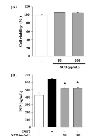
본 연구에서도 앞선 결과를 바탕으로 산유자 잎 에탄올 추출물 의 미백 효능뿐 만 아니라 다양한 활성 능력을 확인하기 위해 먼저, 산유자 잎 에탄올 추출물의 주름개선 활성 능력을 procollagen 합성 시험을 통해 분석하였다. 산유자 잎 에탄올 추 출물 50 µg/mL과 100 µg/mL에서 세포독성이 나타나지 않는 것 을 확인하였고(Fig 2A), TGF-β를 처리하였을 경우 음성대조군 에 비해 procollagen 합성이 150% 증가하였으며 산유자 잎 에
탄올 추출물을 50, 100 µg/mL 처리 하였을 때 음성대조군에 비 해 각각 120, 122%의 procollagen 합성이 증가하였으며, 통계 적으로 유의한 결과를 나타내었다(Fig 2B). 본 결과를 통하여 산유자 잎 에탄올 추출물이 양성대조군 만큼은 아니지만, CCD- 986SK human fibroblast 세포의 procollagen 합성을 증가시킴으 로써 주름개선 활성을 가지는 것을 확인 할 수 있었다.
산유자 잎 에탄올 추출물의 항염증 활성. 상기의 결과를 통해 서 산유자 잎 에탄올 추출물의 미백활성과 주름개선 활성이 확 인됨에 따라 또다른 생리활성을 확인하기 위해 항염증 활성을 분석하였다. 본 연구에서는 항염증 활성 분석에 많이 사용되는 RAW 264.7 murine macrophage를 이용하여 NO 생성 억제능 분석을 통하여 산유자 잎 에탄올 추출물의 항염증 활성 정도를 측정하였다. 먼저, 산유자 잎 에탄올 추출물을 25, 50, 그리고 100µg/mL 농도로 처리하고 세포 생존율을 관찰한 결과, RAW 264.7 cell에서 세포독성이 나타나지 않는 것을 확인하였다 (Fig 3A). 다음으로 산유자 잎 에탄올 추출물의 항염증 활성을 확인 하기 위해 LPS로 RAW 264.7 cell의 NO 생성을 유도하여 Griess reagent를 이용하여 NO 생성량을 측정한 결과, Fig 3B 에 제시된 바와 같이 LPS에 의해 유도된 NO 생성이 산유자 잎 에탄올 추출물의 농도 의존적으로 (25–100 µg/mL) 감소하는 Fig. 2 Effects of XCO on cell viability (A) and procollagen synthesis (B)
in CCD-986SK cells. (A) Cells were treated with or without XCO (50, 100µg/mL) for 24 h. (B) Cells were treated with XCO for 24 h, and procollagen synthesis was determined as described in Materials and Methods. TGF-β (10 ng/mL) was used as a positive control. Values represent the mean ± SD of three independent experiments. Statistical significance is indicated (*p <0.05, compared with untreated control cells).
Fig. 3 Effects of XCO on cell viability (A) and NO synthesis (B) in RAW 264.7 cells. (A) Cells were treated with or without XCO (25–100 µg/mL) for 24 h. (B) Cells were treated with XCO and lipopolysaccharide (LPS, 0.5µg/mL) as described in Materials and Methods. Values represent the mean ± SD of three independent experiments. Statistical significance is indicated (*p <0.05, **p <0.01, compared with LPS- treated cells).
결과를 얻을 수 있었다. 본 실험 결과는 Yang 등(2009)이 발표 한 논문과 유사하게 산유자 잎 에탄올 추출물이 RAW 264.7 cell에서 세포독성 없이 LPS에 의해 유도된 NO 생성을 억제하 는 것을 확인하였으며, NO생성 억제에 따른 높은 항염증 활성 을 가지는 것을 알 수 있었다.
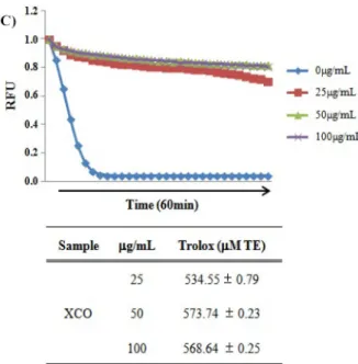
산유자 잎 에탄올 추출물의 항산화 활성. 산유자 잎 에탄올 추 출물의 항산화 활성을 분석하기 위해 DPPH, ABTS, 그리고
ORAC assay를 수행하였다. DPPH 라디칼 소거능 측정은 시료 내의 항산화 물질과 자유 라디칼인 DPPH 시약이 반응하여 시 료의 항산화 작용에 의한 자유라디칼 소거능에 따라 보라색이 노란색으로 탈색되는 원리를 이용하여 항산화 활성을 측정하는 방법으로 널리 사용된다(Bondet 등, 1997). 산유자 잎 에탄올 추출물의 DPPH 라디칼 소거능을 측정한 결과, Fig 4A에서와 같이 산유자 잎 에탄올 추출물의 농도에 따라 DPPH 라디칼 소 거능이 증가하는 것이 확인되었고, 100 µg/mL보다는 오히려 50µg/mL의 농도에서 가장 높은 75.2%의 소거능이 관찰 되었 다. 산유자 잎 에탄올 추출물의 DPPH 라디칼에 대한 IC50값은 22.8µg/mL로 측정 되었다. ABTS 라디칼 소거능은 시료내 항 산화 물질이 ABTS 시약과 potassium persulfate의 반응에 의해 생성된 ABTS 자유 라디칼을 소거시킴으로써 청록색이 탈색되 는 원리로(Re 등, 1999), DPPH 라디칼 소거능과 유사하게 측 정방법이 간단하여 널리 이용된다. 산유자 잎 에탄올 추출물에 의한 ABTS 라디칼 소거능은 DPPH 라디칼 소거능 결과와 유 사하게 산유자 잎 에탄올 추출물의 농도에 따라 소거능이 증가 하는 것이 확인 되었고, DPPH 라디칼 소거능 결과와 유사하게 50µg/mL의 농도에서 가장 높은 99.7%의 ABTS 라디칼 소거 능이 관찰 되었다 (Fig 4B). 산유자 잎 에탄올 추출물의 ABTS 라디칼 소거능에 대한 IC50값은 16.4 µg/mL로 측정되었다. 마지 막으로 ORAC assay를 통해 산유자 잎 에탄올 추출물의 항산 화 활성을 측정하였다. DPPH나 ABTS 라디칼 소거능과 달리 ORAC assay는 라디칼 소거능이 아닌 퍼옥시라디칼 생성제인 AAPH에 의한 산화반응이 시료내의 항산화 물질로 인하여 형 광의 정도가 감소하는 것을 이용 한다(Ou 등, 2002). 대조군으 로 trolox를 사용하여 형광물질과 결합시킴으로써 항산화 활성 을 평가하였다. ORAC activity assay kit를 이용하여 60분간 2 분마다 fluorescence의 감소율을 측정한 결과, Fig 4C에서와 같 이 산유자 잎 에탄올 추출물의 농도에 따라 높은 항산화 활성 이 관찰되었고, ORAC를 Trolox equivalent (TE)값으로 산출한 결과 DPPH 라디칼 소거능과 ABTS 라디칼 소거능 결과와 마 찬가지로 50 µg/mL의 농도에서 573.74±0.79 µM TE/g으로 가장 Fig. 4 Anti-oxidant activity of XCO. (A and B) Free radical scavenging
activity and (C) oxygen radical absorbance capacity of XCO (3.125–100 µg/mL) were determined as described in Materials and Methods. (A and B) Butylated hydroxyanisole (BHA) was used as a positive control. IC50 values represent the concentration required for 50% inhibition of radical scavenging. (C) Relative ORAC values are expressed as Trolox equivalents (µM TE/g). Values represent the mean ± SD of three independent experiments.
Fig. 4 Continued.
높은 항산화 활성을 나타내었다. 본 결과를 통하여 산유자 잎 에탄올 추출물이 높은 항산화 활성을 나타내는 것을 확인 할 수 있었다.
상기의 결과들을 통해 산유자 잎 에탄올 추출물이 높은 미백 과 주름개선, 항염증 및 항산화 활성을 보유하는 것을 확인 할 수 있었으며, 이러한 결과는 천연물 유래 다양한 생리활성을 보 유한 기능성 화장품 소재로서의 활용을 위한 기초자료로 이용 될 수 있을 것으로 사료된다.
초 록
본 연구는 제주도 해변의 벌판에서 드물게 자라는 산유자 (Xylosma congesta) 잎 에탄올 추출물의 화장품 기능성 소재로 서의 활용 가능성을 알아보기 위하여 cell culture model 및 in vitro assay system을 이용하여 미백, 주름개선, 항염증 및 항산 화 활성을 분석하였다. 그 결과 산유자 잎 에탄올 추출물은 B16F10 세포에 세포독성 없이 효과적으로 α-MSH에 의해 유 도된 melanin 합성을 억제함으로써 높은 미백 활성을 가지는 것이 관찰되었다. CCD-986SK 세포를 이용하여 산유자 잎 에 탄올 추출물의 주름개선 활성 능력을 procollagen 합성시험을 통해 분석한 결과, 양성 대조군으로 사용한 TGF-β 만큼은 아 니지만 농도별로 각각 120, 122%의 procollagen 합성을 증가시 키는 것을 통하여 산유자 잎 에탄올 추출물의 주름개선 활성을 확인하였다. 또한 RAW 264.7 murine macrophage를 이용하여 NO 생성 억제능을 분석함으로써 산유자 잎 에탄올 추출물의 항염증 활성 정도를 측정한 결과, LPS에 의해 유도된 NO 생 성이 산유자 잎 에탄올 추출물의 농도 의존적으로 감소하는 결 과를 얻을 수 있었다. DPPH 법, ABTS 법, ORAC 법을 이용 하여 항산화 활성을 분석한 결과, 50 µg/mL의 농도에서 DPPH 및 ABTS 라디칼 소거능이 각각 75.2, 99.1% 증가하는 것이 확인 되었다. ORAC activity assay kit를 이용하여 항산화 활성 을 측정한 결과 산유자 잎 에탄올 추출물의 농도에 따라 높은 항산화 활성이 관찰 되었고, DPPH와ABTS 라디칼 소거능 결 과와 유사하게 50 µg/mL의 농도에서 가장 높은 항산화 활성 (573.74±0.79µM TE/g)을 나타내었다. 이러한 결과를 통하여 산 유자 잎 에탄올 추출물이 높은 미백과 주름개선, 항염증 및 항 산화 활성을 나타내는 것을 확인 할 수 있었으며, 기능성 화장 품 소재로서의 활용가능성을 제시 할 수 있었다.
Keywords 멜라닌합성·산유자·피부주름·항산화활성·항 염증활성
감사의 글 본 연구는 산업통상자원부에서 시행한 “지역특화기술 융복합 연 구지원사업(헬스케어 지향 Happy Drug 제품 및 서비스 적용기술 개발, R0001028)”의 지원으로 이루어진 결과로 이에 감사드립니다.
References
Ames BN (1983) Dietary carcinogens and anticarcinogens. Oxygen radicals and degenerative diseases. Science 221, 1256–64.
Aroca P, Urabe K, Kobayashi T, Taskamoto K, and Hearing VJ (1993) Melanin biosynthesis patterns of following hormonal stimulation. J Biol Chem 268, 25650–5.
Blois MS (1958) Antioxidant determinations by the use of a stable free
radical. Nature 181, 1198–200.
Bondet V, Brand-Williams W, and Berset C (1997) Kinetics and mechanisms of antioxidant activity using the DPPH. free radical method. LWT-Food Sci Technol 30, 609–15.
Cabanes J, Chazara S, and Garcia CF (1994) Kojic acid, a cosmetic skin whitening agent, is slow-binding inhibitor of catecholase activity of tyrosinase. J Pharm Pharmacol 46, 982–5.
Callender VD, St Surin-Lord S, Davis EC, and Maclin M (2011) Postinflammatory hyperpigmentation: etiologic and therapeutic considerations. Am J Clin Dermatol 12, 87–99.
Chen JS, Wei C, and Marshall MR (1991) Inhibition mechanism of Kojic acid on polyphenol oxidase. J Agric Food Chem 39, 1897–901.
Costin GE and Hearing VJ (2007) Human skin pigment-ation: melanocytes modulate skin color in response to stress. FASEB J 21, 976–94.
DeLange RJ and Glazer AN (1989) Phycoerythrin fluorescence-based assay for peroxy radicals: a screen for biologically relevant rotective agents.
Anal Biochem 28, 300–6.
Dudonne S, Vitrac X, Coutiere P, Woillez M, and Merillon JM (2009) Comparative study of antioxidant properties and total phenolic content of 30 plant extracts of industrial interest using DPPH, ABTS, FRAP, SOD, and ORAC assays. J Agric Food Chem 57, 1768–74.
Fridovich I (1986) Biological effects of the superoxide radical. Arch Biochem Biophys 247, 1–11.
Ghiselli A, Serafini M, Maiani G, Azzini E, and Ferro-Luzzi A (1995) A fluorescence-based method for measuring total plasma antioxidant capability. Free Radic Biol Med 18, 29–36.
Jimenze-Cervants C, Solano F, Kobayashi T, Urabe K, Hearing VJ, Lozano J et al. (1994) A new enzymatic function in the melanogenic pathway. The 5,6-dihydroxyindole-2-carboxylic acid oxidase activity of tyrosinase- related protein-1 (TRP1). J Biol Chem 269, 17993–8000.
Jin KS, Oh YN, Park JA, Lee JY, Jin SJ, Hyun SK et al. (2012) Anti-Oxidant, Anti-Melanogenic, and Anti-Inflammatory Activities of Zanthoxylum schinifolium Extract and its Solvent Fractions. Korean J Microbiol Biotechnol 40, 371–9.
Jung SW, Lee NK, Kim SJ, and Han D (1995) Screening of tyrosinase inhibitor from plants. Kor J Food Sci Technol 27, 891–6.
Kang MC, Lee JY, Ko RK, Kim HB, Hong SH, and Kim GO (2009) Melanin inhibitory effect abd anti-inflammatory effects of Dictyota corlacea extracts derived from adjacent sea of the Jeju island. Korean J Biotechnol Bioeng 23, 311–6.
Kawabata T, Cui MY, Hasegawa T, Takano F, and Ohta T (2011) Anti- inflammatory and anti-melanogenic steroidal saponin glycosides from Fenugreek (Trigonella foenum-graecum L.) seeds. Planta Med 77, 705–
10.
Kawamata H, Ochiai H, Mantani N, and Terasawa K (2000) Enhanced expression of inducible nitric oxide synthase by Juzen-taiho-to in LPS- activated RAW 264.7 cells, a murine macrophage cell line. Am J Chin Med 28, 217–26.
Lee KH, Kong JH, Cho YL, Joo CG, Kwon SS, Hwang JS et al. (2012) Anti- microbial and Anti-wrinkle effect of kaempferol and kaempferol rhamnosides isolated from Hibiscus cannabinus L. Korean J Medicinal Crop Sci 20, 454–60.
Lerner AB and Fitzpatrick TB (1950) Biochemistry of melanin formation.
Physiol Rev 30, 91–126.
Miller NJ, Rice-Evans C, Davies MJ, Gopinathan V, and Milner A (1993) A novel method for measuring antioxidant capacity and its application to monitoring the antioxidant status in premature neonates. Clin Sci (Lond) 84, 407–12.
Ou B, Huang D, Hampsch-Woodill M, Flanagan JA, and Deemer EK (2002) Analysis of antioxidant activities of common vegetables employing oxygen radical absorbance capacity (ORAC) and ferric reducing antioxidant power (FRAP) assays: a comparative study. J Agric Food Chem 50, 3122–8.
Ozgen M, Reese RN, Tulio AZ, Scheerens JC, and Miller AR (2006) Modified 2,2-azino-bis-3-ethylbenzothiazoline-6-sulfonic acid (ABTS) method to measure antioxidant capacity of selected small fruits and comparison to ferric reducing antioxidant power (FRAP) and 2,2'- diphenyl-1-picrylhydrazyl (DPPH) methods. J Agric Food Chem 54,
1151–7.
Panich U, Tangsupa-a-nan V, Onkoksoong T, Kongtaphan K, Kasetsinsombat K, Akarasereenont P et al. (2011) Inhibition of UVA-mediated melanogenesis by ascorbic acid through modulation of antioxidant defense and nitric oxide system. Arch Pharm Res 34, 811–20.
Re R, Pellegrini N, Proteggente A, Pannala A, Yang M, and Rice-Evans C (1999) Antioxidant activity applying an improved ABTS radical cation decolorization assay. Free Radic Biol Med 26, 1231–7.
Regoli F and Winston GW (1999) Quantification of total oxidant scavenging capacity of antioxidants for peroxynitrite, peroxyl radicals, and hydroxyl radicals. Toxicol Appl Pharmacol 156, 96–105.
Rice-Evans C and Miller NJ (1994) Total antioxidant status in plasma and body fluids. Methods Enzymol 234, 279–93.
Rice-Evans CA, Miller NJ, and Paganga G (1996) Structure-antioxidant activity relationships of flavonoids and phenolic acids. Free Radic Biol Med 20, 933–56.
Song HM, Seo MS, Kim HM, Ahn MS, and Lee YT (2002) Antioxidative activity of barley polyphenol extract (BPE) separated from pearling by-
products. Kor J Food Sci Technol 34, 889–92.
Tatsuno T, Jinno M, Arima Y, Kawabata T, Hasegawa T, Yahagi N et al.
(2011) Anti-inflammatory and anti-melanogenic proanthocyanidin oligomers from peanut skin. Biol Pharm Bull 35, 909–16.
Urabe K, Aroca P, Tsukamoto K, Mascagna D, Paulumbo A, Prota G et al.
(1994) The inherent cytotoxicity of melanin precursors. Biochim Biophys Acta 1221, 272–8.
Winston GW, Regoli F, Dugas AJ Jr, Fong JH, and Blanchard KA (1998) A rapid gas chromatographic assay for determining oxyradical scavenging capacity of antioxidants and biological fluids. Free Radic Biol Med 24, 480–93.
Yang EJ, Yim EY, Song GP, Kim GO, and Hyun CG (2009) Inhibition of nitric oxide production in lipopolysaccharide-activated RAW 264.7 macrophages by Jeju plant extracts. Interdisc Toxicol 2, 245–9.
Zurick KM, Qin C, and Bernards MT (2012) Adhesion of MC3T3-E1 cells bound to dentin phosphoprotein specifically bound to collagen type I. J Biomed Mater Res A 100, 2492–8.