Anti-cancer effects of enzyme-digested fucoidan extract from seaweed Mozuku
Kiichiro Teruya
1,2,3*Sakiko Matsuda
2Ayumi Nakano
2Takuya Nishimoto
2Masashi Ueno
2Akitono Niho
3Makiko Yamashita
3Hiroshi Eto
4Yoshinori Katakura
1,2,3Sanetaka Shirahata
1,2,31ABSTRACT
Fucoidan is a uniquely-structured sulfated fucose-rich polysaccharide derived from brown algae.
Recently, the abalone glycosidase-digested fucoidan extract (fucoidan extract) derived from seaweed Cladosiphon novae-caledoniae Kylin (Mozuku) draws much attention because of its clinical anti-cancer effect in Japan. Here, we report the cancer cells-specific apoptosis inducing effects of the fucoidan extract. The fucoidan extract suppressed the growth of various anchorage-dependent and -independent cancer cells. The fucoidan extract contained low molecular weight components, which induced apoptosis of human leukemic HL 60 cells but not of human lymphocytes. It was shown that the fucoidan extract lead caspase 3/7 activation and loss of mitochondrial membrane potential in HL 60 cells. Another function of the fucoidan extract was also observed. It has been known that sugar chain expression on the surface of cancer cell membrane changes dependent on their malignancy. The analysis on sugar chain expression profiling using FITC-labeled lectins revealed that the expression of concanavalin A (Con A) binding sugar chain was enhanced by the treatment of human lung adenocarcinoma A549, human uterine carcinoma HeLa and human fibrosarcoma HT1080 cells with the fucoidan extract. Con
Received: Sept. 8, 2008; Revised: Jan. 29, 2009; Accepted: Jun. 3, 2009
1 Department of Genetic Resources Technology, Faculty of Agriculture, Kyushu University
2 Graduate School of Bioresource and Bioenvironmental Sciences, Kyushu University
3 Graduate School of Systems Life Sciences, Kyushu University
4 Daiichi Sangyo Co. Ltd.
* Correspondence: Kiichiro Teruya(E-mail: [email protected])
A-induced apoptosis of cancer cells was stimulated in a dose-and time-dependent manner by the treatment with the fucoidan extract but not of human normal fibroblast TIG-1 cells.
Keywords: apoptosis, cancer, concanavalin A (Con A), fucoidan, seaweed
INTRODUCTION
Fucoidan is a uniquely-structured sulfated polysaccharide found in the cell walls of several types of brown seaweed. Recently, fucoidan has attracted a lot of clinical attention due to its strong anti-tumor potential, which has been intensively investigated. Fucoidan suppresses the growth of tumor cells in vivo and activates the immune system against tumors [1-6]. Sulfation of fucoidan enhanced its antitumor activity [2].
Koyanagi et al. reported that fucoidan inhibited tube formation following migration of human umbilical vein endothelial cells [7]. Ye et al.
reported that enzyme-digested fucoidan extract inhibited invasion and angiogenesis of tumor cells [8]. Aisa et al. reported that fucoidan induced apoptosis HS-Sultan cells accompanied by caspase-3 activation and down-regulation of ERK pathway [9].
In this study, we examined that anti-cancer effects of enzyme-digested fucoidan extract derived from Mozuku of Cladosiphon novae- caledoniae Kylin, which originates in the Kingdom of Tonga, inducing the apoptosis of tumor cells.
MATERIALS AND METHODS
Preparation of Fucoidan and Treatment The abalone glycosidase-digested fucoidan extract prepared from seaweed Mozuku of Cladosiphon novae-caledoniae Kylin from the Kingdom of Tonga, commercially available as a product named ‘Power fucoidan’, was donated for the study by the Daiichi Sangyo Corporation (Osaka, Japan) [8]. An undiluted solution (pH3.7) was neutralized to pH 7.0 with NaOH.
The precipitants were removed by centrifugation at 2,200×g for 15 min. The supernatants were then autoclaved, and stored as ‘fucoidan extract (2.8mg/ml)’ at 4°C. The fucoidan extract was mixed with culture medium, and treatments to various cells were performed.
Cell Culture
The human fibrosarcoma cell line HT1080, human uterine carcinoma cell line HeLa, human lung adenocarcinoma and human normal fibroblast TIG-1 were cultured in Minimum Eagle’s medium (MEM: Nissui Pharmaceutical Co., Ltd., Tokyo, Japan) supplemented with 10%
fetal bovine serum (FBS), 10 mM HEPES, 100
units/ml penicillin and 100 µg/ml streptomycin
(10% FBS/MEM). Human leukemic cell lines
HL60, U937 and K562 cells were cultured in RPMI 1640 medium (Nissui) supplemented with heat-inactivated 10% FBS, 10 mM HEPES, 100 units/ml penicillin and 100 µg/ml streptomycin. All cell culture was performed in a humidified atmosphere with 5% CO
2at 37°C.
Anti-proliferative Assay
Cells were inoculated at a final concentration of 1 × 10
5cells/ml in culture medium in 35 mm culture dishes. After 24 h pre-incubation, the cells were exposed to various concentrations of the fucoidan extract. After cultivation, the cell number in each culture dish was measured using automated hematology analyzer (Sysmex Co., Kobe, Japan).
Caspase-3/7 Activity Assay
To examine caspase activity, HL60 cells were inoculated at a density of 2.0 × 10
4cells in 100 µl culture medium with the fucoidan extract and cultured in a 96-well microplate. After 24 h culture, caspase-3/7 activity was measured with Apo-ONE
™Homogeneous Caspase-3/7 Assay kit (Promega Co., Madison, WI). Briefly, Apo- ONE
™reagent, profluorescent substrate containing the DEVD sequence, was added to the microplate. The sample plate was incubated for 4 h, and then a fluorescent dye rhodamine 110 was released through cleavage of the profluorescent substrate by the caspases-3/7 activity in the samples. The fluorescence was measured by a fluorometer (excitation at 498nm;
emission at 521nm). The amount of fluorescent
product generated is representative of the amount of active caspase-3/7 present in the sample.
Analysis of Cell Cycle and Apoptosis Cell cycle was determined by PI staining and flow cytometry as described [10]. Briefly, HL60 cells (2 × 10
5cells/ml) were incubated in 10%
FBS/RPMI 1640 containing the fucoidan extract for 24 h. After one gentle rinse by PBS, the cells were collected by trypsinization and centrifugation at 200 × g for 5 min. Then the cell pellets were gently resuspended in 150 µl of PBS, and added 350 µl of cold ethanol for 30 min at 4°C. Fixed cells were collected by centrifugation at 200×g for 5 min, and were resuspended in 500 μl of PBS. The cell pellets were incubated in 500 µl of PI/RNase A/PBS staining solution (10 μg/ml PI and 10 μg/ml RNase A in PBS) at 25°C for 20 min in a dark place. Stained cells were analyzed by a flowcytometer (excitation: 488 nm and emission:
610nm). Ten thousand cells were evaluated for
each sample. In some experiments, HL60 cells
was pretreated with 20 µM Z-VAD-FMK
(Promega) for 5 h followed by a 24 h incubation
with the fucoidan extract before analysis of
apoptosis. Mitochondrial membrane potential was
determined by Rhodamine 123 staining using a
flowcytomer, as previously reported [11]. Briefly,
the cells were washed twice with PBS and
incubated with 1 µg/ml rhodamine 123 (Sigma-
Aldrich Inc., St. Louis, MO) at 37°C for 30 min
to assess the mitochondrial membrane potential
by a flowcytometer. A higher intensity of
rhodamine 123 indicated a higher mitochondrial membrane potential [11].
Sugar Chain Expression Profiling Using FITC-Lectins
Lectin-staining of cells was performed. Briefly, Cells were inoculated at a density of 2.0 × 10
5cells into a 6-well culture plate. After pre- incubation, the cells were treated with the fucoidan extract. After treatment, cells were washed with PBS and incubated in 50 µl of PBS containing 2 µg of FITC-conjugate lectins (J-OIL MILLS Inc., Tokyo, Japan) at 25°C for 15 min. After wash with PBS, cells were suspended in 500 µl of PBS, and then the analysis was performed by a flowcytometer.
Stimulation of Con A-induced Apoptosis of Cancer Cells
Cells were inoculated at a density of 1.0 × 10
5cells into a 6-well culture plate. Afterpre- incubation, the cells were treated with the fucoidan extract for 48 h. Then, cells were washed with PBS and incubated in 50 µl of PBS containing 2 µg of Con A at 25°C for 30 min.
After wash with PBS, cell cycle analysis was performed.
RESULTS
Anti-proliferative activity of the fucoidan extract to cancer cells
The treatment of the enzyme-digested fucoidan
extract suppressed the proliferations of various cancer cells (Figure 1). The fucoidan extract suppressed the proliferations of anchorage- dependent human cancer cells such as cervix adenocarcinoma HeLa, connective tissue fibrosarcoma HT1080 and lung adenocarcinoma A549 cells. Anchorage-independent human cancer cells, typical leukemic cells, such as acute promyelocytic leukemia HL-60, histiocytic lymphoma U937 and chronic myelogenous leukemia K562 cells were also suppressed the proliferations by the fucoidan extract treatment.
The anti-proliferation effect of cancer cells was enhanced by treatment with the fucoidan extract in a dose-dependent manner.
On the other hand, the fucoidan extract did not induce apoptosis to human peripheral blood lymphocytes even at the highest dose of 560 µg/ml. At the highest dose, HL-60 and U937 cells were induced 33.2% and 34.5% sub-G1 population, respectively (data not shown). It was suggested that the fucoidan extract specifically suppressed the proliferation of cancer cells.
The fucoidan extract induced apoptosis in HL60 cells
The cell cycle analysis of human leukemic HL60 cells was analyzed using a flowcytometer.
The percentage of sub-G1 fraction was increased
in a time- and dose- dependent manner after
treatment with the fucoidan extract (data not
shown). Pretreatment of HL60 cells with pan-
caspase inhibitor, Z-VAD-FMK, inhibited the
Figure 1. Anti-proliferative activity of the fucoidan extract to human cancer cells. HeLa (cervix adenocarcinoma), (B) HT1080 (connective tissue fibrosarcoma), (C) A549 (lung adenocarcinoma), (D) HL-60 (acute promyelocytic leukemia), (E) U937 (histiocytic lymphoma), (F) K562 (chronic myelogenous leukemia).
augmentation of sub-G1 population induced by the fucoidan extract (Figure 2 A). This result indicated that the caspase activity played the important role for the augmentation of sub-G1 population in the fucoidan extract treated HL60
cells. To evaluate the involvement of the
activation of caspase-3/7, the activity of
caspase-3/7 was determined. After 3 h treatment
of the fucoidan extract, the caspase-3/7 activity
reached the maximum value (Figure 2 B). The
Figure 2. The fucoidan extract induced apoptosis in human leukemic HL60 cells. (A) Detection of sub-G cells treated with or without the fucoidan extract and pan-caspase inhibitor Z-VAD-FMK. (B) Detection of caspase-3/7 activation in the fucoidan extract treated HL60 cells. (C) Analyses of mitochondrial electric potential. Control; a dotted line and gray filling. Fucoidan extract treatment (280 µg/ml, 24 h); a solid line.
mitochondrial membrane potential of HL60 cells was decreased by the fucoidan extract treatment for 24 h (Figure 3 C). These results showed that the treatment of the fucoidan
extract induced apoptosis in HL60 cells
viacaspase-3/7activationandthemitochondrialdepol
arization.
The fucoidan extract changed sugar chain expression on the surface of cancer cell membrane and stimulated Con A-induced apoptosis of cancer cells
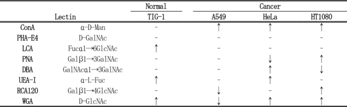
We treated various cancer cells with the fucoidan extract, Con A binding was enhanced in human cancer-derived A549, HeLa and HT1080 cells, but human normal fibroblast TIG-1 cells did not (Table 1). The binding profiles with other lectins were also changed.
The binding of 3 cancer cells with Con A was increased depending upon their malignancy (data not shown). The malignancy is considered to become stronger in order of A549, HeLa and HT1080 cells.
When HT1080 cells were pretreated with the fucoidan extract, apoptosis-inducing ability of Con A was enhanced. However, normal TIG-1
Table 1. Sugar chain expression profiling using FITC-labeled lectins after treatment with the fucoidan extract
Normal Cancer
Lectin TIG-1 A549 HeLa HT1080
ConA α-D-Man – ↑ ↑ ↑
PHA-E4 D-GalNAc – – – –
LCA Fucα1→6GlcNAc ↑ – – –
PNA Galβ1→3GalNAc – – ↓ ↑
DBA GalNAcα1→3GalNAc – – ↑ ↓
UEA-I α-L-Fuc ↑ – ↑ –
RCA120 Galβ1→4GlcNAc – ↓ – ↑
WGA D-GlcNAc ↑ ↓ ↑ ↑
Con A; Concanavalin A (Lectin from Canavalia ensiformis, jack beans), PHA-E4; Phaseolus vulgaris agglutinin (Erythroagglutinin, Lectin from Phaseolus vulgaris, red kidney bean), LCA; Lens culinaris agglutinin (Lectin from Lens culinaris, lentil), PNA; Peanut agglutinin (Lectin from Arachis hypogaea, peanut), DBA; Dolichos biflorus agglutinin (Lectin from Dolichos biflorus, horse gram), UEA-I; Ulex europaeus agglutinin-I (Lectin from Ulex europaeus, gorse, furze), RCA120; Ricinus communis Agglutinin (Lectin from Ricinus communis, castor bean), WGA; Wheat germ agglutinin (Lectin from Triticum vulgaris, wheat).–; no change,↑; increase of expression, ↓; decrease of expression.