Horner’s Syndrome with Abducens Nerve Palsy
4
0
0
전체 글
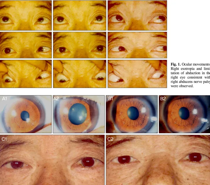
(2) Korean J Ophthalmol Vol.25, No.6, 2011. Fig. 1. Ocular movements. Right esotropia and limitation of abduction in the right eye consistent with right abducens nerve palsy were observed.. A1. A2. C1. B1. B2. C2. Fig. 2. (A1,B1) Before topical application of 0.5% apraclonidine in room light. Pupil right eye (OD) measured 2.5 mm (A1), pupil left eye (OS) measured 3.0 mm (B1). (A2,B2) After topical application of 0.5% apraclonidine in bright light. Pupil OD measures 5.0 mm (A2), pupil OS measured 4.0 mm (B2). Reversal of pupillary dilatation under topical application of 0.5% apraclonidine was detected. (C1,C2) Note also the improvement of the mild ptosis in the right side after topical application of 0.5% apraclonidine.. right cavernous sinus with encasement of the internal carotid artery (ICA) (Fig. 3). The patient underwent bilateral functional endoscopic sinus surgery with biopsy. Pathology was consistent with poorly differentiated invasive squamous cell carcinoma (Fig. 4). Seven weeks later, the esotropia worsened and the limitation of abduction, anisocoria, and right ptosis persisted. No other neurologic abnormalities were found up to the last visit. The patient died of cancerous cachexia 15 months after the diagnosis of nasopharyngeal cancer.. Discussion Neither abducens nerve palsy nor Horner’s syndrome provides an indication of the location of the lesion. However,. 460. when both are present, it is important to localize the site of the lesion [16]. When these rare clinical features are manifested, we have to consider paratrigerminal syndrome or a lesion of the posterior cavernous sinus [1]. Sympathetic nerve fibers travel over the wall of the carotid artery and in the cavernous portion the fibers leave the ICA and accompany the abducens nerve for only a few millimeters in the posterior portion of the sinus [4,10,12,16] (Fig. 5). Therefore, we could consider a posterior cavernous sinus lesion due to combined abducens nerve palsy. Furthermore, incomplete Horner’s syndrome without anhydrosis, as in the case reported here, indicates the involvement of the periarterial sympathetic plexus at the third angulation point [17]. To date, only 13 cases with this combination of symptoms secondary to metastatic.
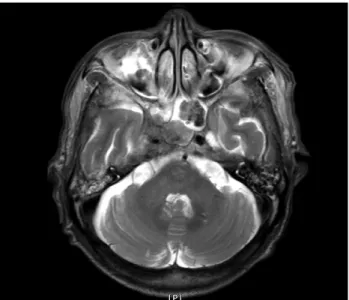
(3) NH Kang, et al. Horner’s Syndrome with Abducens Nerve Palsy. Fig. 3. Axial T1-weighted contrast-enhanced magnetic resonance images. Ill-defined right cavernous sinus lesion extending into the sphenoidal sinus and encasement of the internal carotid were found.. Fig. 4. A photomicrograph of the frozen biopsy during the functional endoscopic sinus surgery showing the moderately to poorly differentiated squamous cells in the right sphenoid sinus (H&E, ×200).. neoplasm within the cavernous sinus have been reported [4,10-16]. Regarding the primary lesions in these cases, there were 1 small cell lung cancer [10], 1 parotid cancer [10,12], 1 breast cancer [11], 1 gastric cancer [10], 1 uterine cancer [4], 3 undetermined origins [13,14] and 3 nasopharyngeal cancers [15]. Unlike in other three nasopharyngeal cancer cases reported by Hirao et al. [15], the cancer only invaded the 6th nerve and sympathetic fiber without involvement of the 5th nerve in this case. Malignant tumors that affect the cavernous sinus commonly result in the 5th and the 6th nerve injuries [3,8]. The more medial location of the abducens nerve has been postulated as the reason for its frequent involvement when confronted with the compressive forces in a confined space or direct invasion [8]. In this case, the nasopharyngeal cancer extended through the sphenoidal sinus demonstrated. Fig. 5. Schematic drawing demonstrating the anatomy of the posterior portion of the cavernous sinus on the right side. Oculosympathetic nerve fibers leave the internal carotid artery, and then join the abducens nerve for a short distance in the posterior portion of the cavernous sinus (arrow). CN III = oculomotor nerve; CN IV = trochlear nerve; CN V = trigeminal nerve; ICA = internal carotid; CN VI = abducens nerve; Sym = oculosympathetic nerve.. by brain MRI and positron emission tomography (Fig. 3). Therefore, the cancer affected only the 6th nerve without involvement the 3rd nerve, which occupies a superior position at the lateral wall of the sinus, in addition to the 4th and 5th nerve [1]. Although autopsy was not permitted and there was a limitation of anatomical access, we suggest that tumor cells in the sphenoidal sinus had spread to the 6th nerve in the cavernous sinus, and then perineurally metastasized to the oculosympathetic nerve fibers before joining the first division of the 5th nerve [3]. A plausible alternative to cocaine or hydroxyamphetamine in the diagnosis of Horner’s syndrome is apraclonidine, a direct α-receptor agonist with strong α2 and weak α1 activity [18]. In patients with Horner’s syndrome, reversal of anisocoria and improvement of ptosis occur after application of apraclonidine due to denervation supersensitivity of the α1 receptor [18]. With respect to the length of survival in patients with tumor invasion of the cavernous sinus region as reported in the literature, most individuals live no longer than six months and the ultimate outcome is a dismal 75% to 85% expected mortality within 2 years [3,8]. The patient described in this report died 5 months after the presentation of combination abducens nerve palsy with Horner’s syndrome. In other words, this manifestation may be a significant predictor of survival in addition to the location of the lesion. This is the first reported case of abducens nerve palsy with Horner’s syndrome due to tumor invasion into the cavernous sinus in a Korean patient. The presence of Horner’s syndrome and the 6th nerve palsy should be cautiously investigated to confirm the existence of the cavernous sinus lesions.. Conflict of Interest No potential conflict of interest relevant to this article was reported.. 461.
(4) Korean J Ophthalmol Vol.25, No.6, 2011. References 1. Silva MN, Saeki N, Hirai S, Yamaura A. Unusual cranial nerve palsy caused by cavernous sinus aneurysms. Clinical and anatomical considerations reviewed. Surg Neurol 1999;52:143-8. 2. Rhoton AL Jr. The cavernous sinus, the cavernous venous plexus, and the carotid collar. Neurosurgery 2002;51(4 Suppl):S375410. 3. Ampil FL, Heldmann M, Ibrahim AM, Balfour EL. Involvement of the cavernous sinus by malignant (extracranial) tumour: palliation in six cases without surgery. J Craniomaxillofac Surg 2000;28:161-4. 4. Tsuda H, Yorinaga Y, Tamada Y, et al. Combination of abducens nerve palsy and ipsilateral postganglionic Horner syndrome as an initial manifestation of uterine cervical cancer. Intern Med 2009;48:1457-60. 5. George A, Haydar AA, Adams WM. Imaging of Horner's syndrome. Clin Radiol 2008;63:499-505. 6. Parkinson D. Bernard, Mitchell, Horner syndrome and others? Surg Neurol 1979;11:221-3. 7. Kano H, Niranjan A, Kondziolka D, et al. The role of palliative radiosurgery when cancer invades the cavernous sinus. Int J Radiat Oncol Biol Phys 2009;73:709-15. 8. Curry MP, Newlon JL, Watson DW. Cavernous sinus metastasis from laryngeal squamous cell carcinoma. Otolaryngol Head Neck Surg 2001;125:567-8. 9. Djalilian HR, Tekin M, Hall WA, Adams GL. Metastatic head and neck squamous cell carcinoma to the brain. Auris Nasus Larynx 2002;29:47-54.. 462. 10. Tsuda H, Ishikawa H, Kishiro M, et al. Abducens nerve palsy and postganglionic Horner syndrome with or without severe headache. Intern Med 2006;45:851-5. 11. Gutman I, Levartovski S, Goldhammer Y, et al. Sixth nerve palsy and unilateral Horner's syndrome. Ophthalmology 1986;93:913-6. 12. Tsuda H, Ishikawa H, Asayama K, et al. Abducens nerve palsy and Horner syndrome due to metastatic tumor in the cavernous sinus. Intern Med 2005;44:644-6. 13. Slamovits TL, Cahill KV, Sibony PA, et al. Orbital fine-needle aspiration biopsy in patients with cavernous sinus syndrome. J Neurosurg 1983;59:1037-42. 14. Wilhelm H, Ochsner H, Kopycziok E, et al. Horner's syndrome: a retrospective analysis of 90 cases and recommendations for clinical handling. Ger J Ophthalmol 1992;1:96-102. 15. Hirao M, Oku H, Sugasawa J, et al. Three cases of abducens nerve palsy accompanied by Horner syndrome. Nihon Ganka Gakkai Zasshi 2006;110:520-4. 16. Kurihara T. Abducens nerve palsy and ipsilateral incomplete Horner syndrome: a significant sign of locating the lesion in the posterior cavernous sinus. Intern Med 2006;45:993-4. 17. Ozveren MF, Uchida K, Erol FS, et al. Isolated abducens nerve paresis associated with incomplete Horner's syndrome caused by petrous apex fracture: case report and anatomical study. Neurol Med Chir (Tokyo) 2001;41:494-8. 18. Freedman KA, Brown SM. Topical apraclonidine in the diagnosis of suspected Horner syndrome. J Neuroophthalmol 2005;25:83-5..
(5)
수치
관련 문서
36 ADNP 증후군, 헬스무르텔-반데르아 증후군 ADNP syndrome, Helsmoortel-VanDerAa syndrome 37 KIFIA유전자돌연변이에의한신경병증 Neuropathy due to KIF1A
-The square module stands free in the garden of the house as a canopy -consisted of a thin shell supported at each of its corners by equally thin reinforced concrete piers.
The purpose of this study was to analyze the impaction pattern of the impacted mandibular third molar and the relationship with the inferior alveolar nerve
Through an empirical analysis of employability of persons with cerebral palsy this study aims to examine the factors affecting their employability and
Serum uric acid levels and risk for vascular disease in patients with metabolic syndrome... Prevalence if the metabolic syndrome in a Turkish
The study on the clinical efficacy of korean red ginseng extract on postmenopausal syndrome..
If we picked the n th element, then it replaces one of the s elements in the sample S, picked uniformly at random. Claim: This algorithm maintains a sample S with
"Architecture is the art and science of making sure that our cities and buildings actually fit with the way we want to live our lives: the proces s of manifesting our
