• 검색 결과가 없습니다.

Poor Prognostic Factors in Surgically Resected Stage I Non-small Cell Lung Cancer: Histopathologic and Immunohistochemical Analysis

N/A
N/A
Protected

Academic year: 2021

Share "Poor Prognostic Factors in Surgically Resected Stage I Non-small Cell Lung Cancer: Histopathologic and Immunohistochemical Analysis"

Copied!
9
0
0

로드 중.... (전체 텍스트 보기)

전체 글

(1)

http://dx.doi.org/10.5090/kjtcs.2012.45.2.101 ISSN: 2233-601X (Print) ISSN: 2093-6516 (Online)

Department of Thoracic and Cardiovascular Surgery, Kyungpook National University Hospital, Kyungpook National University School of Medicine

†This work was supported by a grant from the Industry-Academic Cooperation Foundation of Kyungpook National University.

Received: September 9, 2011, Revised: September 28, 2011, Accepted: October 16, 2011

Corresponding author: Sukki Cho, Department of Thoracic and Cardiovascular Surgery, Kyungpook National University Hospital, Kyungpook National University School of Medicine, 130 Dongdeok-ro, Jung-gu, Daegu 700-721, Korea

(Tel) 82-53-420-5676 (Fax) 82-53-426-4756 (E-mail) [email protected]

C

The Korean Society for Thoracic and Cardiovascular Surgery. 2012. All right reserved.

CC

This is an open access article distributed under the terms of the Creative Commons Attribution Non-Commercial License (http://creative- commons.org/licenses/by-nc/3.0) which permits unrestricted non-commercial use, distribution, and reproduction in any medium, provided the original work is properly cited.

Poor Prognostic Factors in Surgically Resected Stage I Non-small Cell Lung Cancer: Histopathologic and

Immunohistochemical Analysis

Sukki Cho, M.D., Tae In Park, M.D., Eung-Bae Lee, M.D., Ph.D., Shin-Ah Son, M.D.

Background: A better understanding of the histopathology and molecular biology of lung cancer might improve our capability to predict the outcome for any individual patient. The purpose of this study was to evaluate several his- topathologic and molecular markers in order to assess their prognostic value in stage I non-small cell lung cancer.

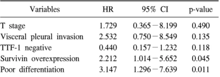
Materials and Methods: One hundred ten patients at the Kyungpook National University Hospital were enrolled in the study. Histopathologic factors and molecular markers were selected. Results: Univariate analysis showed that the T stage, differentiation, visceral pleural invasion, and survivin expression were significantly associated with recurrence. Multivariate analysis demonstrated that differentiation and survivin overexpression emerged as in- dependent prognostic factors of recurrence. Conclusion: In resected stage I non-small cell lung cancer, poor differ- entiation and survivin overexpression have been identified as independent predictors of poor disease-free survival.

Key words: 1. Lung neoplasms 2. Prognosis 3. Pathology

4. Immunohistochemistry

INTRODUCTION

Lung cancer is the most common cause of cancer mortality worldwide. Non-small cell lung cancer (NSCLC) accounts for approximately 80% of lung cancer cases and pathologic stage I represents the fastest growing segment due to the use of low-dose computed tomography for screening. Despite the po- tential benefits of surgical resection, the 5-year survival rate is only 60% to 70% in stage I patients, predominantly as a result of the development of distant metastasis [1,2].

Variation in survival largely reflects the heterogeneity of tu-

mor biology, with some tumors having more aggressive growth and greater metastatic potential than others; therefore, current tumor stage alone cannot exactly establish the prog- nosis for these patients. New prognostic factors must be iden- tified to help clinicians better assess the probability of surviv- al and to optimize therapeutic strategies for each individual patient with pathologic stage I lung cancer. Several studies have already demonstrated possible prognostic roles for sev- eral biological factors in NSCLC and have found helpful tools for identifying patients with a poor prognosis [3-6].

Among those factors, thyroid transcription factor 1 (TTF-1)

(2)

expression, especially in adenocarcinoma, was known to be a prognostic factor as well as a differential diagnostic factor be- tween primary lung cancer and other adenocarcinoma [7].

Nuclear survivin expression might be an independent bio- marker for disease recurrence and survival for NSCLC [8].

Epidermal growth factor receptor (EGFR) overexpression may predict shorter survival in patients with resected stage I-IIIA NSCLC, although this is under debate [9]. E-cadherin is known to play a role in tumor progression and distant meta- stasis; therefore, reduced E-cadherin expression could poten- tially affect tumor differentiation and prognosis [10,11]. As a result, the stratification of patients without lymph node in- volvement, according to prognostic risk, might aid in select- ing a group of high-risk patients who would benefit from ad- juvant therapy.

The purpose of this study is to evaluate several histopatho- logic variables and a panel of molecular markers-TTF-1, sur- vivin, EGFR, and E-cadherin expression-in order to assess their prognostic value and their combined effects on re- currence in patients with resected stage I NSCLC.

MATERIALS AND METHODS 1) Patient characteristics

Between January 2003 and December 2006, a total of 110 patients (84 male, 26 female) with resected stage I NSCLC including squamous cell carcinoma (SCC), adenocarcinoma (AC), and bronchioalveolar carcinoma (BAC) were enrolled in the study. All patients in the study underwent potentially curative surgery consisting of lobectomy including sleeve re- section and bilobectomy (n=104), pneumonectomy (n=4), or segmentectomy (n=2) and complete mediastinal lymph node dissection. None of the patients had neoadjuvant therapy.

Patients who died within one month after surgery were ex- cluded from the study to avoid the bias of perioperative mortality. The age of the patients ranged from 41 to 79 years (mean, 62.3 years). Postsurgical pathologic tumor-node-meta- stasis (TNM) staging was determined according to the guide- lines of the American Joint Cancer Committee (AJCC) 6th edition. There were 38 cases with stage IA (T1N0M0) and 72 cases with stage IB (T2N0M0). Follow-up data on the study population were obtained by direct contact. Follow-up oc-

curred at 3-month intervals for the initial 2 years and at 4-month intervals thereafter. Recurrences were detected by computed tomography scans or positron emission tomography and, if necessary, confirmed by pathologic examination of bi- opsy specimens. Patients were categorized as alive with evi- dence of disease or alive without disease. No patient in this series died of cancer-unrelated causes. The time from the date of the operation to the date of follow-up or death was recorded. Local recurrence was defined as tumor recurrence at the ipsilateral lung or lymph node, and distant recurrence was defined as tumor recurrence at the contralateral lung or lymph node and a distant organ such as the liver, brain, or bone.

2) Pathologic criteria

One pathologist (TIP) reviewed all the histologic slides in a blind fashion. Tumor samples were fixed in 10% buffered formalin, dehydrated, and embedded in paraffin. Then, 4 μL- thick sections were cut and stained with hematoxylin and eosin. Pathologic features were classified according to the his- tologic criteria of the World Health Organization. The degree of differentiation was divided into three groups: good, moder- ate, and poor. Tumor necrosis (negative, <10%; positive, ≥ 10%) and visceral pleural invasion (absent vs. present) were noted. Tumor size (≤2 cm, 2< & ≤3 cm, 3< & ≤5 cm, 5<

& ≤7 cm, >7 cm) was also recorded.

3) Immunohistochemical methods

Briefly, tissues were deparaffinized in xylene and rehy- drated in graded alcohols and water. Endogenous peroxidase was blocked by soaking in 3% H

2

O

2

at 45

o

C for 4 minutes.

The slides were microwaved in citrate buffer (2.1 g/L, pH

6.0) at 120

o

C for 15 minutes to unmask the antigen and were

then treated with a protein-blocking reagent before incubation

at 4

o

C overnight with primary antibodies at a 1:50 dilution,

as recommended by the supplier. After extensive washing, the

sections were incubated at room temperature for 10 minutes

with biotinylated anti-mouse immunoglobulin antibodies (Zymed,

San Francisco, CA, USA) at a 1:20 dilution and subsequently

with streptavidin-biotin peroxidase complexes at a 1:25 dilu-

tion. The reaction products were visualized by immersing the

slides in 3,3'-diaminobenzidine tetrahydrochloride. Countersta-

ining was performed with hematoxylin. All series included

(3)

Table 1. Histopathologic findings in resected stage I non-small cell lung cancer (n=110)

Characteristics No. of patients (%) Cell type

SCC AC BAC T stage

1 2

Tumor size (cm)

≤3

>3 Differentiation

Good Moderate Poor Necrosis

Absent Present

Visceral pleural invasion Absent

Present

69 (62.7) 37 (33.6) 4 (3.7)

38 (34.5) 72 (65.5)

59 (53.6) 51 (46.4)

33 (30.0) 60 (54.5) 17 (15.5)

77 (70.0) 33 (30.0)

58 (52.7) 52 (47.3)

SCC=squamous cell carcinoma; AC=adenocarcinoma; BAC=

bronchioalveolar carcinoma.

positive and negative controls. The negative controls were prepared by omitting the primary antibodies and known pos- itive controls were included in each run.

4) Evaluation of immunohistochemical data

TTF-1 staining was assessed by the intensity relative to the strong staining intensity of type II pneumocytes as: negative (absent staining, 0), low expression (weak staining intensity, 1), medium expression (intermediate staining intensity, 2), or high expression (strong staining intensity, 3), and only nu- clear staining was considered positive staining. Positivity was defined as a staining intensity of 2 and 3. EGFR expression was assessed by an intensity of staining from 0 to 3 and graded as normal (0 and 1) and overexpressed (2 and 3).

E-cadherin was assessed by the percentage of positive tumor cells as follows: 0, negative; 1+, <10%; 2+, 10%−50%;

3+, >50%, and was regarded as lost (<10% of cytoplas- mic staining) or preserved (≥10%). Survivin staining was as- sessed in 5 to 10 high powered fields at 400× magnification.

Cytoplasmic immunoreactivity was evaluated semiquantitia- tively based on the intensity of staining. The percentage of positive tumor cells was evaluated as negative, no survivin cytoplasmic staining; 1+ (weak), <25% staining; 2+ (mo- derate), 25%−50% staining; and 3+ (intense), more than 50% cytoplasmic staining. Positive survivin immunoreactivity was accepted as a positive staining area of more than 25%.

5) Statistical analysis

The chi-square test and Fisher’s exact test were used to an- alyze the association between histopathologic variables, mo- lecular variables, and recurrence. The time to relapse was de- fined as the period ranging from the date of surgery to the date when relapse was diagnosed. The specific time to re- currence curves were plotted using the Kaplan-Meier method, whereas the log-rank test was used to assess the statistical significance of differences between groups. Multivariate anal- yses were performed using the Cox proportional hazards model to identify independent prognostic factors. The crite- rion for significance was p<0.1.

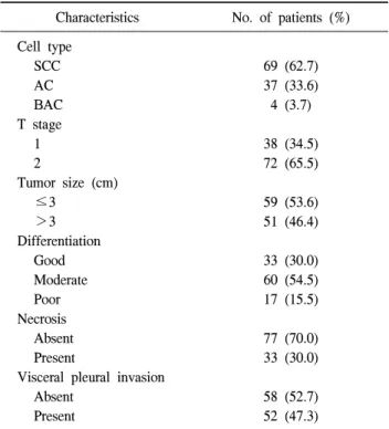
RESULTS 1) Histopathology (Table 1)

The histological type of NSCLC was as follows: 69 cases of SCC; 37 cases of AC; 4 cases of BAC. The average tu- mor size was 35.1±22.9 mm with a range of 3−130 mm and that of 51 (46.4%) patients was over 30 mm. Thirty-three tumors (30.0%) were good-, 60 (54.5%) were moderate-, and 17 (15.5%) were poor-differentiated. Visceral pleural invasion was found in 52 tumors (47.3%) and 33 tumors (30.0%) showed necrosis. Thirty-one (28.2%) patients had tumors of at least 3 cm in size as well as visceral pleural invasion tumors.

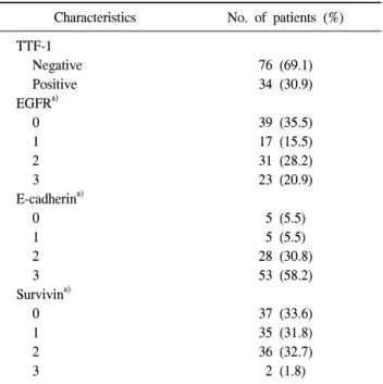
2) Immunohistochemical staining (Table 2)

TTF-1 expression was detected in 34 (30.9%) patients.

EGFR overexpression was found in 54 (49.1%) of the

patients. E-cadherin was lost in 10 (9.1%) cases. Overex-

pression of survivin was observed in 38 (34.5%) patients.

(4)

Table 2. Immunohistochemical staining in resected stage I non-small cell lung cancer (n=110)

Characteristics No. of patients (%) TTF-1

Negative Positive EGFR

a)

0 1 2 3 E-cadherin

a)

0 1 2 3 Survivin

a)

0 1 2 3

76 (69.1) 34 (30.9)

39 (35.5) 17 (15.5) 31 (28.2) 23 (20.9)

5 (5.5) 5 (5.5) 28 (30.8) 53 (58.2)

37 (33.6) 35 (31.8) 36 (32.7) 2 (1.8)

TTF-1=thyroid transcription factor 1; EGFR=epidermal growth factor receptor.

a)

According to the positive area: 0 (<25%), 1 (≥25% &

<50%), 2 (≥50% & <75%), and 3 (≥75%).

Table 3. Association between histopathologic and molecular factors

Cell type Differentiation

SCC AC p-value Good/moderate Poor p-value

TTF-1 Negative Positive EGFR Normal Overexpression E-cadherin Loss Preserved Survivin Normal Overexpression

63 (91.3) 6 (8.7)

31 (44.9) 38 (55.1)

7 (12.5) 49 (87.5)

44 (63.8) 25 (36.2)

12 (33.3) 24 (66.7)

20 (55.6) 16 (44.4)

3 (9.7) 28 (90.3)

25 (69.4) 11 (30.6)

0.000 -

0.076 -

0.760 -

0.783 -

65 (69.9) 28 (30.1)

44 (47.3) 49 (52.7)

7 (9.0) 71 (91.0)

63 (67.7) 30 (32.3)

11 (64.7) 6 (35.3)

12 (70.6) 5 (29.4)

3 (23.1) 10 (76.9)

9 (52.9) 8 (47.1)

0.670 -

0.078

0.151 -

0.238 - Values are presented as number (%).

SCC=squamous cell carcinoma; AC=adenocarcinoma; TTF-1=thyroid transcription factor 1; EGFR=epidermal growth factor receptor.

3) Association between histopathologic and molecular factors (Table 3)

TTF-1 expression was significantly high in several factors

such as AC (p=0.000) and visceral pleural invasion (p=0.032);

however, it was not significantly associated with differ- entiation (p=0.670). The EGFR overexpression was more fre- quent in patients with SCC (p=0.076). Loss of E-cadherin was correlated with larger tumor size (p=0.061) and survivin (p=0.006, no expression).
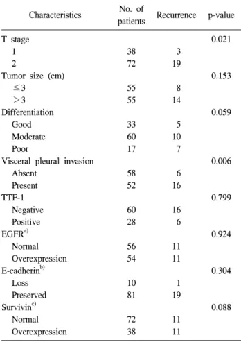
4) Recurrence

The disease free interval was determined as the interval from the date of surgery to the date of first recurrence. Those without recurrence past 31 August 2010 were classified as censored.

Median follow-up was 55.0 (2.3−87.9) months and the 5-year disease free survival rate was 67.1%, with a mean dis- ease free time of 65.1 months. Twenty-two patients (20.0%) had recurrent disease and 15 patients (13.6%) died from dis- ease-related causes. Seven of the 22 patients had local re- currence and 15 patients had distant recurrence. Of the 15 pa- tients with distant metastasis, 5 patients had recurrence in the contralateral lung, 2 patients in the bone, 1 patient in the liv- er, 1 patient in the brain, and 6 patients in multiple sites.

Table 4 shows that histopathologic factors including differ-

entiation (p=0.007), visceral pleural invasion (p=0.006), T

stage (p=0.021), and molecular markers consisting of TTF-1

negativity (p=0.799) and survivin expression (p=0.088) were

associated with recurrence. Table 5 lists the results of uni-

(5)

Table 4. Recurrence according to histologic factors and molecular markers

Characteristics No. of

patients Recurrence p-value T stage

1 2

Tumor size (cm)

≤3

>3 Differentiation

Good Moderate Poor

Visceral pleural invasion Absent

Present TTF-1

Negative Positive EGFR

a)

Normal Overexpression E-cadherin

b)

Loss Preserved Survivin

c)

Normal Overexpression

38 72

55 55

33 60 17

58 52

60 28

56 54

10 81

72 38

3 19

8 14

5 10 7

6 16

16 6

11 11

1 19

11 11

0.021

0.153

0.059

0.006

0.799

0.924

0.304

0.088

TTF-1=thyroid transcription factor 1; EGFR=epidermal growth factor receptor.

a)

Negative=grade 0,1; positive=grade 2,3.

b)

Loss=grade 0,1; pre- served=grade 2,3.

c)

Normal=grade 0,1; overexpression=grade 2,3.

variate analysis for the prognostic significance of each histo- pathologic factor and molecular marker in relation to the time to recurrence. The log rank test showed that the T stage, dif- ferentiation, visceral pleural invasion, and survivin expression were prognostic factors (Fig. 1). By multivariate analysis us- ing the Cox proportional hazards regression model, poor dif- ferentiation and survivin expression emerged as independent prognostic factors of recurrence (Table 6).

DISCUSSION

The prognosis for patients with NSCLC was highly varia- ble and a better understanding of the biology of tumors

should provide useful information for predicting clinical out-

comes and for individualizing treatment. Because lung cancer

is a heterogeneous disease resulting from the acquisition of

multiple somatic mutations, the prognostic role of histopatho-

logic and molecular markers is difficult to determine [12]. As

a significant fraction of the patients at stage I experience dis-

ease recurrence and die after a curative resection, the differ-

entiation of patients with a high risk of early recurrence and

the identification of patients who must receive adjuvant ther-

apy are topics of great interest in the study of early stage

NSCLC. However, stratification of patient prognoses using

only the TNM staging system does not allow one to differ-

entiate between patients with stage I lung cancer that are and

are not at risk of tumor recurrence. It has been demonstrated

that adjuvant therapy is not effective in all patients with stage

I disease; it is only effective in specific subsets of patients. It

is important to select a subgroup of patients with stage I dis-

ease that might benefit from adjuvant therapy and for this

reason, investigators have attempted to pinpoint factors that

predict poor prognosis through analysis of clinical and histo-

pathologic factors [13-16]. Therefore, the goals of this study

were to identify these prognostic factors and to evaluate the

association between histopathologic and molecular markers. A

multitude of histopathologic factors associated with survival

have been described in the literature through retrospective

series studies. Harpole et al. [3] identified vascular invasion,

visceral pleural invasion, a high mitotic index, and a tumor

size greater than 3 cm as risk factors associated with re-

currence in 289 resected stage I lung cancer patients. In this

study, histological factors including tumor size, differentiation,

and T stage were identified as significant factors. The degree

of differentiation of the primary tumor is known to be a sig-

nificant prognostic factor and poor differentiated tumors are

defined as negative prognostic factors in patients with stage I

NSCLC [17]. Tumor size was rearranged in the 7th edition of

the guidelines of the AJCC and this detailed size rearrange-

ment was more effective in predicting recurrence than the 6th

edition guidelines (data not shown). Although observation da-

ta based on clinical and histopathologic description aids in di-

viding patients into risk groups, much variation exists in the

interpretation of these qualitative variables. Therefore, many

molecular biologic markers have recently been studied in lung

(6)

Table 5. Association between histopathologic and molecular factors

Factors Variables Univariate analysis

Age (yr) Sex Location Differentiation Histology

Visceral pleural invasion Necrosis

Tumor size (cm) T stage

TTF-1 EGFR E-cadherin Survivin

<65, ≥65 Male/female Central/periphery/mid Good/moderate/poor SCC/AC/BAC Absent/present Absent/present

≤2, 2< & ≤3, 3< & ≤5, 5< & ≤7, >7 1/2

Negative/positive Normal/overexpressed Loss/preserved Normal/overexpressed

0.497 0.332 0.136 0.026 0.402 0.000 0.284 0.210 0.023 0.304 0.924 0.332 0.049

SCC=squamous cell carcinoma; AC=adenocarcinoma; BAC=bronchioalveolar carcinoma; TTF-1=thyroid transcription factor 1; EGFR=

epidermal growth factor receptor.

Fig. 1. Kaplan-Meier analysis of disease free survival for (A) visceral pleural invasion (VPI) (B) T-factor (C) differentiation, (D) survivin.

(7)

Table 6. Multivariate analysis of disease-free survival by Cox pro- portional hazards regression model

Variables HR 95% CI p-value

T stage

Visceral pleural invasion TTF-1 negative

Survivin overexpression Poor differentiation

1.729 2.532 0.440 2.212 3.147

0.365−8.199 0.750−8.549 0.157−1.232 1.014−5.652 1.296−7.639

0.490 0.135 0.118 0.045 0.011 HR=hazard ratio; CI=confidence interval; TTF-1=thyroid tran- scription factor 1.

cancer to determine their prognostic importance. Gene copy number and mutation analyses are now being explored in prospective clinical studies in enriched populations. These as- says are very promising, but they are costly and are not widely available. In contrast, protein expression studies by immunohistochemistry are rapid, reproducible, and routinely performed in many hospitals. During the last decade, the ag- gressive phenotypes of lung cancer have been characterized by a number of molecular abnormalities, so multiple algo- rithms were created based on relative protein expression pro- files using up to 15 factors with possible significance.

In this study, we have evaluated factors such as TTF-1 and surviving, which have not been extensively studied. TTF-1 is mainly expressed in the thyroid and lung during embryo- genesis, and it plays a physiological role in their development and morphogenesis. Positive TTF-1 staining by immunohisto- chemistry has been described in 25% to 80% of primary ad- nenocarcinoma; however, the vast majority of squamous cell carcinoma of the lung lacks TTF-1 expression [7]. In our study, TTF-1 positivity was 8.7% in squamous cell carcinoma and 66.7% in adenocarcinoma. TTF-1 positive adenocar- cinoma had significant relationships with the prevalence of female, nonsmoker, negative staining of p53, less frequent retinoblastoma protein loss, and preserved expression of p27 [18]. Yatabe et al. [19] demonstrated that TTF-1 was con- sistently expressed during development and this pattern was preserved in transformed cells and metastatic lymph node.

Expression was not affected by the differentiation state.

However, Anagnostou et al. [7] showed that patients with stage I adenocarcinoma with TTF-1 expression had a longer median overall survival than those without expression and re-

vealed TTF1-expression as an independent lower risk factor for patients with stage I adenocarcinoma.

EGFR is a member of the receptor tyrosine kinase (TK) family, and TKs regulate signaling pathways that control crit- ical cellular activities [20]. The clinical importance of EGFR has increased with the development of EGFR TK inhibitors (TKI). EGFR mutations were observed more frequently in women, never-smokers, and in adenocarcinoma, and EGFR TKIs were more effective in patients with EGFR mutations.

Suzuki et al. [21] compared mutation with expression and re- ported that EGFR overexpression was significantly higher in cases where EGFR mutation was positive rather than nega- tive. In this study, EGFR overexpression was more common in squamous cell carcinoma, men, and ever-smokers, which is in contrast with the findings of EGFR mutation. Therefore, the exact mechanism remains unclear whether EGFR mutation is the major contributor to EGFR overexpression.

E-cadherin and catenins are components of the adherens junction protein, with crucial roles in maintaining intercellular junctions in epithelial cells; reduced E-cadherin expression could potentially affect tumor differentiation, metastasis, and prognosis [10,11]. Although the number of patients who have demonstrated the loss of E-cadherin is small, it still repre- sents a significant prognostic factor.

Survivin is a recently identified protein that functions as an

inhibitor of apoptosis, suppressing programmed cell death and

regulating cell division. The expression of survivin is un-

detectable or found at very low levels in normal tissues but

at high levels in various malignancies and also embryonic

and fetal tissues [22]. The overexpression of survivin may

overcome apoptotic checkpoints and promote aberrant pro-

gression of the transformed cell through mitosis. A high level

of survivin expression in malignancies is considered to be an

important indicator of poor prognosis and associated with

high tumor grade, tumor progression, and chemoresistance

[23]. Survivin staining was detected in both the nucleus and

the cytoplasm of NSCLC. However, it was shown that nu-

clear survivin, rather than cytoplasmic staining, was predictive

of poor survival in patients with NSCLC and esophageal

cancer. Shinohara et al. [8] showed patients who had nuclear

staining for survivin had a significantly increased risk of dis-

ease recurrence and concluded that the nuclear presence of

(8)

survivin may be an independent biomarker for disease re- currence and overall survival in patients with resected stage I and II NSCLC. Atikcan et al. [24] evaluated the prognostic significance of both nuclear and cytoplasmic survivin ex- pression in NSCLC and showed that cytoplasmic staining was found to be significantly increased in squamous cell carcino- ma and that nuclear survivin expression might predict prog- nosis in NSCLC, whereas cytoplasmic survivin has no prog- nostic significance.

This study had limitations. First, immunohistochemical scoring is at best a semi-quantitative exercise for which no standard criteria have yet been proposed or adopted. The scoring methods that have been used include estimating the grade of staining intensity, the percentage of tumor cells stained, the cellular localization of the antigen, and systems that combined these parameters. Second, a more important confusion is the method of determining cut-offs for dichoto- mizing scores in log-rank tests. Cut-offs are often arbitrary and are sometimes selected to obtain the desired effect using the minimum p-value approach. Therefore, different anti- bodies and grading systems should also be taken into ac- count; the counting of positive tumor cells and grading of staining intensity may be subject to interobserver variability, which should be evaluated.

CONCLUSION

In resected stage I non-small cell lung cancer, poor differ- entiation and survivin overexpression have been identified as independent predictors of poor disease-free survival.

REFERENCES

1. Mountain CF, Lukeman JM, Hammar SP, et al. Lung cancer classification: the relationship of disease extent and cell type to survival in a clinical trials population. J Surg Oncol 1987;35:147-56.

2. Nesbitt JC, Putnam JB Jr, Walsh GL, Roth JA, Mountain CF. Survival in early-stage non-small cell lung cancer. Ann Thorac Surg 1995;60:466-72.

3. Harpole DH Jr, Richards WG, Herndon JE 2nd, Sugarbaker DJ. Angiogenesis and molecular biologic substaging in pa- tients with stage I non-small cell lung cancer. Ann Thorac Surg 1996;61:1470-6.

4. Strauss GM, Kwiatkowski DJ, Harpole DH, Lynch TJ, Skarin AT, Sugarbaker DJ. Molecular and pathologic mark- ers in stage I non-small-cell carcinoma of the lung. J Clin Oncol 1995;13:1265-79.

5. Duarte IG, Bufkin BL, Pennington MF, et al. Angiogenesis as a predictor of survival after surgical resection for stage I non-small-cell lung cancer. J Thorac Cardiovasc Surg 1998;115:652-8.

6. Quinlan DC, Davidson AG, Summers CL, Warden HE, Doshi HM. Accumulation of p53 protein correlates with a poor prognosis in human lung cancer. Cancer Res 1992;52:

4828-31.

7. Anagnostou VK, Syrigos KN, Bepler G, Homer RJ, Rimm DL. Thyroid transcription factor 1 is an independent prog- nostic factor for patients with stage I lung adenocarcinoma.

J Clin Oncol 2009;27:271-8.

8. Shinohara ET, Gonzalez A, Massion PP, et al. Nuclear sur- vivin predicts recurrence and poor survival in patients with resected nonsmall cell lung carcinoma. Cancer 2005;103:

1685-92.

9. Selvaggi G, Novello S, Torri V, et al. Epidermal growth factor receptor overexpression correlates with a poor prog- nosis in completely resected non-small-cell lung cancer. Ann Oncol 2004;15:28-32.

10. Liu D, Huang C, Kameyama K, et al. E-cadherin expression associated with differentiation and prognosis in patients with non-small cell lung cancer. Ann Thorac Surg 2001;71:949-54.

11. Kase S, Sugio K, Yamazaki K, Okamoto T, Yano T, Sugi- machi K. Expression of E-cadherin and beta-catenin in hu- man non-small cell lung cancer and the clinical significance.

Clin Cancer Res 2000;6:4789-96.

12. Poleri C, Morero JL, Nieva B, et al. Risk of recurrence in patients with surgically resected stage I non-small cell lung carcinoma: histopathologic and immunohistochemical analysis.

Chest 2003;123:1858-67.

13. Macchiarini P, Fontanini G, Hardin MJ, et al. Blood vessel invasion by tumor cells predicts recurrence in completely re- sected T1 N0 M0 non-small-cell lung cancer. J Thorac Car- diovasc Surg 1993;106:80-9.

14. Thomas P, Rubinstein L. Cancer recurrence after resection:

T1 N0 non-small cell lung cancer. Lung Cancer Study Group. Ann Thorac Surg 1990;49:242-6.

15. Takise A, Kodama T, Shimosato Y, Watanabe S, Suemasu K. Histopathologic prognostic factors in adenocarcinomas of the peripheral lung less than 2 cm in diameter. Cancer 1988;61:2083-8.

16. Pairolero PC, Williams DE, Bergstralh EJ, Piehler JM, Bernatz PE, Payne WS. Postsurgical stage I bronchogenic carcinoma: morbid implications of recurrent disease. Ann Thorac Surg 1984;38:331-8.

17. Kobayashi N, Toyooka S, Soh J, et al. Risk factors for re-

currence and unfavorable prognosis in patients with stage I

(9)

non-small cell lung cancer and a tumor diameter of 20 mm or less. J Thorac Oncol 2007;2:808-12.

18. Nishio M, Koshikawa T, Yatabe Y, et al. Prognostic sig- nificance of cyclin D1 and retinoblastoma expression in combination with p53 abnormalities in primary, resected non-small cell lung cancers. Clin Cancer Res 1997;3:1051-8.

19. Yatabe Y, Mitsudomi T, Takahashi T. TTF-1 expression in pulmonary adenocarcinomas. Am J Surg Pathol 2002;26:

767-73.

20. Blume-Jensen P, Hunter T. Oncogenic kinase signalling.

Nature 2001;411:355-65.

21. Suzuki M, Shigematsu H, Hiroshima K, et al. Epidermal

growth factor receptor expression status in lung cancer cor- relates with its mutation. Hum Pathol 2005;36:1127-34.

22. Ambrosini G, Adida C, Altieri DC. A novel anti-apoptosis gene, survivin, expressed in cancer and lymphoma. Nat Med 1997;3:917-21.

23. Falleni M, Pellegrini C, Marchetti A, et al. Survivin gene ex- pression in early-stage non-small cell lung cancer. J Pathol 2003;200:620-6.

24. Atikcan S, Unsal E, Demirag F, Koksal D, Yilmaz A.

Correlation between survivin expression and prognosis in

non-small cell lung cancer. Respir Med 2006;100:2220-6.

수치

Table 1. Histopathologic findings in resected stage I non-small  cell lung cancer (n=110)
Table 2. Immunohistochemical staining in resected stage I  non-small cell lung cancer (n=110)
Table 4. Recurrence according to histologic factors and molecular  markers
Fig. 1. Kaplan-Meier analysis of disease free survival for (A) visceral pleural invasion (VPI)  (B) T-factor (C) differentiation, (D) survivin.
+2

참조

관련 문서

however, such scaffolds could have poor mechanical strengths. 19) For these reasons, multi-pore sized scaffolds have been announced to increase the mechanical

Not “orthogonal to each other” Inventory Waiting Poor flow – Waste of Customer’s time. Poor use of capacity – Waste of

Rapid turning and capsize accident of ro-ro ferry Sewol occurred due to the very poor stability small rapid turning, and its objective turning characteristics

Results: Poorly differentiated colorectal cancer was frequently located at right colon and in advanced stage.. During follow-up, double primary cancer had occurred

- In patients presenting with end-stage organ failure, sequential solid organ (kidney heart liver) transplant followed by ASCT has been successfully. J

a prospective analysis of 50 cases. Preoperative staging of non-small-cell lung cancer with positron-emission tomography. Staging of non- small-cell lung cancer

Patients with breast cancer; ovarian cancer; renal cell carcinoma; pancreatic neuroendocrine cancer; colorectal cancer; head and neck cancer; non-small cell lung

According to the result of this study, to do effective nursing that improve health promoting behaviors of early-stage lung cancer patients, nurses should