대 한 생 식 의 학 회 지 : 제 36 권 제 1 호 2009
탯줄유래 줄기세포의 계대배양에 따른 특성 변화의 분석
서울여자대학교 자연과학대학 생명공학과 박세아·강현미·허진영·윤진아·김해권*
Characterization of Umbilical Cord-derived Stem Cells during Expansion in Vitro
Seah Park, Hyun Mi Kang, Jinyeong Heo, Jinah Yoon, Haekwon Kim*Department of Biotechnology, College of Natural Science, Seoul Women's University, Seoul, Korea
Objectives: Mesenchymal stem cells (MSC) comprise a promising tool for cellular therapy. It is known that long-term in vitro culture of human bone marrow and adipose tissue derived-MSCs lead to a reduction of life span and a change of stem-like characters.
The aim of our study was to examine whether stem cell properties of human umbilical cord-derived stem cells (HUC) could be affected by in vitro expansion.
Methods: HUC were isolated from human umbilical cord and cultured for 10 passages in vitro. Morphology and population doubling time (PDT) were investigated, and changes of stem cell properties were examined using RT-PCR and immunocytochemistry during serial subcultures.
Results: Morphology and PDT of HUC began to change slightly from the 7th passage (p7). Expression level of nestin and vimentin mRNAs increased along with the culture period from p4 until p10. In contrast, expression level of SCF mRNA decreased during the same culture period. Expression level of Oct-4 and HNF-4α mRNAs was not significantly changed throughout the culture period until p10. Expression level of BMP-4, FGF-5, NCAM and HLA-ABC mRNAs appeared to increase as the culture continued, however, the difference was not significant. Immunocytochemical studies showed that HUC at p3, p6 and p9 positively were stained with antibodies against SSEA-3 and SSEA-4 proteins. Interestingly, staining intensity of HUC for ICAM-1 and HLA-ABC gradually increased throughout the culture period. Intensity against thy-1 and fibronectin antibodies increased at p9 while that against TRA-1-60 and VCAM-1 antibodies began to decrease at p6 until p9.
Conclusions: These results suggest that HUC change some of their stem cell characteristics during in vitro culture. Development of culture system might be needed for the maintenance of characteristics. [Korean. J. Reprod. Med. 2009; 36(1): 23-34.]
Key Words: Umbilical cord-derived stem cells, Stem cell characteristics, RT-PCR, Immunocytochemistry
성체줄기세포는 자가증식 (self-renewal)할 수 있 으며 지방세포, 골세포, 연골세포, 심장세포, 간세 포, 신경세포 등 다양한 조직세포로 분화할 수 있 는 미분화 상태의 줄기세포로서 골수유래 중간엽 줄기세포 (bone marrow-derived mesenchymal stem
cells, BM-MSC)가 그 대표적 예이다.1~4 골수유래 중간엽 줄기세포는 심장, 골, 연골 등의 조직 재생 을 위한 세포치료제 뿐만이 아니라 임상에서 조혈 모세포 (hematopoietic stem cells, HSC)의 이식 후 생 착 (engraftment)을 증가시키기 위한 도구로도 이용 되고 있다.5~7 중간엽 줄기세포의 공급원으로는 골 수가 가장 잘 알려져 있는데 제공자의 나이가 증가 함에 따라 줄기세포의 수 및 증식 능력이 감소하며, 골수를 채취하기 위해서는 고통이 수반된다.8,9 이
주관책임자: 김해권, 우) 139-774 서울특별시 공릉2동 126번지, 서울여자대학교 자연과학대학 생명공학과
Tel: (02) 970-5665, Fax: (02) 970-5974 e-mail: [email protected]
*이 논문은 2008학년도 서울여자대학교 교내학술연구비의 지원을 받았음.
에 따라 최근에는 지방, 탯줄, 태반, 탯줄 혈액 등 을 포함한 다양한 조직 등으로부터 얻어지는 중간 엽 줄기세포가 새로운 세포치료제의 공급원으로 관 심을 끌고 있다.10~12
중간엽 줄기세포를 임상에 적용하기 위해서는 체 외 배양을 통한 세포증식 과정이 필요하며 이 과정 동안 줄기세포로서의 특성이 유지되어야 한다. 그 러나 오랜 기간 동안 체외 배양을 하게 되면 사람 의 골수 및 지방유래 줄기세포는 노화되어 특성이 변하고 분화 능력 또한 감소하게 된다.13 따라서 현재까지는 초기 계대배양의 세포만이 임상에 적 용되고 있는 실정이다. 따라서 줄기세포의 특성 변 화 없이 체외증식 기간을 늘릴 수 있는 방법이 연 구되고 있다. 사람의 골수유래 줄기세포의 체외 배 양 시 배양액에 fibroblast growth factor-2 (FGF-2)를 첨가하여 배양하면 세포의 tumor growth factor-β2 (TGF-β2)의 발현을 억제시킴으로써 세포의 노화를 억제한다고 보고된 바 있으며, hTERT 등의 유전자 를 삽입하는 유전자 조작방법에 의해서도 줄기세 포의 노화를 방지할 수 있다고 알려져 있다.14~16
탯줄 정맥은 Wharton's jelly 층으로 둘러싸여 있으며 이는 perivascular zone, intervascular zone, subamnion으로 구성되어 있는데 perivascular zone은 분화한 근섬유세포로 되어 있는 반면 intervascular zone과 subamnion에는 증식 능력을 가진 줄기세 포가 존재한다.17,18 이 탯줄유래 줄기세포 (human umbilical cord-derived stem cells, HUC)는 골수 및 다 른 조직유래 중간엽 줄기세포와 유사하게 CD73, CD90, CD105, CD10, CD13, CD29, CD44, HLA-ABC 유전자를 발현하며, 조혈모세포 표지 인자인 CD- 34, CD45와 조직적합성항원인 CD14, CD31, CD33, HLA-DRα 유전자는 발현하지 않는다.18~21 탯줄유래 줄기세포는 골세포, 연골세포, 지방세포로 분화할 수 있고11 체외 배양 시 골수나 지방유래 줄기세포 보다 분열 능력이 뛰어나며, 또한 primitive stem cells 표지 인자인 ABCG2 transporter를 발현하고 Hoechst dye를 배출하는 능력이 있어 지방이나 골수에서 유 래된 줄기세포보다 비교적 미분화한 세포로 여겨지
기도 한다.18,22~27 이러한 특징들은 탯줄유래 줄기세 포를 세포치료제로 이용하는 것을 용이하게 한다.
현재까지 계대배양 횟수의 증가에 따른 탯줄유 래 줄기세포의 분열 능력 및 유전자와 단백질 발 현 변화는 알려져 있지 않다. 따라서 본 연구에서 는 사람의 탯줄유래 줄기세포를 대상으로 하여 체 외에서 계대배양 횟수가 증가함에 따른 세포의 형 태 및 성장 속도를 조사하고 역전사 중합효소 연쇄 반응과 면역세포화학 염색법을 사용하여 유전자와 단백질의 변화 여부를 조사하였다.
연구대상
및 방법 1. 탯줄로부터 줄기세포의 채취본 연구에 사용된 탯줄은 경기도 수원에 소재한 아주대학교 의과대학 산부인과에 2007년 3월부터 2008년 3월까지 내원한 정상 산모의 제왕절개 또는 질식분만 시 버려지는 탯줄을 산모의 동의 하에 채 취 사용하였다. 약 5~10 cm의 탯줄을 생리식염수 가 들어 있는 50 ml 시험관에 담아 운반하였으며 채취 후 24시간 내에 실험에 사용하였다. 본 실험 은 서울여대 기관윤리위원회의 허가를 받아 실행 하였다. 시료는 서로 다른 3개의 탯줄로부터 각각 세포를 얻어 사용하였다.
2. 탯줄유래 줄기세포의 분리 및 배양
탯줄유래 줄기세포의 분리 및 배양은 이전의 실 험과 동일하게 진행하였다.28,29 탯줄 외부의 혈액을 100 U/mL의 penicillin과 100 μg/mL의 streptomycin이 첨가된 Ca2+과 Mg2+이 포함되지 않은 DPBS로 제 거한 후 외부 양막을 벗기고 동맥 2개를 제거하였 다. 남은 조직을 1 mm3 크기로 자른 후 같은 부피 의 0.5%의 crude type collagenase (Gibco)로 37℃에서 20~24시간 처리하였다. 이 후 같은 부피의 Ca2+과 Mg2+이 포함되지 않은 DPBS를 첨가하고 실온에서 600 ×g로 10분간 원심분리하여 상층액을 제거하고 DMEM-LG로 2번 세척하였다. 분리된 세포를 100 U/mL의 penicillin, 0.1 μg/mL의 streptomycin, 3.7 mg/
mL의 sodium bicarbonate, 10%의 FBS (Hyclone)가 포함된 DMEM-LG에서 37℃ 온도에서 5%의 CO2 가 공급되는 배양기 내에서 배양하였다. 배양 3일 후 배양용기의 바닥에 붙지 않은 세포를 제거하고 1주에 2번 배양액을 교체하였다. 배양용기의 70%에 서 80% 정도의 confluence로 세포가 자라면 0.125%
의 trypsin과 1 mM의 EDTA가 첨가된 HBSS로 3분 간 처리하여 세포를 떼어낸 후 배양접시 1 cm2 당 2×10³의 세포를 넣어 배양하였다.
3. Total RNA 분리 및 역전사 중합효소 연쇄반 응 (reverse transcription polymerase chain reaction, RT-PCR)
세포 pellet을 Ca2+과 Mg2+이 포함되지 않은 DPBS 를 이용하여 세척하고 500 μL의 Tri-reagent (Sigma) 를 첨가한 다음 제조회사 사용설명서에 따라 total RNA를 분리하였다. 5 μg의 RNA는 reaction buffer, 1 mM의 dNTP mixture, 0.5 μg/μL의 oligo(d)T15, 20 U의 RNase inhibitor (Takara, Japan), 20 U의 M-MuLV reverse transcriptase (Fermentas, Burlington, Canada)가 혼합된 20 μL 반응 용액에서 역전사 시켰다. 반응 은 42℃에서 60분간 진행되었다. 얻어진 RT pro- ducts (cDNAs)는 2 mM의 MgCl2, 1×의 Taq buffer, 0.25 U의 Taq polymerase (Fermentas), 10 pM의 sense 와 antisense gene-specific primers가 혼합된 10 μL 반응 용액으로 PCR을 수행하였다. 증폭은 총 35 cycles을 수행하였으며 각 cycle은 94℃에서 30초 간의 denaturation 과정, 30초간의 annealing 과정, 72℃에서 30초간의 extension 과정으로 구성되었다.
각 유전자의 annealing 과정의 온도는 Table 1에 표 기하였다. 반응 종결 후 PCR 생성물은 0.25%의 bromophenol blue과 0.25%의 xylene cyanol과 40%
sucrose가 포함된 6×의 loading buffer과 혼합한 다 음 2% agarose gel에 loading하여 전기영동 하였다.
전기영동 후 ethidium bromide로 염색하고 ultraviolet light를 이용하여 DNA의 영상을 얻었다. 얻은 영상 을 TL100 software (Hofer, USA)로 각 유전자의 발현 양을 분석하였다. 이 후 각각 GAPDH에 대한 상대
적인 값 (%)을 얻어 이를 비교 분석하였다.
4. 면역세포화학적 특성 분석
탯줄유래 줄기세포를 8-well slide chamber (Nunc)에 서 배양한 후 2% paraformaldehyde가 함유된 DPBS 용액으로 4℃에서 2시간 동안 고정하였다. 고정 후 DPBS로 5분간 3번 세척하였다. 그리고 실온에서 10분 동안 0.5%의 Triton X-100가 함유된 DPBS를 처리 후 세척한 다음 endogenous peroxidase 활성을 제거하기 위하여 3%의 hydrogen peroxide (Dako)로 15분간 반응시킨 후 세척하였다. 이 후 2%의 bovine serum albumin (BSA, Sigma)을 함유한 DPBS를 실온 에서 1시간 동안 반응시킨 후 각 mouse monoclonal antibody인 Oct-4 (1:200; Chemicon), TRA-1-60 (1:20;
Chemicon), SSEA-3 (1:50; R&D System), Thy-1 (1:20;
Chemicon), fibronectin (1:200; Novo castra), CD54 (1:
40; Novo castra), CD106 (1:50; Novo castra), HLA-ABC (1:200; Novo castra), HLA-DR (1:50; Novo castra)을 4℃에서 17~24시간 동안 반응시킨 후 세척하였다.
이 후 biotinylated goat anti-mouse IgG와 anti-rabbit IgG를 실온에서 20분간 처리하였다. 3번 세척한 다음 horseradish peroxidase-conjugated streptavidin (Dako)을 20분간 처리하고 3,3'-diaminobenzidine tetrahydrochloride (DAB, Dako) 용액으로 발색하였 다. 발색이 된 세포는 DPBS로 세척하고 Mayer's Haematoxylin (Sigma)으로 대조 염색한 후 광학 현 미경 (LSM410; Carl Zeiss, Oberkochen, Germany) 하 에서 관찰하였다.
5. 다분화능 시험
다분화능의 시험은 Kim 등30의 방법을 원용하여 행하였다.
1) 지방세포로의 분화 유도
1 cm2 당 2×10³의 탯줄유래 줄기세포를 8-well slide chamber (Nunc)에 넣고 3일간 배양한 후 DMEM-LG에 10%의 FBS와 1 μM의 dexamethasone, 0.5 μM의 3-isobutyl-1-methylxanthine, 0.05 mg/L의 human insulin, 200 μM의 indomethacin (Sigma)이 포
함된 분화 배양액으로 교체하였다. 그리고 1주에 2번 배양액을 교체하였다. 3주 배양 후 oil red O 염 색을 시행하여 지질이 축적된 지방세포의 존재 여 부를 관찰하였다.
2) 골세포로의 분화 유도
1 cm2 당 2×10³의 탯줄유래 줄기세포를 8-well slide chamber (Nunc)에 넣고 3일간 배양한 후 DMEM-LG에 10%의 FBS와 0.1 μM의 dexamethasone, 100 mM의 β-glycerol phosphate, 50 μM의 ascorbic Table 1. Primers used for the RT-PCR analysis
Gene Primer sequence Size (bp) Temp. (℃) Accession number 5'- aca act ttg gta tcg tgg aa -3'
GAPDH
5'- aaa ttc gtt gtc ata cca gg -3' 456 53 NM_002046 5'- cgt gaa gct gga gaa gga gaa gct g -3'
Oct-4
5'- caa ggg ccg cag ctc aca cat gtt c -3' 245 55 AF268617 5'- cca ttg atg cct tca agg ac -3'
SCF 5'- ctt cca gta taa ggc tcc aa -3' 275 55 M59964 5'- gct gtg tct cag ggg att gta gga ata -3'
FGF-5
5'-tat cca aag cga aac ttg agt ctg ta -3' 434 55 NM_004464 5'- gag ggg gaa gat gcc gtg atg tg -3'
NCAM
5'-ata ttc tgc ctg gcc cgg atg gta g -3' 269 60 NM_000615 5'- agc cat gct agt ttg ata cc -3'
BMP-4
5'- tca ggg atg ctg ctg agg tt -3' 383 55 D30751 5'- ttg cgg ctg ctc agc atg tt -3'
BMP-2
5'- ttg cga gaa cag atg caa gat g -3' 315 57 BC069214 5'- cca gaa act caa gca cca c -3'
Nestin
5'- ttt tcc act cca gcc atc c -3' 398 54 X65964 5'- aga ttc aga tga ggc tca aa -3'
Pax-6
5'- aat tgg ttg gta gac act gg -3' 313 60 AY707088 5'- gag cag gaa tgg gaa gaa tg -3'
HNF-4α
5'- ggc tgt cct ttg gga tga ag -3' 205 62 NM_178849 5'- cct tcg tga ata cca cg acc tgc -3'
Vimentin
5'- taa tat atc gcc tgc cac tga g -3' 321 56 Z19554 5'- gta ttt ctt cac atc cgt gtc ccg -3'
HLA-ABC
5'- gtc cgc cgc ggt cca aga gcg cag -3' 394 70 L18898 5'- ctg atg agc gct cag gaa tca tgg -3'
HLA-DRα
5'- gac tta ctt cag ttt gtg gtg agg gaa g -3' 220 60 X06079
Seah Park. Characterization of Umbilical Cord-derived Stem Cells during Expansion in Vitro. Korean J Reprod Med 2009.
acid-2-phosphate가 포함된 분화 배양액으로 교체하 였다. 그리고 1주에 2번 배양액을 교체하였다. 3주 배양 후 von Kossa 염색을 시행하여 칼슘이 침착된 세포를 관찰하였다.
3) 연골세포로의 분화 유도
1 cm2 당 2×10³의 탯줄유래 줄기세포를 8-well slide chamber (Nunc)에 넣고 3일간 배양한 후 DMEM-HG에 0.1 μM의 dexamethasone, 50 μg/mL
의 ascorbic acid-2-phosphate, 100 μg/mL의 sodium pyruvate, 40 μg/mL의 proline (Sigma), 10 ng/mL의 transforming growth factor-β3 (TGF-β3 R&D Systems), 50 mg/mL의 ITS plus premix (각 6.25 μg/mL의 insulin, transferring, selenious acid와 1.35 mg/mL의 BSA, 5.35 mg/mL의 linoleic acid가 포함되어 있음; BD Bio- sciences Becton Dickinson)가 포함된 분화 배양액으 로 교체하였다. 그리고 1주에 2번 배양액을 교체하 였다. 3주 배양 후 alcian blue 염색을 시행하여 분화 여부를 관찰하였다.
결
과 1. 형태학적 분석서로 다른 3종류의 탯줄로부터 줄기세포를 얻어 시험한 결과 초기배양 시에는 방추체 모양의 세포 와 크고 편평한 세포로 구성된 이질적인 형태를 나 타냈으나 계대배양 2번째 (p2) 이후부터는 거의 대 부분이 방추체 모양의 세포로 구성된 단일 형태의 세포만이 관찰되었다. 그러나 p7 이 후 세포질이 넓어지고 크기가 커진 형태적 변화가 나타났으며 이러한 변화는 p10 시기에 더욱 두드러지게 관찰 되었다 (Figure 1). 3개의 서로 다른 탯줄유래 줄기 세포는 10번의 계대배양 동안 41.1±2.1번의 분열을 하는 것으로 나타났으며, 분열 속도를 측정한 결과 p1 세포는 평균 43.2±5.5 h의 분열 속도를 나타내 었고 p2부터 p6까지는 평균 36±1.9 h의 분열 속도 를 나타내었으나 p7부터 p10까지는 48±5.3 h로 느 려졌다 (Figure 2).
2. mRNA 발현 분석
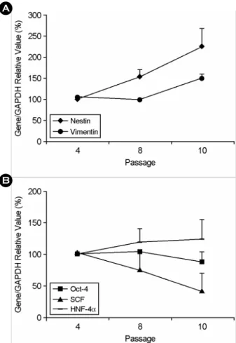
탯줄유래 줄기세포의 p4, 8, 10 시기의 mRNA 분 석 결과, nestin과 vimentin mRNA는 배양이 지속됨 에 따라 발현 양이 증가하였다 (Figure 3A). 이와는 달리 SCF mRNA는 지속적으로 발현이 감소하였고 Oct-4와 HNF-4α mRNA는 p10 시기까지 발현 양에 변화가 없었다 (Figure 3B). 한편 BMP-4, FGF-5, NCAM, HLA-ABC mRNA들은 배양 동안 발현 양
A B
C D
E F
G H
Figure 1. Morphology of HUC during expansion in vitro.
In the primary passages HUC appeared microscopically heterogeneous (A and B). These cells formed a morpho- logically homogenous population of fibroblast-like cells at p3 (C and D). At p7 morphology of HUC slightly began to change (E and F) and showed bigger with flat shape at p10 (G and H). Magnification, A, C, E, G, × 40; B, D, F, H, ×100.
Seah Park. Characterization of Umbilical Cord-derived Stem Cells during Expansion in Vitro. Korean J Reprod Med 2009.
이 증가하였으나 유의한 차이가 없었다. HLA-DRα, Pax-6, BMP-2 mRNA는 이들 세포에서 발현되지 않 았다 (data not shown).
3. 면역세포화학적 특성 분석
탯줄유래 줄기세포의 p3, p6, p9 시기의 단백질 발현을 분석한 결과 배아줄기세포의 표지 물질로 알려진 SSEA-3와 SSEA-4 단백질이 p3부터 p9 시 기까지 지속적으로 발현되었다. 세포연접 물질인
ICAM-1 (intracellular cell adhesion molecule-1, CD54) 과 조직적합성항원인 HLA-ABC 단백질 또한 p3부 터 p9 시기 모두에서 발현되었으나 계대배양 수가 증가함에 따라 발현 정도가 증가되었다. 중간엽 줄 기세포 표지 물질인 Thy-1 (CD90)과 세포 외부 기 질인 fibronectin은 p3와 p6 시기에서 같은 발현 정 도를 나타냈으나 p9 시기에 발현이 증가되었다. 이 와 반대로 배아줄기세포의 표지 물질인 TRA-1-60 와 세포연접 물질인 VCAM-1 (vascular cell adhesion
B A
Figure 3. RT-PCR analysis of HUC during expansion in vitro. The mRNA expression of HUC was examined at p4, 8 and 10 and analyzed the intensity of mRNA expression by densitometry software. The graph showed change of expression of nestin and vimentin mRNAs (A) and SCF, Oct-4 and HNF-4α mRNAs (B) during serial subculture. Expressions of all mRNAs are presented as relative values against the intensity of GAPDH mRNA expression.
Seah Park. Characterization of Umbilical Cord-derived Stem Cells during Expansion in Vitro. Korean J Reprod Med 2009.
A
B
Figure 2. Cumulative doubling number and doubling time of HUC during expansion in vitro. Cumulative doubling number (A) and doubling time (B) of HUC are showed during serial subculture. Cumulative population doubling was calculated in relation to the cell numbers at the first passage. Results are presented as the mean cumulative population doubling (A) and the mean dou- bling time (B) ± SEM of HUC derived from 3 different donors.
Seah Park. Characterization of Umbilical Cord-derived Stem Cells during Expansion in Vitro. Korean J Reprod Med 2009.
molecule-1, CD106)은 p6 시기에 발현 정도가 감소 되었으며 p9 시기에도 약하게 발현되었다. 그러나 조직적합성항원인 HLA-DR 단백질은 p3부터 p9 시기 모두에서 발현되지 않았다 (Figure 4).
4. 골세포, 지방세포, 연골세포로의 분화 유도
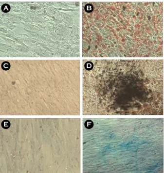
p3 시기의 탯줄유래 줄기세포를 지방세포로 분 화 유도 후 oil red O로 염색한 결과 분화 유도한 세포에서 lipid droplet이 염색된 것이 관찰되었다 (Figure 5B). 골세포로 분화 유도한 후에는 von Kossa 염색법을 시행한 결과 침적된 칼슘이 검은색 으로 염색된 것이 관찰되었으며 (Figure 5D), 연골세 포로 분화 유도한 후에는 alcian blue 염색을 시행한 결과 분화 유도한 세포에서 염색되었다 (Figure 5F).
고
찰중간엽 줄기세포는 체내 및 체외에서 다양한 세 포로 분화가 가능하기 때문에 세포치료제로 이용 될 수 있다. 그러나 임상에서 성체줄기세포를 치 료제로 사용하기 위해서는 많은 수가 필요하기 때 문에 체외증식 과정을 필요로 한다. 이 때 증식된 세포들은 줄기세포로의 특성이 유지되어야 한다.
그러나 사람의 골수유래 중간엽 줄기세포는 일반 체세포와 마찬가지로 체외 배양 시 계대배양 횟 수가 증가함에 따라 증식률이 감소하며 분화 능 력이 감소되는 등의 특성 변화가 나타난다고 알려 져 있다.13,31,32 또한 계대배양 후반 시기에는 mean telomere restriction fragment (mTRF) 길이가 감소하 며 senescence-associated β-galactosidase (SA β-gal)로 염색되는 세포 수가 증가하고,33~35 세포의 성장과 증식 그리고 세포 주기와 관련된 유전자 및 단백질 Figure 4. Immunocytochemical analysis of HUC during expansion in vitro. Immunocytochemical studies showed that HUC at p3, p6 and p9 positively were stained with antibodies against SSEA-3 and SSEA-4 proteins. Staining intensity of HUC for ICAM-1 and HLA-ABC gradually increased throughout the culture period. Intensity against thy-1 and fibronectin antibodies increased at p9 while that against TRA-1-60 and VCAM-1 antibodies began to decrease at p6 until p9. Cells at all passages showed negative staining with antibody against HLA-DR protein.
NC, negative control; scale bar, 100 μm.
Seah Park. Characterization of Umbilical Cord-derived Stem Cells during Expansion in Vitro. Korean J Reprod Med 2009.
발현 변화가 관찰된다.36 그러나 현재까지 탯줄유래 줄기세포의 체외 배양 시 계대배양 증가에 따른 세 포의 특성 연구는 거의 이루어지지 않았다. 따라서 본 연구에서는 사람의 탯줄에서 성체줄기세포를 분리하여 체외에서 배양하면서 증식 속도, 줄기세 포 특이 유전자 및 단백질의 발현 분석 등을 통하 여 계대배양 증가에 따른 세포의 특성 변화를 조 사하였다.
탯줄로부터 줄기세포를 분리하여 10회의 계대배 양을 시행한 결과 p7부터 부분적인 형태 변화가 나타나기 시작하였으며 p10 시기에 이르러서는 거 의 모든 세포들이 넓고 평평해진 형태를 나타내 었다. 이러한 변화는 사람의 골수유래 중간엽 줄 기세포,31 지방유래 중간엽 줄기세포,37 양막유래
중간엽 줄기세포 등30을 계대배양할 때 관찰되는 형태와 유사하였다. 탯줄유래 줄기세포의 계대배양 의 증가에 따른 증식 능력을 관찰하기 위하여 매 계대배양 시기에 population doubling time (PDT)을 조사한 결과 p7 시기부터 증가되는 것으로 나타났 다. 지방유래 줄기세포의 경우 총 10번의 계대배 양 중 p6부터 PDT가 증가하며,38 골수유래 중간엽 줄기세포는 총 10번의 계대배양 중 p3부터 p5 시 기까지 population doubling number가 2.5번 또는 2.6 번이었다가 p10 시기에 1.2번으로 감소한다.36 이 러한 결과들은 탯줄유래 줄기세포 역시 다른 중간 엽 줄기세포와 마찬가지로 체외에서 오랜 기간 배 양할 경우 계대배양 횟수가 늘어남에 따라 세포 분 열 속도가 느려진다는 것을 의미한다.
사람의 골수유래 줄기세포는 체외 배양 시 senescence passage 시기에 골세포의 분화와 기능 을 조절하는 human glycoprotein NMB 및 근육세포 를 재생시키는 역할을 하는 regeneration-associated muscle protease homolog 유전자의 증가가 관찰된 다.13 또한 총 20번의 계대배양 중 p15 시기 세포에 서 p7 시기에 비해 senescence 관련 유전자 26개의 증가하는데 이 중 세포 주기와 관련된 CDKN2b와 노화 및 세포 사멸로부터 세포를 보호하는 것으로 알려진 JUND 등이 포함된다고 보고된 바 있다.34 본 연구에서 체외에서 배양된 탯줄유래 줄기세포 p4, 8, 10 시기의 mRNA 분석 결과 세포 내 골격 물질인 vimentin과 외배엽과 췌장세포에서 발현하 는 nestin mRNA는 배양이 지속됨에 따라 발현이 증가하였으며, 배아줄기세포와 중간엽 줄기세포에 서 발현하는 SCF mRNA는 배양이 지속됨에 따라 발현 양이 감소하였다. 한편, 배아줄기세포 및 배아 종양세포 표지 유전자인 Oct-4 mRNA와 내배엽 세 포 표지 유전자인 HNF-4α mRNA는 p10까지 발현 양의 차이 없이 지속적으로 발현되었다. 중배엽 세 포 표지 유전자인 BMP-4와 외배엽 세포 표지 유전 자인 FGF-5, NCAM, 조직적합성항원인 HLA-ABC mRNA는 배양이 지속됨에 따라 발현 양이 증가하 는 것으로 보였으나 유의한 차이가 없었다. 중간엽
A B
C D
E F
Figure 5. Adipogenic, osteogenic, and chondrogenic differentiation of HUC. HUC at p3 were cultured for 3 weeks in each differentiation medium specific to adipo- genic (B), osteogenic (D) and chondrogenic (F). HUC was cultured in DMEM contained 10% FBS for 3 weeks (A, C, E). HUC stained with oil red O stain for adipocytes (A and B). HUC stained with von Kossa stain for osteocytes (C and D). HUC stained with alcian blue for chondrocytes (E and F). Magnification, ×100.
Seah Park. Characterization of Umbilical Cord-derived Stem Cells during Expansion in Vitro. Korean J Reprod Med 2009.
줄기세포의 체외 배양 시 계대배양에 따른 단백질 변화를 조사한 연구를 살펴보면, 사람의 탯줄유래 줄기세포의 p8 시기는 p4 시기보다 CD49e와 CD- 105 단백질의 발현이 감소하였다는 보고가 있으 며,39 사람의 골수유래 줄기세포의 p12 시기는 p3 시기에 비해 단백질의 발현 여부는 변화하지 않으 나 발현하는 세포 수가 감소한다는 보고가 있다.13 지방유래 줄기세포는 계대배양 수가 증가할수록 CD166 단백질의 발현이 감소하고 CD105 단백질 이 증가한다고 보고되었다.38 본 연구에서 p3, 6, 9 시기의 단백질 발현 변화를 관찰한 결과 계대가 증가할수록 세포연접 물질인 ICAM-1과 VCAM-1, 세포 외부 기질인 fibronectin의 발현이 증가한 반면 배아줄기세포 표지 물질인 TRA-1-60의 발현이 감 소된 것이 관찰되었다. 세포의 연접물질 및 외부 기질 단백질의 변화는 체외에서의 세포 배양이 세 포의 변화를 일으켰다는 것을 짐작할 수 있다. 그 러나 배아줄기세포 및 배아종양세포 표지 단백질인 SSEA-3와 SSEA-4은 계대배양 증가와 상관없이 지 속적으로 발현되는 것으로 나타났다. 본 연구 결과 중간엽 줄기세포에서 발현하는 SCF mRNA과 배아 줄기세포에서 발현하는 TRA-1-60 단백질의 발현이 계대배양이 증가함에 따라 감소되는 것으로 나타 났으며 이는 탯줄유래 줄기세포의 분열 능력의 감 소와 관련이 있을 것으로 생각되나 연구가 더 뒷받 침 되어야 할 것으로 사료된다.
이외에 체외증식 과정이 지방세포와 골세포와 연골세포로의 분화 능력에 영향이 있는지의 여부 는 아직 논쟁이 되고 있다. 사람의 골수유래 줄기 세포는 late passage 시기에 골세포로의 분화 능력 과 지방세포로의 분화 능력 모두 감소한다는 보고 가 있는 반면8 지방세포로의 분화 능만 감소된다 는 보고도 있다.13,32 또한 지방유래 줄기세포에서는 p10 시기까지 골세포로의 분화 능력은 차이가 없으 나 p6 이후 지방세포 표지 유전자의 발현이 감소 된다는 보고도 있다.38 이와는 다르게 p16에서 p18 (senescence passage) 시기에 지방세포로의 분화율은 감소하지 않으나 분화 정도가 감소한다는 주장도
있다.37 사람의 탯줄유래 줄기세포는 계대배양 초 기 시기에 지방세포와 골세포와 연골세포로 분화 한다고 알려져 있으나,11,21 계대배양에 따른 분화 능력에 대한 연구는 아직까지 보고된 바 없다. 본 연구에서는 p3 시기에 세포의 골세포로의 분화 능 력, 지방세포로의 분화 능력, 연골세포로의 분화 능 력을 확인하였다. 그러나 senescence passage 시기의 분화 능력에 대한 실험이 뒷받침되어야 할 것으로 사료된다.
결론적으로 탯줄유래 줄기세포의 체외 배양 시 계대배양의 증가에 따른 분열 능력 및 유전자와 단백질 발현 변화는 거의 알려져 있지 않다. 본 연 구에서 탯줄유래 줄기세포의 체외 배양 시 계대배 양에 따른 형태와 증식 능력을 관찰하고 또한 유 전자와 단백질의 변화를 조사한 결과 탯줄유래 줄 기세포는 체외 배양 시 줄기세포 특성이 일부 변하 는 것을 관찰하였다. 따라서 앞으로 줄기세포의 특 성을 유지할 수 있는 체외 배양법의 발달을 위한 연구들이 수행되어야 할 것으로 사료된다.
참 고 문 헌
1. Prockop DJ. Marrow stromal cells as stem cells for non- hematopoietic tissues. Science 1997; 27: 71-4.
2.Xu W, Zhang X, Qian H, Zhu W, Sun X, Hu J, et al.
Mesenchymal stem cells from adult human bone marrow differentiate into a cardiomyocyte phenotype in vitro. Exp Biol Med 2004; 229: 623-31.
3. Lee KD, Kuo TK, Whang-Peng J, Chung YF, Lin CT, Chou SH, et al. In vitro hepatic differentiation of human mesenchymal stem cells. Hepatology 2004; 40: 1275-84.
4. Woodbury D, Schwarz EJ, Prockop DJ, Black IB. Adult rat and human bone marrow stromal cells differentiate into neurons. J Neurosci Res 2000; 61: 364-70.
5.Giordano A, Galderisi U, Marino IR. From the laboratory bench to the patient's bedside: an update on clinical trials with mesenchymal stem cells. J Cell Physiol 2007; 211: 27-35.
6.Abdel-Latif A, Bolli R, Tleyjeh IM, Montori VM, Perin EC, Hornung CA, et al. Adult bone marrow-derived cells for cardiac repair: a systematic review and meta-analysis. Arch Intern Med
2007; 167: 989-97.
7. Bolland BJ, Tilley S, New AM, Dunlop DG, Oreffo RO. Adult mesenchymal stem cells and impaction grafting: a new clinical paradigm shift. Expert Rev Med Devices 2007; 4: 393-404.
8.Stenderup K, Justesen J, Clausen C, Kassem M. Aging is associated with decreased maximal life span and accelerated senescence of bone marrow stromal cells. Bone 2003; 33: 919 -26.
9. Baxter MA, Wynn RF, Jowitt SN, Wraith JE, Fairbairn LJ, Bellantuono I. Study of telomere length reveals rapid aging of human marrow stromal cells following in vitro expansion. Stem Cells 2004; 22: 675-82.
10. Zuk PA, ZhuM, Ashjian P, De Ugarte DA, Huang JI, Mizuno H, et al. Human adipose tissue is a source of multipotent stem cells. Mol Biol Cell 2002; 13: 4279-95.
11. Romanov YA, Svintsitskaya VA, Smirnov VN. Searching for alternative sources of postnatal human mesenchymal stem cells: candidate MSC-like cells from umbilical cord. Stem Cells 2003; 21: 105-10.
12.Fukuchi Y, Nakajima H, Sugiyama D, Hirose I, Kitamura T, Tsuji K. Human placenta-derived cells have mesenchymal stem/progenitor cell potential. Stem Cells 2004; 225: 649-58.
13. Wagner W, Horn P, Castoldi M, Diehlmann A, Bork S, Saffrich R, et al. Replicative senescence of mesenchymal stem cells: a continuous and organized process. PLoS ONE 2008; 3: e2213.
14.Ito T, Sawada R, Fujiwara Y, Seyama Y, Tsuchiya T. FGF-2 suppresses cellular senescence of human mesenchymal stem cells by down-regulation of TGF-beta2. Biochem Biophys Res Commun 2007; 359: 108-14.
15. Lu FZ, Fujino M, Kitazawa Y, Uyama T, Hara Y, Funeshima N, et al. Characterization and gene transfer in mesenchymal stem cells derived from human umbilical-cord blood. J Lab Clin Med 2005; 146: 271-8.
16.Horikawa I, Barrett JC. Transcriptional regulation of the telomerase hTERT gene as a target for cellular and viral oncogenic mechanisms. Carcinogenesis 2003; 24: 1167-76.
17.Nanaev AK, Kohnen G, Milovanov AP, Domogatsky SP, Kaufmann P. Stromal differentiation and architecture of the human umbilical cord. Placenta 1997; 18: 53-64.
18.Karahuseyinoglu S, Cinar O, Kilic E, Kara F, Akay GG, Demiralp DO, et al. Biology of stem cells in human umbilical cord stroma: in situ and in vitro surveys. Stem Cells 2007; 25:
319-31.
19. Lu LL, Liu YJ, Yang SG, Zhao QJ, Wang X, Gong W, et al.
Isolation and characterization of human umbilical cord mesenchymal stem cells with hematopoiesis-supportive function and other potentials. Haematologica 2006; 91: 1017-26.
20.Fu YS, Shih YT, Cheng YC, Min MY. Transformation of human umbilical mesenchymal cells into neurons in vitro. J Biomed Sci 2004; 11: 652-60.
21. Wang HS, Hung SC, Peng ST, Huang CC, Wei HM, Guo YJ, et al. Mesenchymal stem cells in the Wharton's jelly of the human umbilical cord. Stem Cells 2004; 22: 1330-7.
22. Bieback K, Kern S, Klüter H, Eichler H. Critical parameters for the isolation of mesenchymal stem cells from umbilical cord blood. Stem Cells 2004; 22: 625-34.
23. Kern S, Eichler H, Stoeve J, Klüter H, Bieback K. Comparative analysis of mesenchymal stem cells from bone marrow, umbilical cord blood, or adipose tissue. Stem Cells 2006; 24:
1294-301.
24. Baksh D, Yao R, Tuan RS. Comparison of proliferative and multi lineage differentiation potential of human mesenchymal stem cells derived from umbilical cord and bone marrow.
Stem Cells 2007; 25: 1384-92.
25. Lund RD, Wang S, Lu B, Girman S, Holmes T, Sauvé Y, et al.
Cells isolated from umbilical cord tissue rescue photoreceptors and visual functions in a rodent model of retinal disease. Stem Cells 2007; 25: 602-11.
26. Weiss ML, Medicetty S, Bledsoe AR, Rachakatla RS, Choi M, Merchav S, et al. Human umbilical cord matrix stem cells:
preliminary characterization and effect of transplantation in a rodent model of Parkinson's disease. Stem Cells 2006; 24: 781 -92.
27. Carlin R, Davis D, Weiss M, Schultz B, Troyer D. Expression of early transcription factors Oct-4, Sox-2 and Nanog by porcine umbilical cord (PUC) matrix cells. Reprod Biol Endocrinol 2006; 6: 4-8.
28.Seah Park, Hyeon Mi Kang, Eun Su Kim, Jinyoung Kim, Haekwon Kim. Characterization analysis for cardiogenic poten- tial of three human adult stem cells. Dev Reprod 2007; 11:
167-177.
29.Seah Park, Hyeon Mi Kang, Eun Su Kim, Jinyoung Kim, Haekwon Kim. Cardiomyogenic potential of human adipose tissue and umbilical cord derived-mesenchymal like stem cells. The Korean Journal of Reproductive Medicine 2007;
34: 239-252.
30. Kim J, Kang HM, Kim H, Kim MR, Kwon HC, Gye MC, et al.
Ex vivo characteristics of human amniotic membrane-derived stem cells. Cloning Stem Cells 2007; 9: 581-94.
31. Bruder SP, Jaiswal N, Haynesworth SE. Growth kinetics, self- renewal, and the osteogenic potential of purified human mesenchymal stem cells during extensive subcultivation and following cryopreservation. J Cell Biochem 1997; 64: 278-94.
32. Digirolamo CM, Stokes D, Colter D, Phinney DG, Class R, Prockop DJ. Propagation and senescence of human marrow stromal cells in culture: a simple colony-forming assay iden- tifies samples with the greatest potential to propagate and differentiate. Br J Haematol 1999; 107: 275-81.
33. Izadpanah R, Kaushal D, Kriedt C, Tsien F, Patel B, Dufour J, et al. Long-term in vitro expansion alters the biology of adult mesenchymal stem cells. Cancer Res 2008; 68: 4229-38.
34. Ryu E, Hong S, Kang J, Woo J, Park J, Lee J, et al. Identifi- cation of senescence-associated genes in human bone marrow mesenchymal stem cells. Biochem Biophys Res Commun 2008; 371: 431-6.
35.Sun HJ, Bahk YY, Choi YR, Shim JH, Han SH, Lee JW. A proteomic analysis during serial subculture and osteogenic differentiation of human mesenchymal stem cell. J Orthop Res 2006; 24: 2059-71.
36.Bonab MM, Alimoghaddam K, Talebian F, Ghaffari SH, Ghavamzadeh A, Nikbin B. Aging of mesenchymal stem cell in vitro. BMC Cell Biol 2006; 10: 7-14.
37. Noer A, Boquest AC, Collas P. Dynamics of adipogenic pro- moter DNA methylation during clonal culture of human adipose stem cells to senescence. BMC Cell Biol 2007; 8: 18.
38. Wall ME, Bernacki SH, Loboa EG. Effects of serial passaging on the adipogenic and osteogenic differentiation potential of adipose-derived human mesenchymal stem cells. Tissue Eng 2007; 13: 1291-8.
39. Weiss ML, Medicetty S, Bledsoe AR, Rachakatla RS, Choi M, Merchav S, et al. Human umbilical cord matrix stem cells:
preliminary characterization and effect of transplantation in a rodent model of Parkinson's disease. Stem Cells 2006; 24:
781-92.
= 국문초록 =
목 적: 중간엽 줄기세포를 임상에 적용하기 위해서는 체외 배양을 통한 세포증식 과정이 필요하나, 오랜 기간 동 안 체외 배양을 하게 되면 노화되어 특성이 변하고 분화 능력 또한 감소하게 된다. 따라서 현재까지는 초기 계대배양 의 세포만이 임상에 적용되고 있는 실정이며 체외에서의 세포 배양이 세포의 특성에 미치는 영향에 대한 연구와 함 께 세포의 특성 변화 없이 체외증식이 가능하도록 하는 연구들이 골수 및 지방유래 중간엽 줄기세포에서 보고되고 있다. 그러나 현재 탯줄유래 줄기세포의 체외 배양에 따른 특성 변화 분석 연구는 아직 잘 이루어지지 않고 있다. 본 연구의 목적은 탯줄유래 줄기세포의 체외 배양 시 계대배양 증가에 따른 줄기세포의 특성 변화를 분석하고자 하였다.
연구방법: 사람의 탯줄유래 줄기세포 (human umbilical cord-derived stem cells, HUC)를 분리하여 in vitro에서 계대배양하 였다. 계대배양에 따른 세포의 형태와 population doubling time (PDT)을 조사하고 RT-PCR 방법을 이용하여 mRNA 분 석을 하였으며 면역세포화학 염색법을 이용하여 단백질 발현을 분석하였다.
결 과: 탯줄유래 줄기세포는 평균 10번의 계대배양 후 senescence를 나타냈다. 세포의 형태는 7번째 계대배양 이후 세포질이 넓어지고 세포의 크기가 커지는 변화를 나타냈으며 PDT가 증가하기 시작하였다. 계대배양 4, 8, 10번째 시기 의 세포의 mRNA 변화를 분석한 결과 Oct-4, HNF-4α, mRNA는 10번째 계대배양까지 지속적으로 발현하였으나 nestin, vimentin mRNA는 지속적으로 발현이 감소하였고 SCF mRNA는 지속적으로 발현이 감소하였다. 이에 반해 HLA-DRα, Pax-6, BMP-2 mRNA는 모든 계대배양 시기의 세포에서 발현되지 않았다. 면역세포화학 분석법을 통한 3, 6, 9번째 계대 배양 세포의 단백질 발현 분석 결과 SSEA-3와 SSEA-4는 3, 6, 9번째 계대배양 세포 모두에서 발현하였으나 ICAM-1과 HLA-ABC는 계대배양이 증가함에 따라 발현이 증가되었다. Thy-1 단백질은 p9에서 발현이 증가되었으며 이와 반대로 TRA-1-60와 VCAM-1 단백질은 p6과 p9 시기에 발현이 감소되었다. HLA-DR 단백질은 모든 계대배양 시기에 발현되 지 않았다.
결 론: 본 연구결과 탯줄유래 줄기세포는 체외 배양 시 줄기세포 특성이 일부 변하는 것을 관찰하였다. 앞으로 줄기세포의 특성을 유지할 수 있는 체외 배양법의 발달을 위한 연구들이 수행 되야 할 것으로 사료된다.
중심단어: 탯줄유래 줄기세포, 계대배양, RT-PCR, 면역세포화학 염색법