• 검색 결과가 없습니다.

Current perspectives in stem cell therapies for osteoarthritis of the knee

N/A
N/A
Protected

Academic year: 2021

Share "Current perspectives in stem cell therapies for osteoarthritis of the knee"

Copied!
10
0
0

로드 중.... (전체 텍스트 보기)

전체 글

(1)

Introduction

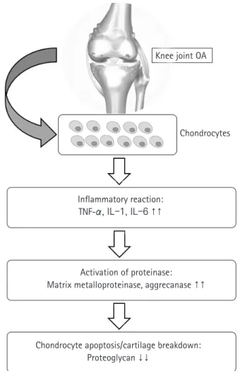
Osteoarthritis (OA) is a degenerative joint disease characterized by loss of cartilage, osteophyte formation, and periarticular bone change, resulting in disability [1,2]. In order to establish the dis- ease-modifying strategies of OA, it is necessary to understand the biomolecular features seen in OA circumstance. Increased proin- flammatory cytokines such as interleukin-1 or tumor necrosis fac- tor-α, decreased growth factors such as transforming growth fac- tor-beta (TGF-β), activated matrix metalloproteinase, and ulti- mate chondrocyte senescence can be observed at the molecular level [3-5] (Fig. 1). Although disease-modifying OA strategies that block inflammatory pathways and enhance cartilage protec- tive function have been developed recently [6,7], their effects on preventing the progression of OA have been still unsatisfactory, and it is particularly difficult to achieve ultimate cartilage regener- ation [8].

Mesenchymal stem cells (MSCs) are emerging as an attractive option for osteoarthritis (OA) of the knee joint, due to their marked disease-modifying ability and chondrogenic potential. MSCs can be isolated from various organ tissues, such as bone marrow, adipose tissue, synovium, um- bilical cord blood, and articular cartilage with similar phenotypic characteristics but different proliferation and differentiation potentials. They can be differentiated into a variety of connective tissues such as bone, adipose tissue, cartilage, intervertebral discs, ligaments, and muscles. Al- though several studies have reported on the clinical efficacy of MSCs in knee OA, the results lack consistency. Furthermore, there is no consensus regarding the proper cell dosage and application method to achieve the optimal effect of stem cells. Therefore, the purpose of this study is to re- view the characteristics of various type of stem cells in knee OA, especially MSCs. Moreover, we summarize the clinical issues faced during the application of MSCs.

Keywords: Knee joint; Mesenchymal stem cells; Osteoarthritis; Stem cells

Current perspectives in stem cell therapies for osteoarthritis of the knee

Gi Beom Kim, Oog-Jin Shon

Department of Orthopedic Surgery, Yeungnam University College of Medicine, Daegu, Korea

Yeungnam Univ J Med 2020;37(3):149-158 https://doi.org/10.12701/yujm.2020.00157

Received: March 12, 2020 Revised: April 1, 2020 Accepted: April 1, 2020 Corresponding author:

Oog-Jin Shon

Department of Orthopedic Surgery, Yeungnam University College of Medicine, 170 Hyeonchung-ro, Nam-gu, Daegu 42415, Korea Tel: +82-53-620-3640 Fax: +82-53-628-4020

E-mail: maestro-jin@hanmail.net

Meanwhile, since articular cartilage has a limited capacity for spontaneous healing, once damaged, it may eventually progress to OA [9]. Numerous attempts for the regeneration of focal cartilage defect have been made. Depending on the degree of defect size, various surgical options have been used, including multiple drill- ing, microfracture, abrasion chondroplasty, autologous chondro- cyte implantation, and osteochondral autologous transplantation [10-13]. However, there has been no optimal regenerative meth- od for cartilage in knee OA.

Recently, mesenchymal stem cells (MSCs) have been in the spotlight for their disease-modifying and chondrogenic potential along with their ease of harvesting, safety, and differentiation po- tential into cartilage [14,15]. Moreover, MSCs have been known to have paracrine and immunomodulatory effects through the se- cretion of cytokines and growth factors [16-19].

Therefore, considering the immunomodulatory and regenera- tive effect, stem cell therapies might be a promising line of treat-

Copyright

©

2020 Yeungnam University College of Medicine

This is an Open Access article distributed under the terms of the Creative Commons Attribution Non-Commercial License (http://creativecommons.org/licenses/by-nc/4.0/) which permits unrestricted non-commercial use, distribution, and reproduction in any medium, provided the original work is properly cited.

(2)

ment for knee OA. The purpose of this study was to review the characteristics of various type of stem cells in knee OA, especially based on MSCs. Moreover, we summarized the clinical issues for application of MSCs (Fig. 2).

General characteristics of stem cells

In general, stem cells can be divided into two major groups: (1) embryonic stem cells (ESCs) from the inner cell mass of blasto- cyst, with totipotency or pluripotency; (2) adult stem cells (tis- sue-specific stem cells) from specific organ, with multipotency [20,21]. Although adult stem cells have usually more restricted differentiation capacity compared to ESCs, they exhibit some ad- vantages including safety, easy derivation, and tissue-specific dif- ferentiation potential [22]. Among them, MSCs appears to be the promising candidates.

MSCs are multipotent progenitor cells that can be obtained from bone marrow, adipose tissue, synovial membrane, and artic- ular cartilage [23]. Several studies report their multidirectional differentiation potential [24,25]. Particularly, autologous MSCs can be easily harvested and applied in clinical settings, and allo- genic cells can be utilized [14,19]. Culture expansion may be re- quired to maximize their clinical effect [14], although that may re- sult in functional deterioration, mutation, and tumorigenesis as passage progresses. Nevertheless, MSCs can be cultivated and amplified while maintaining their potential, and differentiated into a variety of connective tissues such as bone, adipose tissue, Knee joint OA

Chondrocytes

Inflammatory reaction:

TNF-α, IL-1, IL-6 ↑↑

Activation of proteinase:

Matrix metalloproteinase, aggrecanase ↑↑

Chondrocyte apoptosis/cartilage breakdown:

Proteoglycan ↓↓

Fig. 1. Molecular mechanisms of osteoarthritis (OA). TNF, tumor necrosis factor; IL, interleukin.

BMAC BM-MSCs AD-MSCs S-MSCs hUCB-MSCs

Injective treatment

Source?

Dose?

Clinical application?

Complication?

Surgical implantation Scaffold

Tumorigenesis Immune response Calcification Concentrated Culture-expanded

MSCs

Fig. 2. Considerations for clinical application of MSCs. BMAC, bone marrow aspirate concentrate; MSC, mesenchymal stem cell; BM, bone marrow-derived; AD, adipose tissue-derived; S, synovium-derived; hUCB, human umbilical cord blood-derived.

±

(3)

cartilage, intervertebral discs, ligaments, and muscles [26,27].

Owing to their inherent ability for self-renewal, proliferation, and differentiation toward mature tissues, MSCs could have promis- ing applications in cell therapy and regenerative medicine [28].

The Mesenchymal and Tissue Stem Cell Committee of the Inter- national Society for Cellular Therapy proposed the definition of MSC via the following minimal set of criteria: (1) being plas- tic-adherent in standard culture conditions; (2) expressing CD105, CD73, and CD90 at their surface; while (3) lacking CD45, CD34, CD14, CD79α, and HLA-DR; and (4) being able to differentiate into osteoblasts, adipocytes, and chondrocytes in vitro [27].

Bone marrow-derived mesenchymal stem cells

Bone marrow-derived mesenchymal stem cells (BM-MSCs) have been widely studied for OA treatment because of several advan- tages, including high expansion ability, and differentiation poten- tial into cartilage [29-31]. Furthermore, due to easy harvesting from autologous bone marrow, they are cost-effective as com- pared to other types of MSCs. Several studies have reported favor- able clinical outcomes in patients with knee OA who underwent intra-articular injection or surgical implantation using autologous BM-MSCs (Table 1) [31-35]. However, donor site pain, limited number of obtainable cells (0.001% of all nucleated cells in bone marrow), and decreased differentiation potential with increase in donor age are a few limitations while using BM-MSCs [31,36].

Bone marrow aspiration from anterior or posterior iliac crest can be performed with local anesthesia under ultrasonographic or flu- oroscopic guidance to improve accuracy and efficiency [37].

Meanwhile, bone marrow aspirate concentrate (BMAC), which contains abundant nucleated cells and cytokines has been widely applied to the treatment of knee OA. It contains several factors in- volved in the healing process such as platelet-derived growth fac- tor, TGF-β, vascular endothelial growth factor, and fibroblast growth factor [17,38]. BMAC can be easily extracted by via FDA approved commercialized kits and this process condenses the buffy coat containing mononuclear cells and increases the num- ber of MSCs relative to baseline [39]. Recently, it is widely used in various orthopedic fields such as nonunion, osteonecrosis and sports injuries.

Centeno et al. [40] reported encouraging clinical outcomes with a low rate of adverse effects in autologous BMAC for in- tra-articular injection with or without adipose grafts in patients with knee OA. Additionally, significantly better results were ob- served in patients with Kellgren-Lawrence (K-L) grades I or II than in patients with K-L grades III or IV. Other studies have also reported positive clinical outcomes with simple iintra-articular in- jections of BMAC [41,42]. Contrastingly, in a recently conducted prospective randomized controlled trial, Shapiro et al. [42] did not report encouraging outcomes of BMAC as compared to the control group. BMAC studies in OA patients have not yet shown consistent results. Further researches comprising well-designed, randomized, controlled trials with larger sample sizes are needed to elucidate the exact mechanism of BMAC.

Table 1. Details of the clinical studies of BM-MSCs in knee OA Study Study design

(no. of cases) Mean age

(yr) Mean F/U

(mo) Delivery method Cell population

(cells/mL) Additional

factor Outcome Complication

Wakitani et al.

(2011) [33] Case series (45) 50 75 Two-stage surgical im-

plantation 5.0× 10

6

Collagen sheet from porcine tendon

No serious complica- tions such as tumor or infection

-

Davatchi et al.

(2011) [34] Case series (4) 57.8 12 Intra-articular injection 8.0 ‒9.0×10

6

- Pain, walking time, and the number of stairs to climb were improved

-

Wong et al.

(2013) [32] Prospective RCT (28 MSCs+HA

vs. 28 HA)

51 24 Intra-articular injection 1.46± 0.29× 10

7

Microfx-

+MOWHTO Better clinical outcomes and MRI in MSCs group

-

Orozco et al.

(2013) [31] Case series (12) 49 12 Intra-articular injection 4.0× 10

7

- Improvement in pain relief and WOMAC;

improvement of carti- lage quality on MRI

-

Davatchi et al.

(2016) [35] Case series (3) 57.8 60 Intra-articular injection 8.0‒9.0×10

6

- Previous parameters gradually deteriorated, but better than base- line

-

BM-MSCs, bone marrow-derived mesenchymal stem cells; OA, osteoarthritis; F/U, follow-up; RCT, randomized controlled trial; HA, hyaluronic acid;

Microfx, microfracture; MOWHTO, medial open-wedge high tibial osteotomy; MRI, magnetic resonance image; WOMAC, the Western Ontario and

McMaster Universities Osteoarthritis Index.

(4)

Adipose tissue-derived mesenchymal stem cells

Adipose tissue, along with bone marrow, has been the most fre- quently used source for isolating MSCs [43]. Adipose tissue is abundant and easily accessible, making it a reliable site for stem cell isolation. It has copious numbers of MSCs (approximately 0.5× 10

4

–2.0× 10

5

/1 g fat) compared to BM-MSCs and their dif- ferential capacity is relatively less affected by donor age.

The specific premade solution is usually infiltrated into the sub- cutaneous tissue of the abdominal region through a tumescent technique, and then, a conventional abdominal liposuction is per- formed using blunt cannulas [44]. Enzymatic digestion of fat tis- sue is the most used isolation technique to obtain adipose tis- sue-derived mesenchymal stem cells (AD-MSCs) [45]. The ex- tracted fat tissue is digested with enzyme (collagenase, dispase, or trypsin) to generate the stromal vascular fraction (SVF) that con- tains AD-MSCs and other endothelial and hematopoietic cells [46,47]. Among them, only the plastic-adherent, cultured and se- rially passaged multipotent cell populations are termed as AD- MSCs [48]. Meanwhile, non-enzymatic digestion, including me- chanical procedures such as centrifugation and filtration is some- times used. However, the range of yields shows high variation [49].

Several studies have reported encouraging clinical results for in- tra-articular injection of AD-MSCs in knee OA patients (Table 2) [14,50-53]. Lee et al. [50] reported that intra-articular injection of culture expanded AD-MSCs (1× 10

8

cells) showed satisfactory functional improvement and pain relief in patients with knee OA through a prospective randomized controlled trial. Nonetheless, AD-MSCs have some disadvantages such as relatively lower chon- drogenic potential and donor site morbidity [54].

SVF also can be obtained via enzymatic digestion and differen- tial centrifugation of adipose tissue. SVF consists of a heteroge- neous mesenchymal population of cells that includes not only ad- ipose stromal and hematopoietic stem cells but also endothelial cells, erythrocytes, fibroblasts, lymphocytes, monocyte/mac- ro-phages and pericytes (Table 3) [45,55]. Despite a highly heter- ogenous composition and low stem cell proportion, some studies have reported favorable clinical outcomes of SVF in knee OA due to their potential and ease of use [56,57].

Synovium-derived mesenchymal stem cells

Synovium-derived mesenchymal stem cells (S-MSCs) have at-

tracted considerable attention due to their high chondrogenic po- Table 2. Details of the clinical studies of AD-MSCs in knee OA Study Study design (no. of cases)

Mean age (yr) Mean F/U (mo) Delivery method Cell population (cells/mL) Additional factor Outcome Complication Jo et al. (20 14) [

14

]

Case series (dose-depen

-

dent) (3 vs. 3 vs. 12)

60 6 Intra-articular injection Low-dose: 1.0 × 10

7

Mid-dose: 5.0 × 10

7

High-dose: 1.0 × 10

8

Diagnostic arthroscopy

Better clinical outcomes and decreased cartilage defect in high-dose group; Hyaline-like regeneration No treatment-related AEs Most common AEs: naso

-

pharyngitis Serious AEs: urinary stone

Pak et al. (20 13) [

51

] Ca se s eri es (1 00) 51.2 26 Intra-articular injection NA (AD-SVF) PRP+HA+CaCl

2

Pain (VAS) was improved Joint swelling No tumor formation Kim et al. (20 15) [

53

]

Comparative matched-pair analysis (20 vs. 20)

59.2 28.6

Intra-articular injection vs. surgical implanta

- tion

4.0 × 10

6

Injection: PRP Better clinical and second-look arthroscopic outcomes in sur - gical implantation group

NA Surgical implantation: fi - brin glue Second-look arthroscopy Pers et al. (20 16) [

52

]

Prospective (dose-depen

-

dent) (6 vs. 6 vs. 6)

64.6 6 Intra-articular injection Low-dose: 2.0 × 10

6

Mid-dose: 1.0 × 10

7

High-dose: 5.0 × 10

7

-

Improved clinical outcomes in all groups; Limited possible improvement on MRI

- Lee et al. (20 19) [

50

]

Prospective RCT (12 MSCs vs. 12 saline) 62.7 6 Intra-articular injection 1.0 × 10

8

-

Significantly improved clinical outcomes in MSCs group; In

- creased defect in control group

- AD-MSC, adipose tissue-derived mesenchymal stem cell; OA, osteoarthritis; F/U, follow-up; AE, adverse event; NA, non-available; AD-SVF , adipose-derived stromal vascular fraction; PRP , platelet-rich plasma; HA, hyaluronic acid; VAS, visual analogue scale; MRI, magnetic resonance image; RCT , randomized controlled trial; MSCs, mesenchymal stem cells.

(5)

tential and less hypertrophic differentiation than BM-MSCs [58,59]. Embryologically, S-MSC-derived chondrocytes and ar- ticular chondrocytes share similar gene expression profile [60].

They may prove to be the optimal cell source of MSCs as native nature to the joints. Kubosch et al. [58] reported that S-MSCs play an important role in joint homeostasis and possibly in natural cartilage repair. However, most of their evidence was limited to preclinical studies [61]. Only one retrospective study has report- ed the results of S-MSCs in human OA of the knee joint [62].

They reported clinical improvement and secure defect filling con- firmed using second-look arthroscopy and magnetic resonance image, 48 months postoperatively. Further researches are needed to elucidate the interaction of S-MSCs and chondrocytes, and the promising role of S-MSCs in cartilage tissue engineering.

Human umbilical cord blood-derived mesenchymal stem cells

Umbilical cord compartments including Wharton’s jelly, perivas- cular tissue, and umbilical cord blood (UCB) can be utilized to isolate MSCs [43,63]. Umbilical cord-derived MSCs can be ob- tained through pain-free collection methods with fewer ethical is- sues. An experimental comparative study [64] confirmed that UCB-MSCs have biological advantages in comparison to bone marrow or adipose tissue, including higher rate of proliferation and clonality, retardation of senescence, and superior anti-inflam- matory effect.

Recently, clinical outcomes of human UCB-MSCs (hUCB- MSCs) for cartilage regeneration have been reported [65-67], and their medicinal product mixed with hyaluronic acid (Cartistem;

Medipost, Seongnam, Korea) has been widely applied in clinical settings. hUCB-MSCs are also isolated in a non-invasive manner and have the advantage of being hypoimmunogenic. Moreover, they show a hyaline-like histological morphology [67]. Park et al.

[65] reported that an hUCB-MSC-based product appeared safe and effective for the regeneration of hyaline-like cartilage in OA of

the knee after 7 years of follow-up. In our institution, commercial- ized hUCB-MSCs were performed on OA of the knee to obtain fa- vorable clinical outcomes and highly qualified regeneration (Fig. 3).

Clinical issues for application of mesenchymal stem cells

There is no consensus on the optimal dose or cell number to achieve the utmost effect of stem cells. The optimal dose of MSC implantation for cartilage regeneration has not yet been estab- lished. In a dose-dependent prospective study, Jo et al. [14] re- ported that significant clinical improvement was shown only in the high dose group (1× 10

8

cells). Based on this result, culture expansion may be needed to obtain the optimal effect of MSCs.

Treatment strategies for clinical application may also be one of the issues faced by clinicians. Injective treatment is relatively effi- cient because it is easy to apply and does not require hospitaliza- tion, but precise delivery to target site may be difficult [54]. Con- versely, surgical implantation allows direct delivery to the lesion site, but requires hospitalization and is a more invasive approach.

Another option is to mix MSCs with biodegradable scaffolds fol- lowed by surgical implantation. The three-dimensional scaffold maintains the phenotype of differentiated chondrocytes, pro- motes improved chondrogenesis through uniform cell distribu- tion, and reduces the risk of chondrocyte leakage [68]. Scaffold materials include hyaluronic acid, collagen derivatives, agarose, fi- brin glue, and chitosan [69]. However, chondrocyte dedifferenti- ation, apoptotic cell leakage, inadequate cell distribution, and low differentiation have been reported in scaffold-based studies [70].

Potential risks of MSCs in clinical use, such as tumorigenesis, immune response, and heterotrophic calcification are also consid- erable issues [71]. Therefore, it should be recognized that such risk of MSC-mediated abnormal reactions might occur in some cases, and mandating a careful assessment of the patient's condi- tion. Further research is also needed to guarantee the safety of MSCs. Each type of MSCs is summarized in Table 4.

Other advanced techniques

1. Induced pluripotent stem cell

Induced pluripotent stem cells (iPSCs) are also becoming a prom- ising cell source for stem cell-based therapy [72]. They are a kind of stem cells established in the laboratory that can be repro- grammed into somatic cells. Although therapeutic models of neu- rological and cardiovascular diseases using iPSCs have been re- ported [73,74], the research using iPSCs in orthopedic fields is still in its nascent stages, particularly for cartilage regeneration. iP- Table 3. Cell population in stromal vascular fraction

Variable Percentage (%)

Stromal cell 15 ‒30

Hematopoietic-lineage cell

Stem and progenitor cell < 0.1

Granulocyte 10 ‒15

Monocyte 5 ‒15

Lymphocyte 10 ‒15

Endothelial cell 10 ‒20

Pericyte 3 ‒5

(6)

A

E F G H

B C D

Fig. 3. (A, B) Kellgren-Lawrence grade III osteoarthritis is observed in the left knee anteroposterior and 45° flexion standing radiographs in a 49-year-old woman. (C) The scanography image shows neutral alignment of the both lower extremities. (D, E) Coronal T2- weighted fat suppressed magnetic resonance image (MRI) and arthroscopy show the focal chondral defects of International Cartilage Repair Society (ICRS) grade IV in the lateral femoral condyle and lateral tibial plateau. (F) Surgical implantation is performed using a commercialized mixture of human umbilical cord blood-derived mesenchymal stem cells and hyaluronic acid gel (Cartistem). (G, H) The coronal T2-weighted fat suppressed MRI and second-look arthroscopy confirm regenerated cartilage at 18 months after surgery.

Table 4. Summary of advantages and disadvantages of each MSC

BM-MSC AD-MSC S-MSC hUCB-MSC

Advantage High chondrogenic and osteo- genic potential

Good expansion ability

Easily accessible, reliable for isolation Plentiful number of cells

Less affected by donor age

Greater chondrogenic potential (same embryological origin as cartilage)

Direct delivery on defect Hypoimmunogenic

Histology: hyaline-like cartilage Disadvantage Donor site pain

Limited number of cells More affected by donor age

Relatively lower chondrogenic po- tential

Donor site morbidity (hematoma, in- fection)

Less osteogenic potential Not easy to obtain

Evidence still limited to preclini- cal studies

Surgical implantation High cost

Not easy to obtain

MSC, mesenchymal stem cell; BM, bone marrow-derived; AD, adipose-derived; S, synovium-derived; hUCB, human umbilical cord blood-derived.

(7)

SCs exhibit similar proliferation capacity and pluripotency as oth- er tissue-derived stem cells, with no immune rejection and ethical issues [72]. Recently, new methods for producing iPSCs without viral vectors to reduce the risk of tumorigenicity have been devel- oped [75].

Nonetheless, to date, limited data exists regarding the in vitro chondrogenic differentiation of iPSCs and the yield of iPSCs is relatively low.

2. Genetically modified MSCs (engineered MSCs) The efficacy of MSCs in vivo may still be low due to poor survival, retention, and engraftment of the cells. Most MSCs often die within the first few hours after in vivo delivery [76]. Therefore, MSCs need to be genetically modified to improve survival, migra- tion, and secretion of growth factors for their application in regen- erative medicine [76]. Genetic modification of MSCs is usually achieved through viral vectors [76]. The most commonly used vectors include retrovirus, lentivirus, baculovirus, and adenovirus [22]. Viral transduction has improved homing of MSCs to the de- fect or inflammation site through the overexpression of homing receptors. MSCs have been transduced with adenovirus express- ing C-X-C chemokine receptor 4 (CXCR-4) and runt-related transcription factor-2 (Runx-2), and with retrovirus expressing re- ceptor activator of nuclear factor-kB and CXCR-4 [77,78]. Al- though the efficacy of genetically modified MSCs has been demonstrated in preclinical studies, it has not been investigated in clinical trials.

Conclusion

MSCs are the hottest topic in recent stem cell research. The appli- cation of stem cells in cartilage regeneration has been tried a lot, but so far, the effect of cartilage regeneration is not consistent from one study to another. Moreover, the most appropriate cell source is still controversial. Further research is needed to deter- mine which tissue-derived stem cells, which usage and dose will be ideal for the treatment of osteoarthritis. In this review, we brief- ly reviewed the most up-to-date knowledge, including the charac- teristics, types, and clinical issues of MSCs. It is expected that in future, treatment with MSCs will be applied more clinically in the treatment of knee OA.

Acknowledgments

Conflicts of interest

No potential conflict of interest relevant to this article was report- ed.

Author contributions

Conceptualization: GBK, OJS; Data curation: GBK; Formal anal- ysis: GBK; Methodology: GBK; Project administration: OJS; In- vestigation, Resources: GBK; Supervision: OJS; Validation:

GBK; Writing-original draft: GBK; Writing-review & editing:

GBK, OJS.

ORCID

Gi Beom Kim, https://orcid.org/0000-0002-4067-1285 Oog-Jin Shon, https://orcid.org/0000-0002-9123-5694

References

1. Gupta S, Hawker GA, Laporte A, Croxford R, Coyte PC. The economic burden of disabling hip and knee osteoarthritis (OA) from the perspective of individuals living with this condition.

Rheumatology (Oxford) 2005;44:1531–7.

2. de Lange-Brokaar BJ, Ioan-Facsinay A, van Osch GJ, Zuurmond AM, Schoones J, Toes RE, et al. Synovial inflammation, im- mune cells and their cytokines in osteoarthritis: a review. Osteo- arthritis Cartilage 2012;20:1484–99.

3. Vincenti MP, Brinckerhoff CE. Transcriptional regulation of collagenase (MMP-1, MMP-13) genes in arthritis: integration of complex signaling pathways for the recruitment of gene-spe- cific transcription factors. Arthritis Res 2002;4:157–64.

4. Roach HI, Yamada N, Cheung KS, Tilley S, Clarke NM, Oreffo RO, et al. Association between the abnormal expression of ma- trix-degrading enzymes by human osteoarthritic chondrocytes and demethylation of specific CpG sites in the promoter re- gions. Arthritis Rheum 2005;52:3110–24.

5. Martin JA, Buckwalter JA. The role of chondrocyte senescence in the pathogenesis of osteoarthritis and in limiting cartilage re- pair. J Bone Joint Surg Am 2003;85A(Suppl 2):106–10.

6. Chevalier X, Eymard F, Richette P. Biologic agents in osteoar- thritis: hopes and disappointments. Nat Rev Rheumatol 2013;9:400–10.

7. Guler-Yuksel M, Allaart CF, Watt I, Goekoop-Ruiterman YP, de Vries-Bouwstra JK, van Schaardenburg D, et al. Treatment with TNF-α inhibitor infliximab might reduce hand osteoarthritis in patients with rheumatoid arthritis. Osteoarthritis Cartilage 2010;18:1256–62.

8. Hawker GA, Mian S, Bednis K, Stanaitis I. Osteoarthritis year 2010 in review: non-pharmacologic therapy. Osteoarthritis Cartilage 2011;19:366–74.

9. Goyal D, Keyhani S, Lee EH, Hui JH. Evidence-based status of

microfracture technique: a systematic review of level I and II

studies. Arthroscopy 2013;29:1579–88.

(8)

10. Mithoefer K, McAdams T, Williams RJ, Kreuz PC, Mandel- baum BR. Clinical efficacy of the microfracture technique for articular cartilage repair in the knee: an evidence-based system- atic analysis. Am J Sports Med 2009;37:2053–63.

11. Niemeyer P, Porichis S, Steinwachs M, Erggelet C, Kreuz PC, Schmal H, et al. Long-term outcomes after first-generation au- tologous chondrocyte implantation for cartilage defects of the knee. Am J Sports Med 2014;42:150–7.

12. Martincic D, Radosavljevic D, Drobnic M. Ten-year clinical and radiographic outcomes after autologous chondrocyte implanta- tion of femoral condyles. Knee Surg Sports Traumatol Arthrosc 2014;22:1277–83.

13. Gudas R, Gudaite A, Mickevicius T, Masiulis N, Simonaityte R, Cekanauskas E, et al. Comparison of osteochondral autologous transplantation, microfracture, or debridement techniques in articular cartilage lesions associated with anterior cruciate liga- ment injury: a prospective study with a 3-year follow-up. Ar- throscopy 2013;29:89–97.

14. Jo CH, Lee YG, Shin WH, Kim H, Chai JW, Jeong EC, et al. In- tra-articular injection of mesenchymal stem cells for the treat- ment of osteoarthritis of the knee: a proof-of-concept clinical trial. Stem Cells 2014;32:1254–66.

15. Freitag J, Bates D, Boyd R, Shah K, Barnard A, Huguenin L, et al. Mesenchymal stem cell therapy in the treatment of osteoar- thritis: reparative pathways, safety and efficacy. A review. BMC Musculoskelet Disord 2016;17:230.

16. Barry F, Murphy M. Mesenchymal stem cells in joint disease and repair. Nat Rev Rheumatol 2013;9:584–94.

17. Vezina Audette R, Lavoie-Lamoureux A, Lavoie JP, Laverty S.

Inflammatory stimuli differentially modulate the transcription of paracrine signaling molecules of equine bone marrow multi- potent mesenchymal stromal cells. Osteoarthritis Cartilage 2013;21:1116–24.

18. Jeong SY, Kim DH, Ha J, Jin HJ, Kwon SJ, Chang JW, et al.

Thrombospondin-2 secreted by human umbilical cord blood-derived mesenchymal stem cells promotes chondrogenic differentiation. Stem Cells 2013;31:2136–48.

19. Vega A, Martin-Ferrero MA, Del Canto F, Alberca M, Garcia V, Munar A, et al. Treatment of knee osteoarthritis with allogeneic bone marrow mesenchymal stem cells: a randomized con- trolled trial. Transplantation 2015;99:1681–90.

20. Martin DR, Cox NR, Hathcock TL, Niemeyer GP, Baker HJ.

Isolation and characterization of multipotential mesenchymal stem cells from feline bone marrow. Exp Hematol 2002;

30:879–86.

21. Johnson MH, McConnell JM. Lineage allocation and cell polar- ity during mouse embryogenesis. Semin Cell Dev Biol

2004;15:583–97.

22. Airenne KJ, Hu YC, Kost TA, Smith RH, Kotin RM, Ono C, et al. Baculovirus: an insect-derived vector for diverse gene trans- fer applications. Mol Ther 2013;21:739–49.

23. Pittenger MF, Mackay AM, Beck SC, Jaiswal RK, Douglas R, Mosca JD, et al. Multilineage potential of adult human mesen- chymal stem cells. Science 1999;284:143–7.

24. De Bari C, Dell'Accio F, Tylzanowski P, Luyten FP. Multipotent mesenchymal stem cells from adult human synovial membrane.

Arthritis Rheum 2001;44:1928–42.

25. Guilak F, Estes BT, Diekman BO, Moutos FT, Gimble JM. 2010 Nicolas Andry Award: Multipotent adult stem cells from adi- pose tissue for musculoskeletal tissue engineering. Clin Orthop Relat Res 2010;468:2530–40.

26. Trubiani O, Orsini G, Caputi S, Piatelli A. Adult mesenchymal stem cells in dental research: a new approach for tissue engi- neering. Int J Immunopathol Pharmacol 2006;19:451–60.

27. Dominici M, Le Blanc K, Mueller I, Slaper-Cortenbach I, Mari- ni F, Krause D, et al. Minimal criteria for defining multipotent mesenchymal stromal cells. The International Society for Cellu- lar Therapy position statement. Cytotherapy 2006;8:315–7.

28. Ra JC, Shin IS, Kim SH, Kang SK, Kang BC, Lee HY, et al. Safe- ty of intravenous infusion of human adipose tissue-derived mesenchymal stem cells in animals and humans. Stem Cells Dev 2011;20:1297–308.

29. Danisovic L, Lesny P, Havlas V, Teyssler P, Syrova Z, Kopani M, et al. Chondrogenic differentiation of human bone marrow and adipose tissue-derived mesenchymal stem cells. J Appl Biomed 2007;5:139–50.

30. Koga H, Shimaya M, Muneta T, Nimura A, Morito T, Hayashi M, et al. Local adherent technique for transplanting mesenchy- mal stem cells as a potential treatment of cartilage defect. Arthri- tis Res Ther 2008;10:R84.

31. Orozco L, Munar A, Soler R, Alberca M, Soler F, Huguet M, et al. Treatment of knee osteoarthritis with autologous mesenchy- mal stem cells: a pilot study. Transplantation 2013;95:1535–41.

32. Wong KL, Lee KB, Tai BC, Law P, Lee EH, Hui JH. Injectable cultured bone marrow-derived mesenchymal stem cells in varus knees with cartilage defects undergoing high tibial osteotomy: a prospective, randomized controlled clinical trial with 2 years' follow-up. Arthroscopy 2013;29:2020–8.

33. Wakitani S, Okabe T, Horibe S, Mitsuoka T, Saito M, Koyama T, et al. Safety of autologous bone marrow-derived mesenchymal stem cell transplantation for cartilage repair in 41 patients with 45 joints followed for up to 11 years and 5 months. J Tissue Eng Regen Med 2011;5:146–50.

34. Davatchi F, Abdollahi BS, Mohyeddin M, Shahram F, Nikbin B.

(9)

Mesenchymal stem cell therapy for knee osteoarthritis. Prelimi- nary report of four patients. Int J Rheum Dis 2011;14:211–5.

35. Davatchi F, Sadeghi Abdollahi B, Mohyeddin M, Nikbin B. Mes- enchymal stem cell therapy for knee osteoarthritis: 5 years fol- low-up of three patients. Int J Rheum Dis 2016;19:219–25.

36. Medical Advisory Secretariat. Osteogenic protein-1 for long bone nonunion: an evidence-based analysis. Ont Health Tech- nol Assess Ser 2005;5:1–57.

37. Madry H, Gao L, Eichler H, Orth P, Cucchiarini M. Bone mar- row aspirate concentrate-enhanced marrow stimulation of chondral defects. Stem Cells Int 2017;2017:1609685.

38. Caplan AI, Dennis JE. Mesenchymal stem cells as trophic medi- ators. J Cell Biochem 2006;98:1076–84.

39. Hernigou P, Mathieu G, Poignard A, Manicom O, Beaujean F, Rouard H. Percutaneous autologous bone-marrow grafting for nonunions: surgical technique. J Bone Joint Surg Am 2006;88(1 Suppl 2):322-7.

40. Centeno C, Pitts J, Al-Sayegh H, Freeman M. Efficacy of autolo- gous bone marrow concentrate for knee osteoarthritis with and without adipose graft. Biomed Res Int 2014;2014:370621.

41. Sampson S, Smith J, Vincent H, Aufiero D, Zall M, Bot- to-van-Bemden A. Intra-articular bone marrow concentrate in- jection protocol: short-term efficacy in osteoarthritis. Regen Med 2016;11:511–20.

42. Shapiro SA, Kazmerchak SE, Heckman MG, Zubair AC, O'Connor MI. A prospective, single-blind, placebo-controlled trial of bone marrow aspirate concentrate for knee osteoarthri- tis. Am J Sports Med 2017;45:82–90.

43. Jin YZ, Lee JH. Mesenchymal stem cell therapy for bone regen- eration. Clin Orthop Surg 2018;10:271–8.

44. Raposio E, Bonomini S, Calderazzi F. Isolation of autologous adipose tissue-derived mesenchymal stem cells for bone repair.

Orthop Traumatol Surg Res 2016;102:909–12.

45. Bourin P, Bunnell BA, Casteilla L, Dominici M, Katz AJ, March KL, et al. Stromal cells from the adipose tissue-derived stromal vascular fraction and culture expanded adipose tissue-derived stromal/stem cells: a joint statement of the International Feder- ation for Adipose Therapeutics and Science (IFATS) and the International Society for Cellular Therapy (ISCT). Cytothera- py 2013;15:641–8.

46. Patrikoski M, Juntunen M, Boucher S, Campbell A, Vemuri MC, Mannerström B, et al. Development of fully defined xe- no-free culture system for the preparation and propagation of cell therapy-compliant human adipose stem cells. Stem Cell Res Ther 2013;4:27.

47. Thirumala S, Gimble JM, Devireddy RV. Cryopreservation of stromal vascular fraction of adipose tissue in a serum-free freez-

ing medium. J Tissue Eng Regen Med 2010;4:224–32.

48. Baer PC, Geiger H. Adipose-derived mesenchymal stromal/

stem cells: tissue localization, characterization, and heterogene- ity. Stem Cells Int 2012;2012:812693.

49. Gentile P, Calabrese C, De Angelis B, Pizzicannella J, Kothari A, Garcovich S. Impact of the different preparation methods to ob- tain human adipose-derived stromal vascular fraction cells (AD- SVFs) and human adipose-derived mesenchymal stem cells (AD-MSCs): enzymatic digestion versus mechanical centrifu- gation. Int J Mol Sci 2019;20:5471.

50. Lee WS, Kim HJ, Kim KI, Kim GB, Jin W. Intra-articular injec- tion of autologous adipose tissue-derived mesenchymal stem cells for the treatment of knee osteoarthritis: a phase IIb, ran- domized, placebo-controlled clinical trial. Stem Cells Transl Med 2019;8:504–11.

51. Pak J, Chang JJ, Lee JH, Lee SH. Safety reporting on implanta- tion of autologous adipose tissue-derived stem cells with plate- let-rich plasma into human articular joints. BMC Musculoskelet Disord 2013;14:337.

52. Pers YM, Rackwitz L, Ferreira R, Pullig O, Delfour C, Barry F, et al. Adipose mesenchymal stromal cell-based therapy for severe osteoarthritis of the knee: a phase I dose-escalation trial. Stem Cells Transl Med 2016;5:847–56.

53. Kim YS, Kwon OR, Choi YJ, Suh DS, Heo DB, Koh YG. Com- parative matched-pair analysis of the injection versus implanta- tion of mesenchymal stem cells for knee osteoarthritis. Am J Sports Med 2015;43:2738–46.

54. Filardo G, Madry H, Jelic M, Roffi A, Cucchiarini M, Kon E.

Mesenchymal stem cells for the treatment of cartilage lesions:

from preclinical findings to clinical application in orthopaedics.

Knee Surg Sports Traumatol Arthrosc 2013;21:1717–29.

55. Zimmerlin L, Donnenberg VS, Pfeifer ME, Meyer EM, Paault B, Rubin JP, et al. Stromal vascular progenitors in adult human adi- pose tissue. Cytometry A 2010;77:22–30.

56. Yokota N, Hattori M, Ohtsuru T, Otsuji M, Lyman S, Shimomu- ra K, et al. Comparative clinical outcomes after intra-articular injection with adipose-derived cultured stem cells or noncul- tured stromal vascular fraction for the treatment of knee osteo- arthritis. Am J Sports Med 2019;47:2577–83.

57. Fodor PB, Paulseth SG. Adipose derived stromal cell (ADSC) injections for pain management of osteoarthritis in the human knee joint. Aesthet Surg J 2016;36:229–36.

58. Kubosch EJ, Lang G, Furst D, Kubosch D, Izadpanah K, Ro- lauffs B, et al. The potential for synovium-derived stem cells in cartilage repair. Curr Stem Cell Res Ther 2018;13:174–84.

59. Sasaki A, Mizuno M, Ozeki N, Katano H, Otabe K, Tsuji K, et

al. Canine mesenchymal stem cells from synovium have a

(10)

higher chondrogenic potential than those from infrapatellar fat pad, adipose tissue, and bone marrow. PLoS One 2018;13:

e0202922.

60. Kurth TB, Dell'accio F, Crouch V, Augello A, Sharpe PT, De Bari C. Functional mesenchymal stem cell niches in adult mouse knee joint synovium in vivo. Arthritis Rheum 2011;63:

1289–300.

61. Koga H, Muneta T, Ju YJ, Nagase T, Nimura A, Mochizuki T, et al. Synovial stem cells are regionally specified according to local microenvironments after implantation for cartilage regenera- tion. Stem Cells 2007;25:689–96.

62. Shimomura K, Yasui Y, Koizumi K, Chijimatsu R, Hart DA, Yo- netani Y, et al. First-in-human pilot study of implantation of a scaffold-free tissue-engineered construct generated from autol- ogous synovial mesenchymal stem cells for repair of knee chon- dral lesions. Am J Sports Med 2018;46:2384–93.

63. Klontzas ME, Kenanidis EI, Heliotis M, Tsiridis E, Mantalaris A.

Bone and cartilage regeneration with the use of umbilical cord mesenchymal stem cells. Expert Opin Biol Ther 2015;

15:1541–52.

64. Jin HJ, Bae YK, Kim M, Kwon SJ, Jeon HB, Choi SJ, et al. Com- parative analysis of human mesenchymal stem cells from bone marrow, adipose tissue, and umbilical cord blood as sources of cell therapy. Int J Mol Sci 2013;14:17986–8001.

65. Park YB, Ha CW, Lee CH, Yoon YC, Park YG. Cartilage regen- eration in osteoarthritic patients by a composite of allogeneic umbilical cord blood-derived mesenchymal stem cells and hyal- uronate hydrogel: results from a clinical trial for safety and proof-of-concept with 7 years of extended follow-up. Stem Cells Transl Med 2017;6:613–21.

66. Matas J, Orrego M, Amenabar D, Infante C, Tapia-Limonchi R, Cadiz MI, et al. Umbilical cord-derived mesenchymal stromal cells (MSCs) for knee osteoarthritis: repeated MSC dosing is superior to a single MSC dose and to hyaluronic acid in a con- trolled randomized Phase I/II trial. Stem Cells Transl Med 2019;8:215–24.

67. Ha CW, Park YB, Chung JY, Park YG. Cartilage repair using composites of human umbilical cord blood-derived mesenchy- mal stem cells and hyaluronic acid hydrogel in a minipig model.

Stem Cells Transl Med 2015;4:1044–51.

68. Grigolo B, Lisignoli G, Piacentini A, Fiorini M, Gobbi P, Maz- zotti G, et al. Evidence for redifferentiation of human chondro-

cytes grown on a hyaluronan-based biomaterial (HYAff 11):

molecular, immunohistochemical and ultrastructural analysis.

Biomaterials 2002;23:1187–95.

69. Kon E, Verdonk P, Condello V, Delcogliano M, Dhollander A, Filardo G, et al. Matrix-assisted autologous chondrocyte trans- plantation for the repair of cartilage defects of the knee: system- atic clinical data review and study quality analysis. Am J Sports Med 2009;37(Suppl 1):156S–166S.

70. Rai V, Dilisio MF, Dietz NE, Agrawal DK. Recent strategies in cartilage repair: a systemic review of the scaffold development and tissue engineering. J Biomed Mater Res A 2017;105:2343–

54.

71. Breitbach M, Bostani T, Roell W, Xia Y, Dewald O, Nygren JM, et al. Potential risks of bone marrow cell transplantation into in- farcted hearts. Blood 2007;110:1362–9.

72. Zhu Y, Wu X, Liang Y, Gu H, Song K, Zou X, et al. Repair of cartilage defects in osteoarthritis rats with induced pluripotent stem cell derived chondrocytes. BMC Biotechnol 2016;16:78.

73. Miao Q, Shim W, Tee N, Lim SY, Chung YY, Ja KP, et al. iP- SC-derived human mesenchymal stem cells improve myocardi- al strain of infarcted myocardium. J Cell Mol Med 2014;18:

1644–54.

74. Sareen D, Gowing G, Sahabian A, Staggenborg K, Paradis R, Avalos P, et al. Human induced pluripotent stem cells are a nov- el source of neural progenitor cells (iNPCs) that migrate and in- tegrate in the rodent spinal cord. J Comp Neurol 2014;522:

2707–28.

75. Trokovic R, Weltner J, Nishimura K, Ohtaka M, Nakanishi M, Salomaa V, et al. Advanced feeder-free generation of induced pluripotent stem cells directly from blood cells. Stem Cells Transl Med 2014;3:1402–9.

76. Park JS, Suryaprakash S, Lao YH, Leong KW. Engineering mes- enchymal stem cells for regenerative medicine and drug deliv- ery. Methods 2015;84:3–16.

77. Lien CY, Chih-Yuan Ho K, Lee OK, Blunn GW, Su Y. Resto- ration of bone mass and strength in glucocorticoid-treated mice by systemic transplantation of CXCR4 and cbfa-1 co-expressing mesenchymal stem cells. J Bone Miner Res 2009;24:837–48.

78. Cho SW, Sun HJ, Yang JY, Jung JY, An JH, Cho HY, et al. Trans-

plantation of mesenchymal stem cells overexpressing RANK-Fc

or CXCR4 prevents bone loss in ovariectomized mice. Mol

Ther 2009;17:1979–87.

수치

Fig. 1. Molecular mechanisms of osteoarthritis (OA). TNF, tumor  necrosis factor; IL, interleukin.
Table 1. Details of the clinical studies of BM-MSCs in knee OA Study Study design
Fig. 3.  (A, B) Kellgren-Lawrence grade III osteoarthritis is observed in the left knee anteroposterior and 45° flexion standing radiographs  in a 49-year-old woman

참조

관련 문서

Vertical ridge augmentation by means of deproteinized bovine bone block and recombinant human platelet-derived growth factor-BB: a histologic study in a

The purpose of this study was to evaluate the effect of these substances on bone regeneration by applying these materials to bone defects after cyst

Our results also showed that pigment epithelium-derived factor concentration in vitreous was higher than that in serum, suggesting that it is mainly derived from

– Embryonic stem cell : Pluripotent cells that can give rise to all tissue types. – Adult stem cell : Multipotent cells

Determining optimal surface roughness of TiO₂blasted titanium implant material for attachment, proliferation and differentiation of cells derived from

It might also act in conjunction with a growth factor, such as bone morphogenic protein (BMP), insulin-like growth factor, or platelet-derived growth factor.

Approved clinical use of bone marrow stem cells for myocardial infarction treatment... Cardiac

– Water-filling solution in the frequency-time domain is derived – Theoretical limit of data rate in single user OFDM. • Transmit power allocation