사람의 지방 및 제대에서 유래된 유사중간엽 줄기세포로부터 심근세포로의 분화 유도
서울여자대학교 자연과학대학 생명공학과 박세아·강현미·김은수·김진영·김해권*
Cardiomyogenic Potential of Human Adipose Tissue and Umbilical Cord Derived-Mesenchymal Like Stem Cells
Seah Park, Hyeon Mi Kang, Eun Su Kim, Jinyoung Kim, Haekwon Kim* Department of Biotechnology, College of Natural Sciences, Seoul Women's University
Objectives: In the present study, we examined the differentiation potential of human adipose- (HAD) and human umbilical cord-derived mesenchymal like stem cells (HUC) into cardiomyocytes.
Methods: Cells were initially exposed to 5-azacytidine for 24 h cells and then were cultivated in the presence or absence of activin A, TGF-β1, or Wnt inhibitor with various combinations of BMP and FGF. Assessment of cardiomyogenic differentiation was made upon the expression of cardiomyocyte-specific genes using RT-PCR.
Results: HAD that cultivated in control medium for 4 weeks after 5-azacytidine expose showed new expression of TnT gene and increased expression of Cmlc1 and kv4.3 genes. However, HAD cultivated in the presence of combinations of BMP-4/FGF-4 (B4/F4) and BMP-4/FGF-8 (B4/F8) showed new expression of β-MHC gene and more increased expression of Cmlc1, TnT, TnI, Kv4.3 genes. Significantly enhanced expression of Cmlc1, TnT, and Kv4.3 genes were also observed compared to that cultivated in the control medium. Treatment of HUC with either 5-azacytidine or combinations of BMP and FGF did not affect the expression profile of these genes. However, when activin A or TGF-β1 was present in addition to the BMP-2/FGF-8 (B2/F8) after 5-azacytidine exposure, HUC exhibited new expression of β-MHC gene and increased expression of α-CA, TnT and Kv4.3 genes. When Wnt inhibitor was present in addition to BMP and FGF, HUC showed new expression of Cmlc1 gene and increased expression of α-CA, TnT, TnI and Kv4.3 genes.
Conclusions: Based on these observations, it is suggested that HAD and HUC could differentiate into cardiomyocytes which might be used as therapeutic cells for the heart diseases. [Korean. J. Reprod. Med. 2007; 34(4): 239-252.]
Key Words: Mesenchymal stem cells, Adipose tissue-derived stem cells, Umbilical cord-derived stem cells, Cardiomyocyte, Gene expression
심장은 신체의 여러 조직 중 재생되기 어려운 조직 중의 하나로 알려져 있다.1,2 따라서 심근경색 (myocardial infarction) 또는 다른 질병에 의한 심근
의 손실은 심장기능장애 (heart failure)를 일으킨다.3 일단 심근의 많은 부분이 파괴되면 약물 치료로는 병의 진행을 막기 어렵기 때문에 심장 이식 방법을 통해서만이 치료가 가능하나 이 방법은 공급자가 부족하고 면역 거부반응이 일어나는 문제점이 있 다.4 따라서 줄기세포를 이용한 치료법이 연구되 고 있다. 배아줄기세포 (embryonic stem cells, ESC)
주관책임자:김해권, 우) 139-774 서울시 공릉2동 126번지, 서울여자대학교 자연과학대학 생명공학과
Tel: (02) 970-5665, Fax: (02) 970-5974 e-mail: [email protected]
*이 논문은 2007학년도 서울여자대학교 교내학술특별연구비의 지 원을 받았음.
와 성체줄기세포 (adult stem cells, ASC)는 전임상 실험이 진행되고 있으며 네 종류 (skeletal myoblast, bone marrow mononuclear cells, mesenchymal stem cells, endothelial progenitor cells) 이상의 줄기세포에 서 초기단계의 임상 실험이 이루어지고 있다.5,6 배 아줄기세포는 윤리적인 문제점과 발암의 가능성이 있으며, 가장 잘 알려진 성체줄기세포인 골수유래 줄기세포를 얻기 위해서는 고통을 수반해야 하는 등의 단점이 있다. 따라서 새로운 성체줄기세포의 공급원을 찾는 연구들이 진행되어 현재 탯줄혈액, 말초혈액, 지방, 양막, 양수, 탯줄, 태반 등으로부터 줄기세포가 분리된다고 보고되었다.7~14
배아줄기세포는 배아체 (embryoid body, EB) 형성 후에 저절로 심근세포로 분화한다고 알려져 있다.
15,16 그러나 효율성은 1~25% 정도로 낮으며 연구
자들마다 다른 결과를 보고하고 있다. 분화율을 높 이기 위하여 5-azacytidine 및 cytokine을 처리하여 분화를 유도하는 연구도 진행되고 있다.17,18 성체 줄기세포를 심근세포로 분화 유도하기 위하여 가 장 많이 사용하는 방법은 demethylation agent인 5- azacytidine으로 24시간 동안 처리하고 4주 후 분 화된 세포를 얻는 것이다.19,20 이에 대해 Balana 등 은 같은 방법을 사용한 결과 체외에서 심장세포로 의 분화는 일어나지 않고 단지 전기생리학적 특성 만 변한다고 보고하였다.21 한편 5-azacytidine 대신 FGF-2 또는 activin A가 심근세포 특유의 유전자 발현을 일으키며,22 insulin과 transferrin 등의 다양한 물질을 함께 처리할 경우 체외박동을 일으키는 심 근세포로 분화되었다고 보고된 바 있다.23
낭배형성 중인 닭의 배아에 BMP-2를 생산하는 세포를 이식하면 cardiac specification이 발생되며,28 BMP-2와 BMP-4는 각각 FGF-4와 함께 작용하여 조류의 posterior mesoderm을 심장중배엽 (cardiac mesoderm)으로 유도하고 TGF-β와 activin은 상배엽 (epiblast)을 심근세포로 분화 유도한다고 보고되었 다.29 한편 Wnt는 발달과정에 발생되는 paracrine/
autocrine 신호물질로 canonical signal은 β-catenin과 T-cell factor/lymphoid enhancer factor를 활성화시켜
target 유전자를 조절하며, noncanonical signal은 cJun- N-terminal kinase (JNK)을 포함한 여러 경로를 통해 유전자의 활성을 조절한다.33,34 현재 canonical signal 은 심근세포로의 분화를 억제하지만 noncanonical signal은 심근세포로의 분화를 증가시킨다고 알려 져 있다.35 Noncanonical Wnt를 처리하기 위해서는 conditioned medium을 사용해야 하기 때문에 본 연 구에서는 canonical signal을 억제하기 위하여 secre- ted Frizzled Related Protein-3 (sFRP-3)를 처리하였다.
본 연구에서는 사람의 지방유래 줄기세포 (human adipose tissue-derived mesenchymal like stem cells, HAD)와 탯줄유래 줄기세포 (umbilical cord-derived mesenchymal like stem cells, HUC)에 5-azacytidine을 24시간 동안 처리하고 4주간 여러 가지 BMP와 FGF 조합을 처리하여 심근세포로의 분화를 유도 하였다. 또한 HUC의 경우에는 추가로 여러 가지 BMP와 FGF 인자들과 함께 activin A, TGF-β1 또는 Wnt inhibitor를 처리하여 심근세포로의 효과적인 분화 조건을 찾아보았다.
연구대상 및 방법
1. 지방과 탯줄로부터 줄기세포의 채취
본 연구에 사용된 탯줄은 만삭 정상 산모의 제왕 절개 또는 질식 분만 시 산모의 동의 하에 태아로 부터 채취하였다. 지방조직은 환자의 동의 하에 성 형 수술 시 채취하였다. 채취한 조직은 4시간 내에 실험에 사용하였다.
2. 줄기세포의 배양 1) HAD의 배양
지방조직을 잘게 자른 후 0.075% type 1 colla- genase (Gibco, Grand Island, NY)가 포함된 Ca2+, Mg2+-free Dubecco's phosphate-buffered saline (DPBS, Gibco)에 넣어 37℃에서 30분 동안 교반하여 반응 시켰다. 이 후 500 ×g로 3번 세척하고 얻어진 세 포를 100 U/mL penicillin (Gibco), 0.1 µg/mL strepto- mycin (Gibco), 3.7 mg/mL sodium bicarbonate, 그리고
10% fetal bovine serum (FBS, Hyclone, South Logan, UT)이 포함된 low-glucose Dulbecco's Modified Eagle Medium (DMEM-LG, Gibco) 에서 37℃, 5% CO2가 공급되는 배양기 내에서 배양하였다. 배양 3일 후 배양용기의 바닥에 붙지 않은 세포를 제거하고 1 주에 2번 배양액을 교체하였다. 배양용기의 70~
80% 정도로 세포가 자라면 0.125% trypsin (Gibco) 과 1mM ethylenediamine tetraacetic acid (EDTA)가 함 유된 Hanks Balanced Salt Solution (HBSS, Gibco)을 3 분간 처리하여 세포를 떼어낸 후, 2×10³/cm2의 농 도로 세포를 배양하였다.
2) HUC의 배양
탯줄을 100 U/mL의 penicillin과 100 µg/mL의 streptomycin이 첨가된 Ca2+, Mg2+-free DPBS로 탯줄 외부의 혈액을 제거한 후 외부 양막을 벗기고 동 맥 2개를 제거하였다. 남은 조직을 1 mm3 크기로 자른 후 같은 부피의 0.5% crude type collagenase (Gibco)로 37℃에서 20~24시간 처리하였다. 이 후 Ca2+, Mg2+-free DPBS를 40배 첨가하여 실온에서 600 ×g로 10분간 원심분리하여 상층액을 제거하고 DMEM-LG로 2번 세척하였다. 분리된 세포를 100 U/mL penicillin, 0.1 µg/mL streptomycin, 3.7 mg/mL sodium bicarbonate, 그리고 10% FBS가 포함된 DMEM-LG에서 37℃, 5% CO2가 공급되는 배양기 내에서 배양하였다. 배양 3일 후 배양용기의 바닥 에 붙지 않은 세포를 제거하고 1주에 2번 배양액 을 교체하였다. 배양용기의 70~80% 정도로 세포 가 자라면 0.125% trypsin과 1 mM EDTA가 첨가된 HBSS로 3분간 처리하여 세포를 떼어낸 후 2×10³/ cm2의 농도로 배양하였다.
3) Total RNA 분리 및 역전사 중합효소 연쇄반 응 (reverse transcription polymerase chain reaction, RT-PCR)
Cell pellet을 Ca2+, Mg2+-free DPBS를 이용하여 세 척하고 500 µL의 Tri-reagent를 첨가한 다음 manu- facturer의 instruction에 따라 total RNA를 분리하였 다. 5 µg의 RNA는 reaction buffer, 1 mM NTP mixture, 0.5 µg/µL oligo(d)T15, 20 U RNase inhibitor (Takara,
Japan), 20 U M-MuLV reverse transcriptase (Fermentas, Burlington, Canada)가 혼합된 20 µL 반응 용액에서 역전사 시켰다. 반응은 42℃에서 60분간 진행되었 다. 얻어진 RT products (cDNAs)는 2 mM MgCl2, 1×
Taq buffer, 0.25 U Taq polymerase (Fermentas), 10 pM 의 sense와 antisense gene-specific primers가 혼합된 10 µL 반응 용액으로 PCR을 수행하였다. Ampli- fication은 총 35 cycles 수행하였으며, 각 cycle은 94℃에서 30초간의 denaturation, 30초간의 annealing, 72℃에서 30초간의 extension 과정으로 구성되었다.
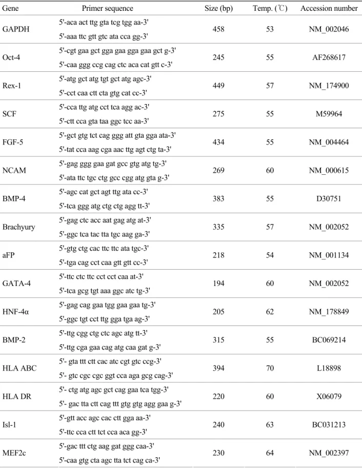
Annealing 과정의 온도는 Table 1에 표기하였다. 반 응 종결 후, PCR 생성물들은 0.25% bromophenol blue, 0.25% xylene cyanol, 40% sucrose가 포함된 6 × loading buffer에 혼합한 다음 2% agarose gel을 이용 하여 전기영동하였다. 전기영동 후 ethidium bromide 로 염색하고 ultraviolet light를 이용하여 DNA의 영 상을 얻었다.
4) 면역세포화학적 특성 분석
HAD와 HUC을 8-well slide chamber (Nunc, Roche- ster, NY)에서 배양한 후 2% paraformaldehyde가 함 유된 DPBS 용액을 이용하여 4℃에서 2시간 동안 고정하였다. 고정 후 DPBS로 5분간 3번 세척하였 다. 그리고 실온에서 10분 동안 0.5% Triton X-100가 함유된 DPBS를 처리 후 세척한 다음 endogenous peroxidase activities를 제거하기 위하여 3% hydrogen peroxide (Dako, Carpinteria, CA)로 15분간 반응시킨 후 세척하였다. 2% bovine serum albumin을 함유한 DPBS에서 실온으로 1시간 동안 반응시켰다. 그 후 각 TRA-1-60 (1:20), SSEA-3 (1:50), SSEA-4 (1:50), Thy-1 (1:20), α-SMA (1:50), vimentin (1:100), CK18 (1:50), CD44 (1:500), CD54 (1:40), CD106 (1:50), FSP (1:500), vWF (1:200), CD31 (1:40), HLA ABC (1:200), HLA DR (1:50) mouse monoclonal antibody를 4℃에서 17~24시간 동안 처리 후, 세척하였다. 이 후 bio- tinylated goat anti-mouse IgG와 anti-rabbit IgG를 실 온에서 20분간 처리하였다. 3번 세척한 다음, hor- seradish peroxidase-conjugated streptavidin (Dako)을 20분간 처리하였다. 3,3'-diaminobenzidine tetrahydro-
Table 1. Primer used for the RT-PCR analysis for stem cell and cardiomyocyte-specific markers
Gene Primer sequence Size (bp) Temp. (℃) Accession number 5'-aca act ttg gta tcg tgg aa-3'
GAPDH
5'-aaa ttc gtt gtc ata cca gg-3' 458 53 NM_002046 5'-cgt gaa gct gga gaa gga gaa gct g-3'
Oct-4
5'-caa ggg ccg cag ctc aca cat gtt c-3' 245 55 AF268617 5'-atg gct atg tgt gct atg agc-3'
Rex-1
5'-cct caa ctt cta gtg cat cc-3' 449 57 NM_174900 5'-cca ttg atg cct tca agg ac-3'
SCF 5'-ctt cca gta taa ggc tcc aa-3' 275 55 M59964 5'-gct gtg tct cag ggg att gta gga ata-3'
FGF-5
5'-tat cca aag cga aac ttg agt ctg ta-3' 434 55 NM_004464 5'-gag ggg gaa gat gcc gtg atg tg-3'
NCAM
5'-ata ttc tgc ctg gcc cgg atg gta g-3' 269 60 NM_000615 5'-agc cat gct agt ttg ata cc-3'
BMP-4
5'-tca ggg atg ctg ctg agg tt-3' 383 55 D30751 5'-gag ctc acc aat gag atg at-3'
Brachyury
5'-ggc tca tac tta tgc aag ga-3' 335 57 NM_002052 5'-gtg ctg cac ttc ttc ata tgc-3'
aFP 5'-tga cag cct caa gtt gtt cc-3' 218 54 NM_001134 5'-ttc ctc ttc cct cct caa at-3'
GATA-4
5'-tca gcg tgt aaa ggc atc tg-3' 194 60 NM_002052 5'-gag cag gaa tgg gaa gaa tg-3'
HNF-4α
5'-ggc tgt cct ttg gga tga ag-3' 205 62 NM_178849 5'-ttg cgg ctg ctc agc atg tt-3'
BMP-2
5'-ttg cga gaa cag atg caa gat g-3' 315 55 BC069214 5'- gta ttt ctt cac atc cgt gtc ccg-3'
HLA ABC
5'- gtc cgc cgc ggt cca aga gcg cag-3' 394 70 L18898 5'- ctg atg agc gct cag gaa tca tgg-3'
HLA DR
5'- gac tta ctt cag ttt gtg gtg agg gaa g-3' 220 60 X06079 5'-gtt acc agc cac ctt gga aa-3'
Isl-1
5'-ttc cca ctt tct cca aca gg-3' 240 63 BC031213 5'-gac ttt ctg aag gat ggg caa-3'
MEF2c
5'-caa gtg cta agc tta tct cag ca-3' 230 64 NM_002397
chloride (DAB, Dako) 용액으로 발색하였다. 발색이 된 세포는 DPBS로 세척하고 Mayer's Haematoxylin (Sigma, St.Louis, MO)으로 대조 염색한 후 광학 현 미경 (LSM410; Carl Zeiss, Oberkochen, Germany)하에 서 관찰하였다.
3. 심근세포로의 분화 유도 1) HAD의 분화 유도
배양 접시에 2×10³/cm의 농도의 세포를 넣어준 후 3일 후에 10 µM 5-azacytidine이 첨가된 DMEM- Table 1. Primer used for the RT-PCR analysis for stem cell and cardiomyocyte-specific markers (continued)
Gene Primer sequence Size (bp) Temp. (℃) Accession number 5'-ggt cta tga act gga gcg gc-3'
Nkx2.5
5'-ata ggc ggg gta ggc gtt at-3' 322 64 AB021133 5'-ccg gca ctg tgg act aca ac-3'
α-MHC
5'-agc ctc tcg tcc ctc att tc-3' 598 59 NM_002471 5'-cta acc tgg aga aga tgt gcc-3'
β-MHC
5'-tcg atc tca ttc tgt agc cg-3' 550 59 NM_000257 5'-cag gtc gag ttt gat gct tcc-3'
Cmlc1
5'-cga agt cct cat agg tgc ctg-3' 300 63 NM_000258 5'-act atc atg gac cag aac ag-3'
Cvmlc2
5'-agt cct tct ctt ctc cgt g-3' 400 59 NM_000432 5'-gct ctt tgg gga gaa gct ca-3'
MLC-2a
5'-cgt ctc cat ggg tga tga tg-3' 239 57 BC027915 5'-tct gct ggc atc cat gaa ac-3'
α-CA
5'-gat gag gaa agg tgg ttt gg-3' 400 59 NM_005159 5'-tca ctg agc gtg gct act cc-3'
α-SA
5'-ctt ggt gat cca cat ctg ctg-3' 500 59 NM_001100 5'-ggc agc gga aga gga tgc tga a-3'
TnT 5'-gag gca cca agt tgg gct tga acg a-3' 152 57 BC002653 5'-cct gcg gag agt gag gat ct-3'
TnI 5'-tag gca gga agg ctc agc tc-3' 218 57 X54163 5'-acg cag acc tga tgg att tc-3'
ANP 5'-aga tga cac gaa tgc agc ag-3' 450 57 NM_006172 5'-ttc agc ctc gga ctt gga aac-3'
BNP 5'-cct tgt gga atc aga agc agg-3' 350 59 NM_002521 5'-cac ccc aga aga gga gca cat-3'
Kv4.3
5'-agt agc tgg cag gtt aga att-3' 322 63 AF048712 5'-cat gac aag aac cag cga cag tgc g-3'
a1c 5'-atc acg atc agg agg gcc aca tag gg-3' 562 68 NM_000719
LG를 24시간 동안 처리하고 4주간 기본배양액에서 배양하거나 10 ng/mL 농도의 각 BMP-2와 BMP-4 에 FGF-4 또는 FGF-8 또는 FGF-10을 첨가하여 4 주간 배양하였다.
2) HUC의 분화 유도
배양 접시에 2×10³/cm2의 농도의 세포를 넣어준 후 3일 후에 10 µM 5-azacytidine이 첨가된 DMEM- LG를 24시간 처리하고 기본배양액으로 4주간 배양 하거나 기본배양액에 10 ng/mL 농도의 각 BMP-2 와 BMP-4에 FGF-4 또는 FGF-8 또는 FGF-10을 첨 가하여 4주간 배양하였다. 또한 BMP와 FGF와 함 께 10 ng/mL 농도의 activin A (Peprotech, Rocky Hill, NJ) 또는 TGF-β1 (Peprotech)를 처리하여 4주간 배 양하였고, BMP와 FGF와 함께 10 ng/mL 농도의 Wnt inhibitor (secreted Frizzled Related Protein 3, sFRP-3, R&D Systems, Minneapolis, MN)를 처리하여 1 또는 4주간 배양하였다.
3) 심근세포 특이 유전자 발현양상 조사
Table 1에서의 antisense cardiomyocyte gene-specific primers를 사용하여 RT-PCR을 시행하였다.
결
과1. HAD와 HUC의 특성 분석 1) 형태학적 분석
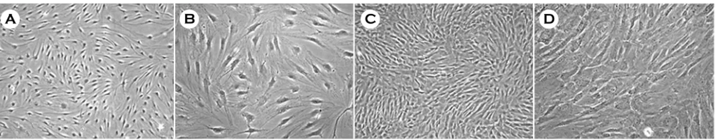
HAD와 HUC은 골수유래 중간엽 줄기세포와 유 사한 섬유아세포의 형태를 나타냈다 (Figure 1). 계 대배양 동안 HAD와 HUC의 분열 속도를 측정한 결과 HAD는 총 116일 (10 계대)의 배양기간 동안
평균 3.9일의 분열 속도로 21번 분열하였고, HUC 은 총 73일 (9 계대)의 배양기간 동안 평균 2.8일의 분열 속도로 19번 분열하였다.
2) 유전자 발현 분석
HAD와 HUC의 줄기세포 관련 유전자 분석 결과 배아줄기세포와 배아종양세포에서 발현하는 유전 자인 Oct-4, 배아줄기세포, 배아종양세포, 조혈모세 포, 중간엽 줄기세포에서 발현하는 유전자인 SCF (stem cell factor), 외배엽 세포에서 발현하는 유전자 인 neural cell adhesion molecule (NCAM)과 fibroblast growth factor-5 (FGF-5), 중배엽 세포에서 발현하는 유전자인 bone morphogenic protein-4 (BMP4), 내배 엽 세포에서 발현하는 유전자인 GATA-4, vimentin, 조직적합성 복합체 유전자인 HLA-ABC와 HLA-DR 을 두 가지 줄기세포에서 모두 발현하였다. 이에 반해 배아줄기세포, 배아종양세포에서 발현하는 유 전자인 Rex-1과 내배엽 세포에서 발현하는 유전자 alpha feto protein (αFP)은 HUC에서 발현하지 않았 고 중배엽 세포에서 발현하는 유전자 Brachyury와 중배엽 및 내배엽 세포에서 발현하는 BMP-2는 두 줄기세포에서 모두 발현하지 않았다 (Table 2).
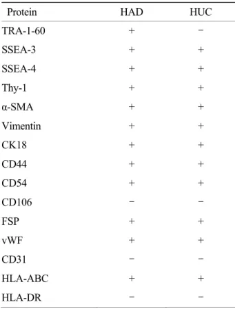
3) 면역세포화학적 특성 분석
HAD와 HUC을 면역세포화학 분석법을 통하여 분석한 결과 두 가지 유사중간엽 줄기세포 모두 발 현하는 단백질은 배아줄기세포의 표지 물질로 알려 진 SSEA-3, SSEA-4, 중간엽 줄기세포 표지 물질인 Thy-1, 세포골격 물질인 vimentin, α-smooth muscle actin (α-SMA), cytokeratin18 (CK18), 세포연접 물질 인 homing cell adhesion molecule (HCAM, CD44),
A B C D
Figure 1. Phase-contrast images of HAD and HUC. A, B, HAD at 3rd passage; C, D, HUC at 4th passage. A, C:
magnification, ×40. B, D: magnification, ×100.
intracellular cell adhesion molecule (ICAM, CD54) 그 리고 조직적합성 복합체 항원인 HLA-ABC로 나타 났다. 이에 반해 배아줄기세포 표지 물질인 TRA- 1-60은 HUC에서 발현하지 않았고, vascular cell adhesion molecule (VCAM, CD106), 내피세포 표지 물질인 platelet endothelial cell adhesion molecule (PECAM, CD31)과 조직적합성 복합체 항원인 HLA-DR은 두 줄기세포 모두에서 발현하지 않았다 (Table 3).
2. HAD와 HUC의 심근세포 특이 유전자 발현 분석
HAD와 HUC의 심근세포 특이 유전자의 발현을 조사하였다 (Figure 2). Cardiac progenitor cell marker 유전자인 myocyte enhancer factor-2C (MEF-2C), Islet1 (Isl-1) 그리고 NK2 transcription factor related, locus5 (Nkx2.5) 유전자 발현을 조사한 결과, MEF-2C는 HAD와 HUC에서 모두 발현하였고, Isl-1은 HUC에 서만 발현하였다. 그러나 Nkx2.5는 HAD와 HUC에
서 모두 발현하지 않았다. Thick filament component 유전자인 cardiac ventricular myosin light chain-1 (Cmlc-1), atrium myosin light chain-2a (MLC-2a), α- myosin heavy chain (α-MHC), β-myosin heavy chain (β-MHC) 그리고 cardiac ventricular myosin light chain- 2 (Cvmlc-2)를 조사한 결과, Cmlc-1은 HAD와 HUC 에서 모두 발현하였으나, HAD에서 보다 강한 발현 양상을 보였으며 MLC-2a는 HUC에서만 발현하였 다. 한편 α-MHC, β-MHC 그리고 Cvmlc-2는 HAD 와 HUC에서 모두에서 발현하지 않았다. Thin fila- ment component 유전자인 α-cardiac actin (α-CA), α- skeletal actin (α-SA), troponin T (TnT), troponin I (TnI) 는 HAD와 HUC에서 모두 발현되었다. Potassium channel 유전자인 Kv4.3는 HAD와 HUC에서 모두 발현하였으나, HUC에서 보다 강하게 발현되었고 calcium channel 유전자인 α1C는 HUC에서만 발현 하였다. 한편, natriuretic peptide인 atrial natriuretic Table 2. RT-PCR analysis of stem cell marker genes
expressed in HAD and HUC
Gene HAD HUC
Oct-4 + +
Rex-1 + -
SCF + +
NCAM + +
FGF-5 + +
BMP-4 + +
Brachyury - -
HNF-4α + +
GATA-4 + +
αFP + -
BMP-2 - -
HLA-ABC + +
HLA-DR + +
Table 3. Immunocytochemical analysis of proteins expressed in HAD and HUC
Protein HAD HUC
TRA-1-60 + -
SSEA-3 + +
SSEA-4 + +
Thy-1 + +
α-SMA + +
Vimentin + +
CK18 + +
CD44 + +
CD54 + +
CD106 - -
FSP + +
vWF + +
CD31 - -
HLA-ABC + +
HLA-DR - -
peptide (ANP)와 brain natriuretic peptide (BNP) 유 전자는 HAD와 HUC 모두에서 발현되지 않았다 (Figure 2).
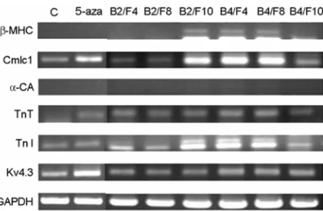
3. HAD의 심근세포로의 분화
HAD는 5-azacytidine을 24시간 처리한 후 4주 동 안 기본배양액으로 배양한 세포 (5-aza)는 5-azacy- tidine을 처리하지 않고 배양한 세포 (대조군) 보다 TnT 유전자 발현이 새롭게 유도되었으며 Cmlc1과 Kv4.3 유전자 발현이 증가하였다 (Figure 3).
5-azacytidine을 24시간 처리한 후 BMP-2에 FGF- 4 (B2/F4) 또는 FGF-8 (B2/F8)을 첨가하여 배양한 세포는 5-aza 보다 TnI의 발현이 증가하였으며, BMP-2와 FGF-10 (B2/F10)을 첨가하여 배양한 세 포는 β-MHC 유전자 발현이 새롭게 유도되었으며, Cmlc1, TnI의 발현이 많이 증가되었다. BMP-4에 FGF-4 (B4/F4) 또는 FGF-8 (B4/F8)을 첨가하여 4주 간 배양한 세포는 β-MHC 유전자 발현이 새롭게 유도되었으며, Cmlc1, TnT, TnI 그리고 Kv4.3 유전자 발현이 증가되었다. BMP-4와 FGF-10 (B4/F10)로 배
양한 세포는 TnI와 Kv4.3의 발현은 더 증가하였지 만 Cmlc1의 발현은 감소하였다 (Figure 3).
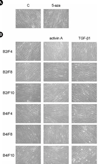
4. HUC의 심근세포로의 분화 1) 형태학적 분석
HUC은 5-azacytidine을 24시간 처리한 후 4주간 배양하였을 때 대조군과 비교하여 형태적 변화는 관찰되지 않았다 (Figure 4A). 또한 5-azacytidine 처 리 후 여러 가지 BMP와 FGF를 첨가하여 배양하 였을 때와 여러 가지 BMP와 FGF 조합과 함께 activin A를 첨가하여 배양하였을 때도 대조군과 비교하여 형태적 변화가 관찰되지 않았다. 그러나 5-azacytidine 처리 후 여러 가지 BMP와 FGF 조합 과 함께 TGF-β1를 첨가하여 배양하였을 때는 세 포질이 다소 넓게 퍼지는 형태적 변화를 나타냈다 (Figure 4B).
2) BMP와 FGF을 처리한 분화 유도
HUC은 5-azacytidine을 24시간 처리한 후 4주간 배양하였을 때 대조군보다 Cmlc1 유전자 발현이 증가였으나 α-CA와 kv4.3의 발현이 감소하였다 (Fi- gure 5).
5-azacytidine 처리 후 BMP-2에 FGF-4 (B2/F4) 또 는 FGF-10 (B2/F10)을 첨가하여 배양한 세포는 대 조군보다 Cmlc1, α-CA, TnT의 발현이 다소 증가하 는 것으로 관찰되었으나 Kv4.3은 유사한 수준의 발 현양상을 나타냈다. BMP-4와 FGF-4 (B4/F4)를 첨가 Figure 3. Gene expression profiles in HAD at 4 weeks after cardiac differentiation. Cells were differentiated with various combinations of BMPs and FGFs.
Figure 2. Expression profiles of cardiomyocyte-specific gene in HAD (3rd passage) and HUC (2nd passage).
하여 배양한 세포는 Cmlc1, α-CA, TnT의 발현이 증 가하였으나 Kv4.3의 발현이 감소하였다. BMP-2와 FGF-8 (B2/F8)을 첨가하여 배양한 세포는 Kv4.3 유 전자만이 발현하였으며, BMP-4와 FGF-8 (B4/F8)을 첨가하여 배양한 세포는 모든 유전자를 발현하지 않았다. BMP-4와 FGF-10 (B4/F10)로 배양한 세포는 낮은 수준의 Kv4.3 발현양상만을 나타냈다 (Figure 5A).
3) BMP와 FGF 조합과 함께 activin A 또는 TGF-β1 처리한 분화 유도
HUC을 BMP와 FGF 조합과 함께 activin A를 첨 가하여 4주 동안 배양하였을 때 BMP-2에 FGF-4 B
A
Figure 4. Phase-contrast images in HUC at 4 weeks after cardiac differentiation. A, Cells were cultured after exposure for 24 hr in serum free DMEM (C, control) or serum free 5-azacytidine (5-aza, 5-azacytidine). B, Cells were differentiated with various combination of BMPs and FGFs without additional factors or in the presence either of activin A or TGF-β1.
B
C A
Figure 5. Gene expression profiles in HUC at 4 weeks after cardiac differentiation. Cells were differentiated with various combination of BMPs and FGFs without addi- tional factors (A) or in the presence either of activin A (B) or TGF-β1 (C).
(B2/F4) 또는 FGF-8 (B2/F8)을 첨가하였을 때와 BMP-4와 FGF-8 (B4/F8)을 첨가된 조합에서 가장 많은 유전자의 발현증가가 있었다. 5-aza 보다 β- MHC 발현이 새로이 유도되었으며, α-CA와 Kv4.3 유전자의 발현이 증가되었으나 Cmlc1, TnT 그리고 TnI의 발현증가가 보이지 않는 것으로 관찰되었다 (Figure 5B). BMP와 FGF 조합과 함께 TGF-β1을 첨 가하여 4주 동안 배양하였을 때는 BMP-2와 FGF-8 (B2/F8) 그리고 BMP-2와 FGF-10 (B2/F10)을 첨가 하였을 때 5-aza 보다 β-MHC 발현이 새로이 유도 되었으며 Cmlc1, α-CA, 그리고 Kv4.3의 발현이 증 가되었다 (Figure 5C).
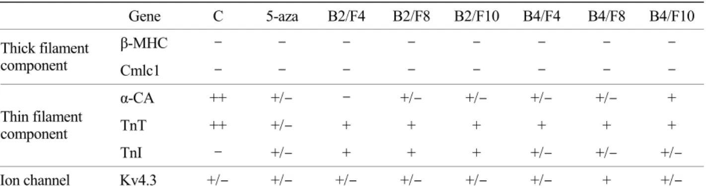
4) BMP와 FGF 조합과 함께 Wnt inhibitor 처 리한 분화 유도
HUC을 BMP와 FGF 조합과 함께 Wnt inhibitor
를 처리하여 1, 4주간 배양한 세포의 심근세포 관련 유전자의 발현을 조사하였다. 5-azacytidine을 처리한 후 1주 동안 분화시켰을 때 BMP-2가 처리된 그룹 에서는 FGF의 모든 종류에서 5-aza 보다 Cmlc1 발 현이 새로이 유도되었다. TnT와 TnI 그리고 Kv4.3 유전자 발현이 증가된 반면 α-CA는 약간 감소하였 다. BMP-4가 처리된 그룹 또한 FGF의 모든 종류 에서 5-aza 보다 Cmlc1 유전자 발현이 새로이 유도 되었고 TnI와 Kv4.3 유전자 발현이 증가하였으나 α-CA와 TnT 유전자 발현은 차이가 없었다. 이 중 BMP-4와 FGF-10 (B4/F10)이 처리된 세포에서 가장 많은 유전자의 발현증가가 나타났다 (Table 4). 그러 나 4주 동안 배양하였을 때 모든 실험군에서 모든 유전자의 발현이 1주째 보다 감소되었다 (Table 5).
Table 4. Gene expression profiles in HUC after differentiation induction with BMP, FGF and Wnt inhibitor for 1 week Gene C 5-aza B2/F4 B2/F8 B2/F10 B4/F4 B4/F8 B4/F10
β-MHC - - - -
Thick filament
component Cmlc1 - - + +/- +/- +/- +/- ++
α-CA ++ +++ ++ ++ ++ +++ +++ +++
TnT ++ ++ +++ +++ +++ ++ ++ ++
Thin filament component
TnI - +/- ++ ++ ++ + + ++
Ion channel Kv4.3 +/- +/- + ++ ++ +++ +++ +++
C, control (cultivated in complete medium for 1 week after exposure in DMEM-LG for 24 h); 5-aza, 5-azacytidine;
B, BMP; F, FGF
Table 5. Gene expression profiles in HUC after differentiation induction with BMP, FGF and Wnt inhibitor for 4 weeks Gene C 5-aza B2/F4 B2/F8 B2/F10 B4/F4 B4/F8 B4/F10
β-MHC - - - -
Thick filament
component Cmlc1 - - - -
α-CA ++ +/- - +/- +/- +/- +/- +
TnT ++ +/- + + + + + +
Thin filament component
TnI - +/- + + + +/- +/- +/-
Ion channel Kv4.3 +/- +/- +/- +/- +/- +/- + +/- C,control (cultivated in complete medium for 4 weeks after exposure in DMEM-LG for 24 h); 5-aza, 5-azacytidine;
B, BMP; F, FGF
고
찰본 연구에서는 HAD와 HUC에 5-azacytidine을 24시간 처리하고, BMP, FGF, activin A, TGF-β1, Wnt inhibitor를 처리한 후 심근특이 유전자의 발현이 증 가하는 것을 관찰하였다. 쥐의 배아줄기세포는 1 µM 농도의 5-azacytidine을 6시간 처리하면 3일 후 에 역분화된 것이 관찰된 바 있으며,24 사람의 배 아줄기세포는 1 µM 및 10 µM 농도의 5-azacytidine 을 6~8일간 처리하고 15일 동안 배양하였을 때 심 근특이 유전자의 발현이 증가한다고 보고되었다.17 골수유래 중간엽 줄기세포와 탯줄 정맥유래 줄기 세포의 경우 각각 6 µM과 10 µM의 5-azacytidine을 24시간 처리하면 4주 후에 심근세포로 분화한다고 보고되었다.19,20 Demethylation agent인 5-azaycytidine 이 어떤 mechanism에 의하여 심근세포로 분화 유도 하는지 아직 밝혀지지 않았으나 최근 5-azaycytidine 처리 후 돼지의 골수유래 줄기세포 단백질을 분석 한 결과 총 1,700개의 단백질 중 11개의 단백질이 증가하고 26개의 단백질이 감소하였는데 이 중 세포의 증식과 분화에 관련된 alpha B-crystallin, annexin A2 그리고 stathmin 1 단백질이 분석되어졌 다.25 이로 미루어보아 5-azaycytidine 처리를 통한 분화는 5-azaycytidine에 의해 세포가 역분화된 후 배양기간 동안 심근세포로의 특이 분화 과정을 진 행되는 것으로 생각할 수 있다.
사람의 배아줄기세포는 5-azacytidine 대신 activin A를 24시간 처리 후 4일 동안 10 ng/ml의 BMP-4 를 처리하였을 때도 심근세포로 분화 유도되었고,18 쥐의 배아줄기세포는 visceral endoderm like cell line (END-2)과 함께 BMP-2를 첨가하여 배양하면 bea- ting하는 세포가 증가하였다고 보고되었다.26 Zebra- fish의 배아는 in vitro 배양 시 BMP-2를 첨가하였 을 때 cardiac contractility가 일어났다.27 조류의 pre- cardiac mesoderm에 인접한 FGF-8을 발현하는 내배 엽을 제거하게 되면 Nkx2.5와 MEF2C를 포함하는 심근특이 유전자들의 발현이 감소하고 FGF-8을 주
입하면 다시 증가하는데 이 때 BMP가 발현되는 곳에서만 유전자의 발현이 증가되는 것으로 보아 early cardiogenesis 과정은 FGF와 BMP 모두를 필 요로 한다고 제안된 바 있다.30,31 또한 FGF-8과 Mesp1 유전자는 쥐의 낭배형성 중 중배엽에서 발 현되었는데 knockout mouse를 이용한 연구 결과 이들 유전자는 심장전구세포의 early specification에 중요한 역할을 하는 것으로 알려져 있다.32 조류의 non-precardiac mesoderm의 cardiogenic induction을 유 도하는 BMP-2/4과 FGF-2/4의 대체물질 연구 결과 activin-A, insulin 그리고 leukocyte inhibitory factor는 BMP-2/4를 대체하지 못했으며 FGF-7, activin A 그 리고 insulin은 FGF-2/4를 대체하지 못했다.39 이러 한 점으로 미루어 심근분화를 유도할 때 BMP와 FGF는 필수적인 것으로 여겨진다.
한편 쥐의 배아종양세포인 P19 세포와 배아줄 기세포는 noncanonical Wnt인 Wnt11 conditioned medium으로 배양하였을 때 심근세포로 분화하였고,
36,37 사람의 endothelial progenitor cells (EPCs)은 neonatal 심근세포와 공배양하여 분화 유도되면 noncanonical Wnt인 Wnt5a, Wnt11가 증가되었다.38 따라서 canonical signal은 억제하고 noncanonical signal은 활성화시킴으로써 심근세포로 분화를 조 절할 수 있을 것으로 보인다.
본 연구에서 HAD는 5-azacytidine을 처리하고 여 러 가지 BMP와 FGF의 조합으로 4주간 배양하였 을 때 BMP-2/FGF-10 (B2/F10), BMP-4/FGF-4 (B4/F4), BMP-4/FGF-8 (B4/F8) 조건에서 심근특이 유전자의 발현이 두드러지게 높아졌다. 그러나 HUC은 5- azacytidine 처리 및 BMP와 FGF에 의한 효과가 없 었으나 BMP와 FGF와 함께 activin A 또는 TGF-β1 를 첨가하여 배양하였을 때, activin A와 TGF-β1 처 리 조건 모두에서 BMP-2와 FGF-8 (B2/F8)이 함께 첨가된 세포의 심근특이 유전자 발현이 가장 많이 증가하였다. 따라서 HUC의 심근 분화를 위해서는 BMP와 FGF와 함께 activin A 또는 TGF-β1의 signal 이 필요한 것으로 관찰되었다. 그러나 Wnt inhibitor 를 처리하여 1주간 배양하였을 때 activin A 또는
TGF-β1을 처리한 것보다 심근특이 유전자의 발현 양이 두드러지게 증가한 것으로 보아 activin A 또 는 TGF-β1보다 Wnt inhibitor의 효과가 더 좋은 것 으로 관찰되었다. 그러나 Wnt inhibitor 처리 후 4주 간 배양하였을 때는 1주 때 증가했던 유전자의 발 현들이 모두 감소된 것으로 보아 1주차에 분화 유 도 되었다가 4주차로 진행되면서 역분화 되었거나 또는 다른 세포로의 분화 가능성이 있는 것으로 보 인다. 따라서 다른 조건 실험에서도 분화 시간을 알맞게 조절하는 것이 필요할 것으로 사료된다.
본 연구 결과 사람의 지방과 제대유래 줄기세포 는 5-azaycytidine 및 BMP, FGF, activin A, TGF-β1, Wnt inhibitor를 사용하여 체외에서 심장근육세포로 분화가 가능한 것으로 관찰되었다. 앞으로 더 많은 연구가 필요하겠지만 두 종류의 줄기세포는 심장 질환 치료를 위한 세포치료법에 이용될 수 있을 것으로 사료된다.
참
고 문 헌1. Soonpaa MH, Field LJ. Assessment of cardiomyocyte DNA synthesis in normal and injured adult mouse hearts. Am J Physiol 1997; 272: 220-6.
2. Pasumarthi KB, Field LJ. Cardiomyocyte cell cycle regulation.
Circ Res. 2002 May 31; 90: 1044-54.
3. Rubart M, Field LJ. Cardiac regeneration: repopulating the heart. Annu Rev Physiol 2006; 68: 29-49.
4. Zimmermann WH, Eschenhagen T. Embryonic stem cells for cardiac muscle engineering. Trends Cardiovasc Med 2007;
17: 134-40.
5. Dimmeler S, Zeiher AM, Schneider MD. Unchain my heart:
the scientific foundations of cardiac repair. J Clin Invest 2005;
115: 572-83.
6. Murry CE, Field LJ, Menasché P. Cell-based cardiac repair:
reflections at the 10-year point. Circulation 2005; 15: 3174-83.
7. Friedenstein AJ, Piatetzky-Shapiro II, Petrakova KV. Osteo- genesis in transplants of bone marrow cells. J Embryol Exp Morphol 1966; 16: 381-90.
8. Prindull G, Prindull B, Meulen N. Haematopoietic stem cells (CFUc) in human cord blood 1978; 67: 413-6.
9. Zvaifler NJ, Marinova-Mutafchieva L, Adams G, Edwards CJ, Moss J, Burger JA, et al. Mesecnchymal precursor cells in the blood of normal individuals. Arthritis Res 2000; 2: 477-88.
10. Zuk PA, ZhuM, Ashjian P, De Ugarte DA, Huang JI, Mizuno H, et al. Human adipose tissue is a source of multipotent stem cells. Mol Biol Cell 2002; 13: 4279-95.
11. Toda A, Okabe M, Yoshida T, Nikaido T. The potential of amniotic membrane/amnion-derived cells for regeneration of various tissues. J Pharmacol Sci 2007; 105: 215-28.
12. In 't Anker PS, Scherjon SA, Kleijburg-van der Keur C, Noort WA, Claas FH, Willemze R, et al. Amniotic fluid as a novel source of mesenchymal stem cells for therapeutic transplan- tation. Blood 2003; 102: 1548-9.
13. Romanov YA, Svintsitskaya VA, Smirnov VN. Searching for alternative sources of postnatal human mesenchymal stem cells: candidate MSC-like cells from umbilical cord. Stem Cells 2003; 21: 105-10.
14. Fukuchi Y, Nakajima H, Sugiyama D, Hirose I, Kitamura T, Tsuji K. Human placenta-derived cells have mesenchymal stem/progenitor cell potential. Stem Cells 2004; 225: 649-58.
15. He JQ, Ma Y, Lee Y, Thomson JA, Kamp TJ. Human embryonic stem cells develop into multiple types of cardiac myocytes:
action potential characterization. Circ Res 2003; 93: 32-9.
16. Leor J, Gerecht S, Cohen S, Miller L, Holbova R, Ziskind A, et al. Human embryonic stem cell transplantation to repair the infarcted myocardium. Heart 2007; 93: 1278-84.
17. Xu C, Police S, Rao N, Carpenter MK. Characterization and enrichment of cardiomyocytes derived from human embryonic stem cells. Circ Res 2002; 9: 501-8.
18. Laflamme MA, Chen KY, Naumova AV, Muskheli V, Fugate JA, Dupras SK, et al. Cardiomyocytes derived from human embryonic stem cells in pro-survival factors enhance function of infarcted rat hearts. Nat Biotechnol 2007; 25: 1015-24.
19. Xu W, Zhang X, Qian H. Mesenchymal stem cells from adult human bone marrow differentiate into a cardiomyocyte pheno- type in vitro. Exp Biol Med 2004; 229: 623-31.
20. Kadivar M, Khatami S, Mortazavi Y, Shokrgozar MA, Taghi- khani M, Soleimani M. In vitro cardiomyogenic potential of human umbilical vein-derived mesenchymal stem cells. Bio- chem Biophys Res Commun 2006; 340: 639-47.
21. Balana B, Nicoletti C, Zahanich I, Graf EM, Christ T, Box- berger S, et al. 5-Azacytidine induces changes in electro- physiological properties of human mesenchymal stem cells.
Cell Res 2006; 16: 949-60.
22. Zhao P, Ise H, Hongo M, Ota M, Konishi I, Nikaido T. Human amniotic mesenchymal cells have some characteristics of cardiomyocytes. Transplantation 2005; 79: 528-35.
23. Shim WS, Jiang S, Wong P, Tan J, Chua YL, Tan YS, et al. Ex vivo differentiation of human adult bone marrow stem cells into cardiomyocyte-like cells. Biochem Biophys Res Commun 2004; 324: 481-8.
24. Andrée B, Duprez D, Vorbusch B, Arnold HH, Brand T.
BMP-2 induces ectopic expression of cardiac lineage markers and interferes with somite formation in chicken embryos.
Mech Dev 1998; 70: 119-31.
25. Ladd AN, Yatskievych TA, Antin PB. Regulation of avian cardiac myogenesis by activin/TGFbeta and bone morpho- genetic proteins. Dev Biol 1998; 204: 407-19.
26. He X. A Wnt-Wnt situation. Dev Cell 2003; 4: 791-7.
27. Dale TC. Signal transduction by the Wnt family of ligands.
Biochem J 1998; 329: 209-23.
28. Solloway MJ, Harvey RP. Molecular pathways in myocardial development: a stem cell perspective. Cardiovasc Res 2003;
58: 264-77.
29. Tsuji-Takayama K, Inoue T, Ijiri Y, Otani T, Motoda R, Naka- mura S, et al. Demethylating agent, 5-azacytidine, reverses differentiation of embryonic stem cells. Biochem Biophys Res Commun 2004; 323: 86-90.
30. Ye NS, Zhang RL, Zhao YF, Feng X, Wang YM, Luo GA.
Effect of 5-azacytidine on the protein expression of porcine bone marrow mesenchymal stem cells in vitro. Genomics Proteomics Bioinformatics. 2006; 4: 18-25.
31. Bin Z, Sheng LG, Gang ZC, Hong J, Jun C, Bo Y, et al.
Efficient cardiomyocyte differentiation of embryonic stem cells by bone morphogenetic protein-2 combined with visceral endoderm-like cells. Cell Biol Int 2006; 30: 769-76.
32. Wang YX, Qian LX, Liu D, Yao LL, Jiang Q, Yu Z, et al. Bone morphogenetic protein-2 acts upstream of myocyte-specific enhancer factor 2a to control embryonic cardiac contractility.
Cardiovasc Res 2007; 74: 290-303.
33. Barron M, Gao M, Lough J. Requirement for BMP and FGF signaling during cardiogenic induction in non-precardiac mesoderm is specific, transient, and cooperative. Dev Dyn 2000; 218: 383-93.
34. Alsan BH, Schultheiss TM. Regulation of avian cardiogenesis by FGF8 signaling. Development 2002; 129: 1935-43.
35. Saga Y, Kitajima S, Miyagawa-Tomita S. Mesp1 expression is the earliest sign of cardiovascular development. Trends Car- diovasc Med. 2000; 10: 345-52.
36. Ilagan R, Abu-Issa R, Brown D, Yang YP, Jiao K, Schwartz RJ, et al. Fgf8 is required for anterior heart field development.
Development. 2006; 133: 2435-45.
37. Koyanagi M, Bushoven P, Iwasaki M, Urbich C, Zeiher AM, Dimmeler S. Notch signaling contributes to the expression of cardiac markers in human circulating progenitor cells. Circ Res 2007; 101: 1139-45.
38. Terami H, Hidaka K, Katsumata T, Iio A, Morisaki T. Wnt11 facilitates embryonic stem cell differentiation to Nkx2.5- positive cardiomyocytes. Biochem Biophys Res Commun.
2004; 325: 968-75.
39. Pandur P, Läsche M, Eisenberg LM, Kühl M. Wnt-11 activa- tion of a non-canonical Wnt signalling pathway is required for cardiogenesis. Nature 2002; 418: 636-41.
= 국문초록 = 목 적: 사람의 HAD와 HUC를 심근세포로 분화 유도하고자 하였다.
연구방법: 사람의 HAD와 HUC를 분리하여 5-azacytidine을 24시간 처리하고 여러 가지 BMP와 FGF을 첨가하여 배양 하였다. 또한 HUC은 BMP와 FGF와 함께 activin A 또는 TGF-β1 또는 Wnt inhibitor를 첨가하여 배양한 후 심근세포 특이 유전자의 발현을 조사하였다.
결 과: HAD를 5-azacytidine 처리하고 기본배양액에서 4주 동안 배양하였을 때 TnT 유전자가 새로이 발현하였으 며 Cmlc1과 kv4.3의 발현 양이 증가하였다. 5-azacytidine 처리 후에 BMP-4와 함께 FGF-4 (B4/F4) 또는 FGF-8 (B4/F8) 을 첨가하여 배양하였을 때는 β-MHC 유전자 발현이 새로이 유도되었으며, Cmlc1, TnT, TnI 그리고 Kv4.3 유전자 발 현 양이 더 많이 증가하였다. HUC은 5-azacytidine 및 BMP와 FGF 처리에 의해 유전자 발현 변화가 없었다. 그러나 BMP와 FGF와 함께 activin A 또는 TGF-β1을 첨가하여 배양하였을 때, BMP-2와 FGF-8 (B2/F8)을 첨가하여 배양한 세 포에서 β-MHC 발현이 새로이 유도되었으며 α-CA, TnT 그리고 Kv4.3 유전자의 발현이 증가하였다. 또한 BMP와 FGF 와 함께 Wnt inhibitor를 처리하여 1주 동안 배양하였을 때 Cmlc1 유전자 발현이 새로이 유도되었으며 α-CA, TnT, TnI 그리고 Kv4.3의 발현이 증가되었다.
결 론: HAD는 BMP와 FGF 처리에 의해 심근세포 특이 유전자의 발현증가를 유도할 수 있었으며 HUC는 BMP와 FGF와 함께 activin A 또는 TGF-β1 또는 Wnt inhibitor를 처리함으로써 심근세포 특이 유전자의 발현증가를 유도할 수 있었다. 따라서 HAD와 HUC는 심장질환 치료를 목적으로 하는 세포 치료에 이용될 수 있을 것으로 사료된다.
중심단어: HAD, HUC, 심근세포, 분화 유도, 유전자 발현