• 검색 결과가 없습니다.

Studies on Conditioned Media in Human Cells: Evaluation Using Various Cell and Culture Conditions, Animal Disease Models

N/A
N/A
Protected

Academic year: 2021

Share "Studies on Conditioned Media in Human Cells: Evaluation Using Various Cell and Culture Conditions, Animal Disease Models"

Copied!
8
0
0

로드 중.... (전체 텍스트 보기)

전체 글

(1)

INTRODUCTION

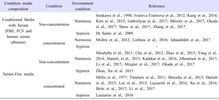
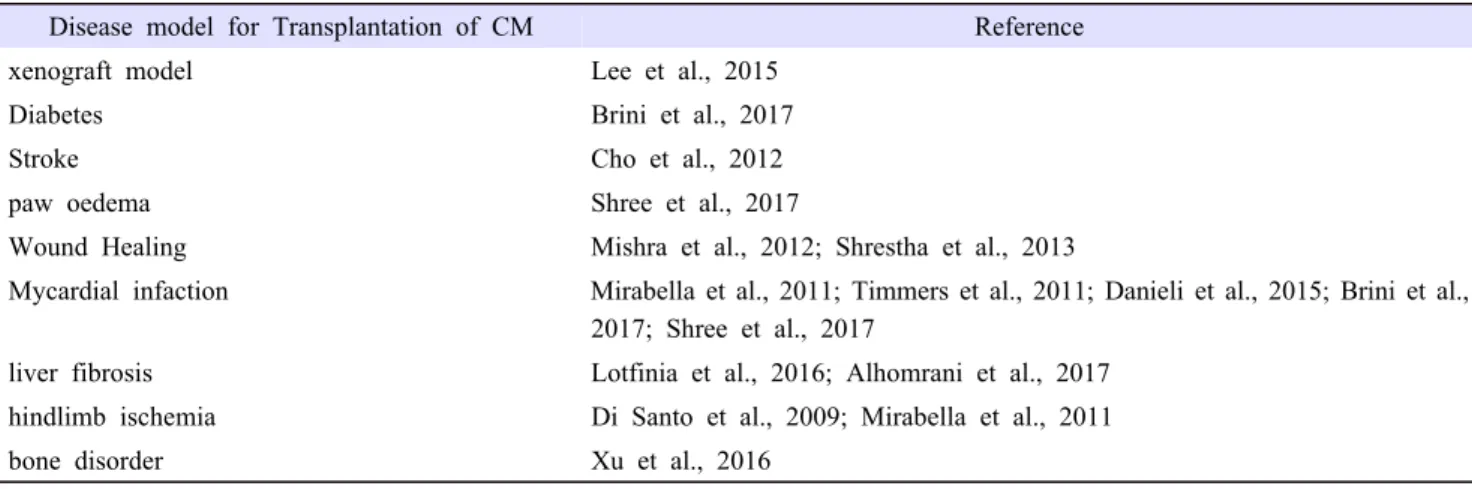
Cell therapy seem ideal because cells are the basic components of tissue. If healthy cells were transplanted in damage area, transplanted cells could replace the damaged part and release factors for treatment. Moreover, since the discovery of stem cell, people have studied how to use stem cell for human diseases. Stem cells have two characteristics: self-renewal and differentiation potency. Theoretically, stem cells could stay in the injury area, release many beneficial factors, differentiate into mature cells, and replace damaged tissue. However, recent studies have reported that transplanted stem cells cannot survive for long in a damaged area, and can be removed through the circulatory system. In addition, there are not many types of stem cells that can be used for cell therapy because most stem cells have the capacity of tumorigenicity (Lin et al., 2013). To utilize the therapeutic advantages of stem cell, some studies use materials derived from stem cell culture because in vitro, stem cells release many beneficial factors that play a role similar to the biological processes in vivo. Conditioned media (CM), supernatant from cell has these proteins and

Studies on Conditioned Media in Human Cells:

Evaluation Using Various Cell and Culture Conditions, Animal Disease Models

Keun Cheon Kim

1,2

and Eun Ju Lee

3,†

1

Department of Agricultural Biotechnology, Seoul National University, Seoul 151-921, Korea

2

Research Institutes of Agriculture and Life Sciences, Seoul National University, Seoul 151-921, Korea

3

Biomedical Research Institute, Seoul National University Hospital, Seoul 03080, Korea

Abstract

In the last several decades, cell therapy research has increased worldwide. Many studies have been conducted on cell therapy, and have revealed that transplanted cells did not survive for long, and implanted cells remained inactive causing immune rejection depending on the patient’s condition. Therefore, studies on cell-free therapy need to be conducted. To overcome these limitations, an alternative is the use of supernatant from cells, called “conditioned media (CM).” During in vitro cell culture, culture media supply nutrients to maintain cell characteristics and viability. In the culture, cells not only consume nutrients but also release beneficial proteins and substances, which are called “secretome.” CM from cells can be stored for a long time and is easy to handle. Moreover, secretome in CM can also be measured;

exact amount of secretome is important to set the standard value for disease treatment. Here, we reviewed studies on CM and confirmed that various secretomes from CM were identified in these studies. Moreover, these findings could benefit cell and animal studies in future. In conclusion, CM could be a potential candidate for an alternative to cell therapy.

Received :22 December 2017 Revised :05 January 2018

Accepted:13 March 2018 Key Words : Conditioned media(CM), Cell-free therapy, Stem cell, Secretome

(2)

substances, called secretome. CM from cell is easy to handle and can be stored for a long time because the cell supernatant does not contain live cells. CM can be modified as a ready-to-use powder (Alhomrani et al., 2017). Many studies identified secretome in CM and compared implanted cell with CM treatment (Di Santo et al., 2009; Mirabella et al., 2011; Cho et al., 2012; Mishra et al., 2012; Zhao et al., 2013; Zhou et al., 2013; Kang et al., 2014; Danieli, et al., 2015; Lotfinia et al., 2016; Gabrielyan et al., 2017; Jahandideh et al., 2017; Mizgier et al., 2017; Okada et al., 2017; Shree et al., 2017). Furthermore, they also established manufacturing method of CM and proven potential therapeutic effect of CM in vivo and in vitro (Di Santo et al., 2009; Mirabella et al., 2011; Timmers et al., 2011; Cho et al., 2012; Mishra et al., 2012; Zhou et al., 2013; Danieli et al., 2015; Lee et al., 2015;

Lotfinia et al., 2016; Xu et al., 2016; Alhomrani et al., 2017; Brini et al., 2017; Shree et al., 2017).

MATERIALS and METHODS

We used the National Center for Biotechnology Information (NCBI) database to find related articles about CM and secretome.

‘Conditioned medium’, ‘cell’, ‘Stem cell’, ‘therapy’ and ‘secretome’ were used as keywords. These articles were reviewed based on source of cell, culture, usage condition of CM, composition and analysis of CM.

Cell source

Many studies on conditioned-media from human tissue-derived stem cell have been conducted since 2000, the use of condition-media to human cell is still done using fibroblast (Millis et al., 1977; Imokawa et al., 1998; Danieli et al., 2015;

Gabrielyan et al., 2017; Metzler et al., 2017). Fibroblast is a type of cell from connective tissue, located in various organs such as skin, lung, and bladder. Moreover, fibroblast can synthesize and secrete many materials such as extracellular matrix and collagen. CM from fibroblast was used mainly in the cosmetic industry.

The discovery of stem cells has increased the applicability of cell therapy. Stem cells are undifferentiated cells that can differentiate into any type of mature cell. Mesenchymal Stem Cell (MSC) is a type of stem cells that can differentiate into mesodermal cell types (adipose, muscle, bone, tendon, and cartilage). MSCs have properties suitable for cell therapy applications.

MSCs have no immune reaction when transplanted, and can be easily obtained from various tissue. Because of these advantages, many studies with CM used MSCs isolated from adult tissues such as adipose (Cho et al., 2012; Ivanova-Todorova et al., 2012;

Zhao et al., 2013; Zhou et al., 2013; Yang et al., 2014; Lee et al., 2015; Brini et al., 2017; Li et al., 2017; Shree et al., 2017), bone marrow (Mishra et al., 2012; Gabrielyan, Neumann et al., 2017), umbilical blood (Lin et al., 2013; Shrestha et al., 2013;

Yang et al., 2014; Kim et al., 2015; Kadekar et al., 2016; Li et al., 2017), and amniotic fluid (Mirabella et al., 2011; Kang et al., 2014; Danieli et al. 2015; Lazzarini et al., 2016; Alhomrani et al., 2017) as basic material. Also some studied used CM from MSCs induced by embryonic stem cells (Timmers et al., 2011; Lotfinia et al,, 2016; Jahandideh et al., 2017). A few studies used CM from other mature cells, which were myotubes (Mizgier et al., 2017), endothelial progenitor cells (Di Santo et al., 2009), cancer cells and retinal pigment epithelial cells (Zhang et al., 2017) (Table 1).

Condition media composition

When cells were cultured in vitro, culture media supplies nutrients and chemicals to survive outside of an organ. FBS, fetal bovine serum, is an important supplement in culture media. All ingredients of FBS is not known yet. Nevertheless, most laboratory used FBS as main growth nutrient for in vitro cell culture. However, for cell therapy applications, using the cell cultured with FBS gives rise to an interspecies problem. In other words, it is difficult to predict the kind of reaction about FBS when cells enter the body since FBS originates from bovine. To overcome this issue, many companies produce Serum-Free supplement as an alternative to FBS, but it is not a perfect replacement of FBS yet (Tangjit et al., 2018; Cimino et al., 2017). Some studies used serum free media before making CM (Millis et al., 1977; Mirabella et al., 2011; Timmers et al., 2011; Cho et al., 2012;

Shrestha et al., 2013; Zhao et al., 2013; Zhou et al., 2013; Yang et al., 2014; Danieli et al., 2015; Lee et al., 2015; Kadekar

(3)

et al., 2016; Lazzarini et al., 2016; Xu et al., 2016; Alhomrani et al., 2017; Brini et al., 2017; Li et al., 2017; Li et al., 2017).

On the other hand, other studies still used culture media with FBS to make CM (Imokawa et al., 1998; Mirabella et al., 2011;

Ivanova-Todorova et al., 2012; Mishra et al., 2012; Kang et al., 2014; Kim et al., 2015; Gabrielyan et al, 2017; Okada et al., 2017; Shree et al., 2017; Zhang et al., 2017). Also, a few studies used low percentage of serum or serum albumin from human to supply least nutrients to cells (Di Santo et al., 2009; Lotfinia et al., 2016; Jahandideh et al., 2017; Metzler et al., 2017) (Table 2).

Table 1. Studies on cell source to make conditioned medium (CM).

Cell type Reference

Human Fibroblast Millis et al., 1977; Imokawa et al., 1998; Danieli et al., 2015; Gabrielyan et al., 2017;

Metzler et al., 2017 Human Adipose derived Mesenchymal

Stem Cell

Cho et al., 2012; Ivanova-Todorova et al., 2012; Zhao et al., 2013; Zhou et al., 2013;

Yang et al., 2014; Lee et al. 2015; Brini et al., 2017; Li et al., 2017; Shree et al., 2017 Human umbilical cord derived

Mesenchymal Stem Cell

Shrestha et al., 2013; Kim et al., 2015; Kadekar et al., 2016; Li et al., 2017

Human bone marrow derived Mesenchymal Stem Cell

Mishra et al., 2012

Human amniotic fluid derived stem cells Mirabella et al., 2011; Kang et al., 2014; Danieli et al., 2015; Lazzarini et al., 2016;

Alhomrani et al., 2017 Human Mesenchymal Stem Cell from

Embryonic stem cells

Timmers et al., 2011; Lotfinia et al., 2016; Jahandideh et al., 2017

Human myotubes Mizgier et al., 2017

Human Myeloid-derived suppressor cells Okada et al., 2017 Human retinal pigment epithelial cell Zhang et al., 2017 Human endothelial progenitor cells Di Santo et al., 2009

Culture period and environment

Other than the presence or absence of serum in the culture medium, incubation period and environment are also important for producing the conditioned medium. If cell culture period was long, cells might consume all nutrients in the medium and secrete harmful substances fatal for the cells (Braun et al., 2011). On the other hand, when culture period was short, cells could not secrete enough beneficial substances for acquisition of many proteins and other biochemical from the cell. Most studies synthesized CM with normal incubation period (Millis et al., 1977; Imokawa et al., 1998; Di Santo et al., 2009; Mirabella et al., 2011; Timmers et al., 2011; Cho et al., 2012; Ivanova-Todorova et al., 2012; Mishra et al., 2012; Lin et al., 2013; Shrestha et al., 2013; Zhao et al., 2013; Zhou et al., 2013; Yang et al., 2014; Danieli et al., 2015; Kim et al., 2015; Lee et al., 2015; Kadekar et al., 2016;

Lazzarini et al., 2016; Lotfinia et al., 2016; Xu et al., 2016; Alhomrani et al., 2017; Brini et al., 2017; Gabrielyan, Neumann et al., 2017; Jahandideh et al., 2017; Li et al., 2017; Li et al., 2017; Metzler et al., 2017; Mizgier et al., 2017; Okada et al., 2017; Shree et al., 2017; Zhang et al., 2017). One study was performed for longer than normal incubation period to obtain the conditioned medium (Kang et al., 2014).

The hypoxia condition is oxygen deficient condition and there have been more cell biology studies with hypoxia recently (Panchision, 2009; Anastasia et al., 2014; Pimton et al., 2015; Senavirathna et al., 2018). These studies showed that cell culture in hypoxia have more proliferation and differentiation ability with lower stress than that observed in cells in normoxia. Thus, hypoxia in cell culture could increase functional abilities. Some studies compared CM in normoxia and hypoxia (Table 2).

Concentration of selected angiogenic growth factors in endothelial progenitor cells (IL-8, SDF-1, HGF, Angiogenin, PDGF-BB and

VEGF-A) was compared between normoxia and hypoxia (Di Santo et al., 2009). In another study, human amniotic fluid-derived

stem cell conditioned medium (hAFS-CM) in normoxia and hypoxia is shown to have therapeutic effects on dox-induced

senescence and apoptosis in H9C2 cells (Lazzarini et al., 2016). These two studies showed that CM from hypoxia have more

(4)

cytokine and are better for anti-apoptosis and anti-senescence than CM from normoxia.

Usage condition for CM

Use of CM in cell biology should be considered carefully because CM had been already incubated in cell. Generally, culture media make cells healthy and maintain their biological function. Though CM can contain many proteins and growth factors from cells, nutrients for cell maintenance in culture media are mostly consumed. When cells were cultured, simply replacing basal medium to CM could cause a lack of essential elements for survival in cell culture. Some studies used CM concentrated by various methods because non-concentrated CM could have lower amounts of effective factors. Lyophilized or concentrated CM could be added to the culture media for better cell survival. This led to many studies to determine the optimal concentration and various method to use CM (Table 2).

Table 2. Studies on culture condition for making CM.

Condition media

composition Condition Environment

condition Reference

Conditioned Media with Serum (FBS, FCS and

human serum albumin)

Non-concentration Normoxia

Imokawa et al., 1998; Ivanova-Todorova et al., 2012; Kang et al., 2014;

Kim et al., 2015; Gabrielyan et al., 2017; Metzler et al., 2017; Okada et al., 2017; Shree et al., 2017; Zhang et al., 2017

hypoxia Di Santo et al., 2009

concentration Normoxia Mishra et al., 2012; Lotfinia et al., 2016; Jahandideh et al., 2017

hypoxia -

Serum-Free media

Non-concentration Normoxia

Mirabella et al., 2011; Cho et al., 2012; Zhao et al., 2013; Yang et al., 2014; Danieli, et al., 2015; Kadekar et al., 2016; Alhomrani et al., 2017;

Li et al., 2017; Mizgier et al., 2017; Okada et al., 2017 hypoxia Zhou, Xu et al. 2013

concentrated Normoxia

Millis et al., 1977; Timmers et al., 2011; Shrestha et al., 2013; Danieli et al., 2015; Lee et al., 2015; Lazzarini et al., 2016; Xu et al., 2016;

Brini et al., 2017; Li et al., 2017 hypoxia Lazzarini et al., 2016

Many studies on cell biology do not use CM directly as it is but use CM modified for cell culture. Nevertheless, some studies used non-concentrated CM for in vitro cell culture instead of basal medium (Di Santo et al., 2009; Mirabella et al., 2011; Timmers et al., 2011; Cho et al., 2012; Mishra et al., 2012; Danieli et al., 2015; Lee et al., 2015; Lotfinia et al., 2016; Xu et al., 2016;

Alhomrani et al., 2017; Brini et al., 2017; Shree et al., 2017). These types of CM were cultured in FBS or other serum before being used as CM, and it did not affect cell survive. To minimize nutrient loss for cell growth, CM was diluted with normal culture media or PBS ranged from 20% to 50% (Cho et al., 2012; Zhao et al., 2013; Kang et al., 2014; Danieli et al., 2015;

Metzler et al., 2017; Shree et al., 2017; Zhang et al., 2017). In other studies, concentrated CM was used with centrifugal filter and freeze-dryer used as the concentration method. Using centrifugal filter is one of the cost-effective methods to concentrate CM.

The use of centrifugal filter tube is simple with centrifuge machine. Centrifugal filters could eliminate low weight protein and unnecessary chemicals from CM and were easily available to buy (Chernokalskaya et al., 2004). Freeze dryer converts the liquid to a powder form and could handle large volumes of CM (Eiró et al., 2014). Powder form of CM could be stored long time and could be diluted to various concentrations. CM was concentrated 10 folds in many studies after which, concentrated CM was treated to cell and animal to each optimal concentration (Timmers et al., 2011; Mishra et al., 2012; Danieli et al., 2015; Lee et al., 2015; Lazzarini et al., 2016; Lotfinia et al., 2016; Xu et al., 2016; Brini et al., 2017; Jahandideh et al., 2017; Li et al., 2017).

Lyophilized CM was diluted with distill water or PBS and used (Millis et al., 1977).

Analysis of CM

(5)

Various proteins and growth factors in CM are present as a mixture and may have potential therapeutic effects in regenerative medicine. However, all components of CM are not known yet because identifying various cell types and culture conditions is difficult. ELISA (enzyme-linked immunosorbent assay), multiplex were performed to analyze components in CM. ELISA is a technique that can quantify substances such as proteins, hormones, and peptides. ELISA was employed in some studies for quantifying secretome components like HGF, FGF, and IGFBP1 in CM (Imokawa et al., 1998; Cho et al., 2012; Ivanova-Todorova et al., 2012; Mishra et al., 2012; Zhao et al., 2013; Zhou et al., 2013; Jahandideh et al., 2017; Metzler et al., 2017; Mizgier et al., 2017; Shree et al., 2017). Cytokine array is a membrane-based analysis technique. Various antibodies were located on array membrane, and secretome in CM could bind to suitable antibodies, and could be detected by immune reaction (Mirabella et al., 2011; Kang et al., 2014; Danieli et al., 2015; Lotfinia et al., 2016; Gabrielyan, et al., 2017). Multiplex is immunoassay based on multiple analysis profiling techniques to detect identification of a variety of secreted proteins by fluorescent microbeads (Di Santo et al., 2009; Mizgier et al., 2017; Okada et al., 2017) (Table 3).

Table 3. Analysis methods for secretome in CM.

Analysis of CM Reference

ELISA Imokawa et al., 1998; Di Santo et al., 2009; Cho et al., 2012; Ivanova-Todorova et al., 2012;

Mishra et al., 2012; Zhao et al., 2013; Zhou et al., 2013; Jahandideh et al., 2017; Mizgier et al., 2017;

Shree et al., 2017

Cytokine Antibody Array Mirabella et al., 2011; Kang et al., 2014; Danieli et al., 2015; Lotfinia et al., 2016; Gabrielyan et al., 2017

multiplex Di Santo et al., 2009; Mizgier et al., 2017; Okada et al., 2017

Transplantation of CM

In vitro, there were many difficulties to evaluate the therapeutic effect of the cell itself. Because cells have to be grown in suitable culture media, changes in composition of culture media can lead to unexpected results. For example, co-culture is a method where different cells are cultured in same plate at the same time and, which can improve therapeutic efficacy on target cells.

However, studies on co-culture cannot reliably determine efficacy of cells if the types of culture media were different. Since cells and conditioned medium are used as materials for animal studies, animal experiments do not have to consider about culture media in vitro. Some studies in animal disease model compared transplantation of cell with transplantation of CM (Mirabella et al., 2011;

Brini et al., 2017; Shree et al., 2017) (Table 4).

Table 4. Animal disease model for evaluating therapeutic efficiency of CM

Disease model for Transplantation of CM Reference

xenograft model Lee et al., 2015

Diabetes Brini et al., 2017

Stroke Cho et al., 2012

paw oedema Shree et al., 2017

Wound Healing Mishra et al., 2012; Shrestha et al., 2013

Mycardial infaction Mirabella et al., 2011; Timmers et al., 2011; Danieli et al., 2015; Brini et al., 2017; Shree et al., 2017

liver fibrosis Lotfinia et al., 2016; Alhomrani et al., 2017

hindlimb ischemia Di Santo et al., 2009; Mirabella et al., 2011

bone disorder Xu et al., 2016

(6)

Once positive effects on condition media were proven in in vitro, further studies like animal study or clinical trial would be required. Various studies used animal disease model such as mouse (Mirabella et al., 2011; Cho et al., 2012; Mishra et al., 2012;

Shrestha et al., 2013; Lee et al., 2015; Lotfinia et al., 2016; Alhomrani et al., 2017; Brini et al., 2017; Shree et al., 2017), rat (Danieli et al., 2015; Xu et al., 2016), or pig (Timmers et al., 2011). Most studies dealing with animal model, determined main factors of CM that positively affected the cells and then selected an animal disease model related to these factors. Xenograft (Lee et al., 2015), diabetes (Brini et al., 2017), stroke (Cho et al., 2012), paw edema (Shree et al., 2017), wound healing (Mishra et al., 2012; Shrestha et al., 2013), myocardial infarction (Mirabella et al., 2011; Timmers et al., 2011; Brini et al., 2017; Shree et al., 2017), liver fibrosis (Lotfinia et al., 2016; Alhomrani et al., 2017), hindlimb ischemia (Di Santo et al., 2009; Mirabella et al., 2011) and distraction osteogenesis (Xu et al., 2016) were used as animal disease models for evaluating therapeutic effects of CM.

CONCLUSION

CM from various types of cell were studied and established. Many studies have confirmed secretome in CM and its proven therapeutic effect in various animal disease models. CM is easy to handle compared to cells and is expected to be used as a cell-free biological medicine for human diseases. However, to apply CM as medicine, establishing optimal standardized methods for making CM and identifying all component need to be performed.

REFERENCES

Alhomrani M, Correia J, Zavou M, Leaw B, Kuk N, Xu R, Saad M I, Hodge A, Greening D W, Lim R and Sievert W. 2017.

The Human Amnion Epithelial Cell Secretome Decreases Hepatic Fibrosis in Mice with Chronic Liver Fibrosis. Front Pharmacol 8: 748.

Anastasia G, Sven K, Michael G, Arnhold S and Rösen-Wolff A. 2014. Hypoxia-conditioned media allows species-specific attraction of bone marrow stromal cells without need for recombinant proteins. BMC Veterinary Research 10(56).

Braun F, Bertin-Ciftci J, Gallouet A S, Millour J and Juin P. 2011. Serum-nutrient starvation induces cell death mediated by Bax and Puma that is counteracted by p21 and unmasked by Bcl-x(L) inhibition. PLoS One 6(8): e23577.

Brini A T, Amodeo G, Ferreira L M, Milani A, Niada S, Moschetti G, Franchi S, Borsani E, Rodella L F, Panerai A E and Sacerdote P. 2017. Therapeutic effect of human adipose-derived stem cells and their secretome in experimental diabetic pain.

Sci Rep 7(1): 9904.

Chernokalskaya E, Gutierrez S, Pitt A M and Leonard J T. 2004. Ultrafiltration for proteomic sample preparation. Electrophoresis 25(15): 2461-2468.

Cho YJ, Song HS, Bhang S, Lee S, Kang BG, Lee JC, An J, Cha CI, Nam DH, Kim BS and Joo KM .2012. Therapeutic effects of human adipose stem cell-conditioned medium on stroke. J Neurosci Res 90(9): 1794-1802.

Cimino M, Goncalves R M, Barrias CC and Martins MCL. 2017. Xeno-Free Strategies for Safe Human Mesenchymal Stem/Stromal Cell Expansion: Supplements and Coatings. Stem Cells Int 2017: 6597815.

Danieli P, Malpasso G, Ciuffreda M C, Cervio E, Calvillo L, Copes F, Pisano F, Mura M, Kleijn L, de Boer R A, Viarengo G, Rosti V, Spinillo A, Roccio M and Gnecchi M. 2015. Conditioned medium from human amniotic mesenchymal stromal cells limits infarct size and enhances angiogenesis. Stem Cells Transl Med 4(5): 448-458.

Di Santo S, Yang Z, Wyler von Ballmoos M, Voelzmann J, Diehm N, Baumgartner I and Kalka C. 2009. Novel cell-free strategy for therapeutic angiogenesis: in vitro generated conditioned medium can replace progenitor cell transplantation. PLoS One 4(5):

e5643.

Eiró N, Sendon-Lago J, Seoane S, Bermúdez M A, Lamelas M L, Garcia-Caballero T, Schneider J, Fernandez R P and Vizoso

(7)

F J. 2014. Potential therapeutic effect of the secretome from human uterine cervical stem cells against both cancer and stromal cells compared with adipose tissue stem cells. Oncotarget 5(21): 10692-10708.

Gabrielyan A, Neumann E, Gelinsky M and Rosen-Wolff A. 2017. Metabolically conditioned media derived from bone marrow stromal cells or human skin fibroblasts act as effective chemoattractants for mesenchymal stem cells. Stem Cell Res Ther 8(1):

212.

Imokawa G, Yada Y, Morisaki N and Kimura M 1998. Biological characterization of human fibroblast-derived mitogenic factors for human melanocytes. Biochem. J. 330(3): 1235–1239.

Ivanova-Todorova E, Bochev I, Dimitrov R, Belemezova K, Mourdjeva M, Kyurkchiev S, Kinov P, Altankova I and Kyurkchiev D. 2012. Conditioned medium from adipose tissue-derived mesenchymal stem cells induces CD4+FOXP3+ cells and increases IL-10 secretion. J Biomed Biotechnol 2012: 295167.

Jahandideh S, Maghsood F, Ghahhari N M, Lotfinia M, Mohammadi M, Johari B and Kadivar M. 2017. The effect of Trimetazidine and Diazoxide on immunomodulatory activity of human embryonic stem cell-derived mesenchymal stem cell secretome. Tissue Cell 49(5): 597-602.

Kadekar D, Rangole S, Kale V and Limaye L. 2016. Conditioned Medium from Placental Mesenchymal Stem Cells Reduces Oxidative Stress during the Cryopreservation of Ex Vivo Expanded Umbilical Cord Blood Cells. PLoS One 11(10): e0165466.

Kang H H, Kang J J, Kang H G and Chung S S. 2014. Urothelial differentiation of human amniotic fluid stem cells by urothelium specific conditioned medium. Cell Biol Int 38(4): 531-537.

Kim E S, Jeon H B, Lim H, Shin J H, Park S J, Jo Y K, Oh W, Yang Y S, Cho D H and Kim J Y. 2015. Conditioned Media from Human Umbilical Cord Blood-Derived Mesenchymal Stem Cells Inhibits Melanogenesis by Promoting Proteasomal Degradation of MITF. PLoS One 10(5): e0128078.

Lazzarini E, Balbi C, Altieri P, Pfeffer U, Gambini E, Canepa M, Varesio L, Bosco M C, Coviello D, Pompilio G, Brunelli C, Cancedda R, Ameri P and Bollini S. 2016. The human amniotic fluid stem cell secretome effectively counteracts doxorubicin-induced cardiotoxicity. Sci Rep 6: 29994.

Lee J H, Park C H, Chun K H and Hong S S. 2015. Effect of adipose-derived stem cell-conditioned medium on the proliferation and migration of B16 melanoma cells. Oncol Lett 10(2): 730-736.

Li M, Zhao Y, Hao H, Dong L, Liu J, Han W and Fu X. 2017. Umbilical cord-derived mesenchymal stromal cell-conditioned medium exerts in vitro antiaging effects in human fibroblasts. Cytotherapy 19(3): 371-383.

Li P, Zhou H, Di G, Liu J, Liu Y, Wang Z, Sun Y, Duan H and Sun J. 2017. Mesenchymal stem cell-conditioned medium promotes MDA-MB-231 cell migration and inhibits A549 cell migration by regulating insulin receptor and human epidermal growth factor receptor 3 phosphorylation. Oncol Lett 13(3): 1581-1586.

Lin H T, Otsu M and Nakauchi H. 2013. Stem cell therapy: an exercise in patience and prudence. Philos Trans R Soc Lond B Biol Sci 368(1609): 20110334.

Lotfinia M, Kadivar M, Piryaei A, Pournasr B, Sardari S, Sodeifi N, Sayahpour F A and Baharvand H. 2016. Effect of Secreted Molecules of Human Embryonic Stem Cell-Derived Mesenchymal Stem Cells on Acute Hepatic Failure Model. Stem Cells Dev 25(24): 1898-1908.

Metzler V M, Pritz C, Riml A, Romani A, Tuertscher R, Steinbichler T, Dejaco D, Riechelmann H and Dudas J. 2017. Separation of cell survival, growth, migration, and mesenchymal transdifferentiation effects of fibroblast secretome on tumor cells of head and neck squamous cell carcinoma. Tumour Biol 39(11): 1010428317705507.

Mirabella T, Cilli M, Carlone S, Cancedda R and Gentili C. 2011. Amniotic liquid derived stem cells as reservoir of secreted angiogenic factors capable of stimulating neo-arteriogenesis in an ischemic model. Biomaterials 32(15): 3689-3699.

Mishra P J, Mishra P J and Banerjee D. 2012. Cell-free derivatives from mesenchymal stem cells are effective in wound therapy.

World J Stem Cells 4(5): 35-43.

Mizgier M L, Cataldo L R, Gutierrez J, Santos J L, Casas M, lanosP L, Contreras-Ferrat A E, Moro C, Bouzakri K and Galgani J E. 2017. Effect of Human Myotubes-Derived Media on Glucose-Stimulated Insulin Secretion. J Diabetes Res 2017: 1328573.

Okada S L, Simmons R M, Franke-Welch S, Nguyen T H, Korman A J, Dillon S R and Gilbertson D G. 2017. Conditioned

(8)

media from the renal cell carcinoma cell line 786.O drives human blood monocytes to a monocytic myeloid-derived suppressor cell phenotype. Cell Immunol 323:49-58.

Panchision D M. 2009. The role of oxygen in regulating neural stem cells in development and disease. J Cell Physiol 220(3):

562-568.

Pimton P, Lecht S, Stabler C T, Johannes G, Schulman E S and Lelkes P I. 2015. Hypoxia enhances differentiation of mouse embryonic stem cells into definitive endoderm and distal lung cells. Stem Cells Dev 24(5): 663-676.

Senavirathna L K, Huang C, Yang X, Munteanu M C, Sathiaseelan R, Xu D, Henke C A and Liu L. 2018. Hypoxia induces pulmonary fibroblast proliferation through NFAT signaling. Sci Rep 8(1): 2709.

Shree N, Venkategowda S, Venkatranganna M V and Bhonde R R. 2017. Treatment with adipose derived mesenchymal stem cells and their conditioned media reverse carrageenan induced paw oedema in db/db mice. Biomed Pharmacother 90: 350-353.

Shrestha C, Zhao L, Chen K, He H and Mo Z. 2013. Enhanced healing of diabetic wounds by subcutaneous administration of human umbilical cord derived stem cells and their conditioned media. Int J Endocrinol 2013: 592454.

Tangjit N, Dechkunakorn S, Anuwongnukroh N, Khaneungthong A and Sritanaudomchai H. 2018. Optimal Xeno-free Culture Condition for Clinical Grade Stem Cells from Human Exfoliated Deciduous Teeth. Int J Stem Cells.

Timmers L, Lim S K, Hoefer I E, Arslan F, Lai R C, van Oorschot A A, Goumans M J, Strijder C, Sze S K, Choo A, Piek J J, Doevendans P A, Pasterkamp G and de Kleijn D P. 2011. Human mesenchymal stem cell-conditioned medium improves cardiac function following myocardial infarction. Stem Cell Res 6(3): 206-214.

Xu J, Wang B, Sun Y, Wu T, Liu Y, Zhang J, Lee W Y, Pan, X Chai Y and Li G. 2016. Human fetal mesenchymal stem cell secretome enhances bone consolidation in distraction osteogenesis. Stem Cell Res Ther 7(1): 134.

Yang C, Lei D, Ouyang W, Ren J, Li H, Hu J and Huang S. 2014. Conditioned media from human adipose tissue-derived mesenchymal stem cells and umbilical cord-derived mesenchymal stem cells efficiently induced the apoptosis and differentiation in human glioma cell lines in vitro. Biomed Res Int 2014: 109389.

Zhang Y, Zhang D, Wei W, Shen B, Wang Y, Zhang Y, Zhang Y, Ji J, Sun H, Luo M and Gu P. 2017. Effects of RPE-conditioned medium on the differentiation of hADSCs into RPE cells, and their proliferation and migration. Exp Ther Med 14(4): 3699-3707.

Zhao J, Hu L, Liu J, Gong N and Chen L. 2013. The effects of cytokines in adipose stem cell-conditioned medium on the migration and proliferation of skin fibroblasts in vitro. Biomed Res Int 2013: 578479.

Zhou B R, Xu Y, Guo S L, Xu Y, Wang Y, Zhu F, Permatasari F, Wu D, Yin Z Q and Luo D. 2013. The effect of conditioned

media of adipose-derived stem cells on wound healing after ablative fractional carbon dioxide laser resurfacing. Biomed Res

Int 2013: 519126.

수치

Table  2. Studies on culture condition for making CM.
Table  4. Animal disease model for evaluating therapeutic efficiency of CM

참조

관련 문서

Approved clinical use of bone marrow stem cells for myocardial infarction treatment... Cardiac

In the tissue culture using leaf tissue of Orostachys japonicus , callus were induced most efficiently in 5-week-after culture in the MS medium supplemented with 10 μ BAP and 4

▪ When we control the potential of the working electrode with respect to the reference, it is equivalent to controlling the energy of the electrons within the

Glutamate excitotoxicity induced by excessive activation of NMDA receptor causes various damage to cells, which leads to cell death.. In previous studies, increased ROS

4 > Effects of Taro on COX-2 expression and iNOS expression(hot water) in human thyroid cancer cells. The cells were pretreated for 48hours with either

PI3K inhibition decreased antioxidants/GD-induced apoptosis in A549 cells, and PI3K inhibitor LY294002 had inhibitory effect on antioxidants/GD-induced caspase-3

Taken together, these results suggested that latex containing the ficin inhibited the cell growth and induced apoptosis by caspase and Bcl-2 family signaling pathway in

Meterials and Methods: Our study involved three stem cell sources-subacromial bursal tissue, bone marrow, and umbilical cord-derived stem cells. Under consent we