doi:http://dx.doi.org/10.5397/CiSE.2012.15.2.65
Volume 15, Number 2, December, 2012
줄기세포의 분화능의 기원에 따른 비교 - 견봉하 점액낭, 골수, 탯줄 혈액 -
조선대학교 의과대학 정형외과학교실
심성우∙문영래∙강정훈
Differential Potential of Stem Cells Following Their Origin - Subacromial Bursa, Bone Marrow, Umbilical Cord Blood -
Sung Woo Sim, M.D., Young Lae Moon, M.D., Jung Hun Kang, M.D.
Departments of Orthopedics, Chosun University Hospital, Republic of Korea
Purpose: To evaluate the differentiation potential of stem cells and their immunophenotype from 3 dif- ferent sources.
Methods: Our study involved three stem cell sources-subacromial bursal tissue, bone marrow, and umbil- ical cord blood. We obtained the subacromial bursal tissue and bone marrow from the patients undergoing shoulder surgery. After collecting the sample, we applied specific induction media for neurogenic, adi- pogenic and osteogenic differentiation. Also, flow-cytometry analysis was done to reveal the cell surface antigens.
Results: We obtained 100% (8 cases) neural and adipogenic differentiation, but 62.5% (5 of 8 cases) osseous differentiation among the subacromial bursal tissue group. Bone marrow derived cells showed 100% neural (6 cases) and adipogenic (5 cases) differentiation, but 80% (4 of 5 cases) osseous differentia- tion. Umbilical cord blood derived cells revealed 97% (65 of 67 cases) neural, 53.7% (29 of 54 cases) adi- pogenic and 68.4% (39 of 57 cases) osseous differentiation. Immunophenotype analysis revealed that sur- face markers of bone marrow, subacromial bursal cell and umbilical cord blood derived mesenchymal stem cells are different from each other.
Conclusions: Mesenchymal stem cells are potential agents in regenerative medicine and are characterized by expression of surface markers and by their differentiation potential. Our study with stem cells from subacromial bursal tissue, bone marrow and umbilical cord discovered that each stem cell has unique dif- ferentiation potential and function based on its origin. Various stem cells show multi-lineage differentia- tions in vitro which can be correlated to in vivo conditions.
Key Words: Differentiation potential, Mesenchymal stem cell, Umbilical Cord blood, Bone marrow, Subacromial bursal cell
※통신저자: 문 영 래
광주광역시 동구 서석동 588 조선대학교 의과대학 정형외과학교실
Tel: 062) 220-3147, Fax: 062) 226-3379, E-mail: [email protected] 접수일: 2012년 3월 15일, 1차 심사완료일: 2012년 7월 6일, 게재 확정일: 2012년 8월 22일
�이 논문은 2011년도 조선대학교 학술연구비의 지원을 받아 연구되었음.
INTRODUCTION
We are still in the early stages of a clinical era where mesenchymal stem cells (MSCs) col- lected from different sources such as umbilical cord blood (UCB), bone marrow (BM) and skeletal muscle are made to differentiate into various tissues lineages such as bone, cartilage, and fat under controlled conditions so that these tissue lineages can serve as cell sources in regenerative medicine.1-3) This is because of their property to self-replicate with high prolif- erative capacity and their ability to differenti- ate into various tissues unlike matured stromal cells.2,4-7)
Recently, a study reported that MSCs are morphologically similar to fibroblasts and play an important role in the regulation of the microenvironment of hematopoietic cell.8) As the MSCs can proliferate into various cell lin- eages, they have a potential to be the agents for regenerative medicine, tissue engineering and cell therapy.9,10)
The character of various MSCs is variable and mainly depends upon which the tissue they are derived from.11) Therefore, researchers have started to isolate MSC from various sources, in order to look for disease-specific cell therapy.
Due to their prospective clinical use, MSCs are being isolated from a number of different tis- sues, e.g., bone marrow, placenta, umbilical cord blood, bursal tissue and many more.
When the rotator cuff is wounded or injured, subacromial bursal tissue exhibits an inflam- matory response for the repair of wounded lesion. Uhthoff and Trudel have described the regenerative function of the subacromial bursal (SB) tissues.12) The current study successfully isolated, characterized and verified the differ- entiation potential of human SB cells.13)
In case of UCB, there are some limitations in collecting and supplying the demand for tissue, so there could be a limitation in its clinical application. Also the tissue being allogenic to the receiver may raise some issues regarding its compatibility. But in case of BM or SB tissue, there are no such limitations. Bone marrow
stem cells may have the potential to contribute to regeneration of injured bony structures while SB cells seem to be an attractive source of cell therapy for damaged musculo-tendinous struc- tures.
The purpose of this research is to evaluate the difference in the differentiation potential of the MSCs from the SB, BM and the UCB and the differences in their immunophenotype.
METHODS 1. SB cell collection
We obtained the SB tissue from patients undergoing the shoulder arthroscopic surgery for the torn rotator cuff or from cases of unstable shoulder undergoing arthroscopic sta- bilization. After identification of the patholo- gy, the arthroscope was redirected into the subacromial space, the SB tissue was visual- ized, and then a small piece of tissue was col- lected using the forceps under the arthroscopic guidance.
2. Cell collection for BM group
We obtained the BM sample from the iliac crest in all the patients undergoing the shoul- der and elbow arthroscopic procedures. After general anesthesia and just before preoperative sterilization, BM sample was collected by a sterile technique using a 12-gauge bone marrow aspiration injection needle.
3. Cell collection for UCB group
Umbilical cord blood was collected from the placenta during (vaginal or cesarean) delivery from gestations carried till term. Donors with congenital or genetic anomalies were excluded from the study. Written informed consent was taken from the parents for inclusion in the study. Cord blood sample was taken after the delivery of the baby and clamping of the umbilical cord but before placental expulsion.
Immediately after clamping the cord, 16 gauge
needle connected to the blood sampling bag was inserted into the engorged umbilical vein and blood was allowed to flow into the bag by gravity. If the patient had excessive blood loss during delivery, sampling was avoided to pre- vent maternal postpartum anemia. The sam- pling bag was stirred gently during blood col- lection for mixing the blood with the anticoag- ulant. Polyvinyl bag containing 3.5 ml CDPA with capacity of 320 ml (Single BSDC-MP-SB3, Green, Korea) was used. Mean collected blood volume was 100 to 130 ml. Blood bag sealed in designed box was immediately transported to the laboratory at room temperature.
This study was evaluated by the institutional review board (IRB) of authors’ hospital and was given the required approval.
4. Stem cell culture
Each group of collected tissue was transferred to 100 mm culture plates (Becton Dickinson), and cultured in MSC-GM (Cambrex) for one week in the proliferative culture media. If con- stant amount of adhesive cells proliferated in the bottom of the culture plate, those cells were harvested and subjected to repeat subcul- ture (subculture) (passage 1=P1). Maintaining the inoculation cell density of MSCs at about 3,000~10,000 cells/cm2 for subculture, the cul- ture media was changed every 48 hours.
5. Differentiation induction
1) Osteogenic differentiation
Culture media used for bone differentiation contained β-glycerol phosphate, ascorbic acid 2- phosphate, and dexamethasone. Culture media was changed twice per week. The chemical analysis was done by using Von Kossa staining to note for the calcium mineralization in the samples after 2 weeks of growth in the osteogenic culture medium.
2) Lipogenic differentiation
Culture media, used to achieve the fatty dif- ferentiation was composed of dexamethasone,
3-isobutyl-1-methylxanthine, indomethacin, and insulin. After 24 hours of induction, the maintenance process was started and after 2-3 weeks of culture, the cultured cells were sub- jected to oil red O staining to assess for fatty differentiation.
3) Neurogenic differentiation
For inducing neurogenic differentiation, the stem cells were cultured in Dulbecco’s modified eagle medium (DMEM) (GIBCO, invitrogen, USA) mixed with basophilic-fibroblast factor (10 ng/ml) and 20% fetal bovine serum (FBS) (GIBCO, Invitrogen, USA). After 5-24 hours of culture cells were analyzed by means of RT- PCR for the assessment of neural-marker gene expression, and primers of nestin, fibrillary acidic protein (GFAP) and microtubule-associ- ated protein (MAP2) were applied in the detec- tion of neurogenic markers. We confirmed the complete neurogenic differentiation into neu- rons under the microscope after six days.
6. Surface marker analysis
Among the bursal derived MSCs that were cultured, surface marker analysis was done by selecting the cells from the fourth to eighth passages which were good in proliferation and culture. Cells were then marked by various surface antigens and were analyzed by using flow cytometry to confirm the character of cul- tured MSCs. Briefly, after harvesting and washing the cell with phosphate buffered salts (PBS) (TAKARA, Japan) surface marker study was done for HLA-ABC, HLA-DR, CD14, CD29 (b1 integrin), CD34, CD44 (HCAM-1), CD45, CD73 (SH3, SH4), CD74 (HLA-DR associated protein), CD90 (Thy-1), CD105 (SH2, endoglin) and CD106 (VCAM-1) combined with FITC (fluorescin isothiocyanate) or PE (phycoerythrin). FITC-combined mouse anti-human IgG1, IgG2a and PE-combined mouse anti-human IgG1 (Becton Dickinson, San Jose, CA, USA) were used to isolate the control group. The marked cells were then analyzed by Cytomics FC450 (Beckman Coul-
ter, Krefeld, Germany).
RESULTS Tissue differentiation
In umbilical cord derived cells, 65 out of the 67 cases (97%) showed neurogenic differentia- tion; 29 among 54 cases (53.7%) showed fatty differentiation (Fig. 1); 39 cases among the studied 57 cases (68.4%) showed osteogenic dif- ferentiation. They were differentiated with each other umbilical cord derived cells from total 178 cases. In BM derived cells, 100%
cases showed neurogenic differentiation and fat differentiation but bone differentiation was noticed only in 4 cases among the 5 cases (80%). On the other hand, in SB derived cells, 100% cases showed neurogenic and fat differ- entiation, 5 cases among 8 cases (62.5%) have revealed bone differentiation (Fig. 2, Table 1).
Immunophenotype analysis
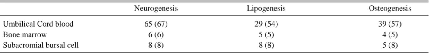
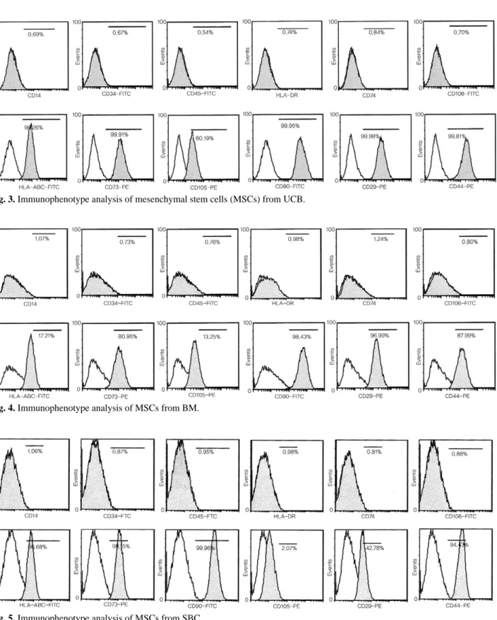
The result of flow cytometry of multipotent cell derived from umbilical cord blood, bone marrow and subacromial bursal at surface antigen testing using, CD 29, CD 44, CD 90/Thy-1, CD 105/SH-2 (generally known as surface antigen of MSC) was strongly expressed. The UCB derived MSC showed posi- tive findings as HLA-ABC 77.32%, CD-73 91.50%, CD-29 98.87%, CD-44 98.43(+), CD-90 90.62%, CD-105 94.36% (Fig. 3). BM derived MSC showed positive findings as CD-29 71.3%, CD-90 99.5%, and CD-105 80.3%.(Fig. 4) The SB derived cell showed positive findings of CD- 73 79.85% , CD-44 91.13%, CD-90 87.01%
(Fig. 5, Table 2). All 3 types of MSCs showed a negative finding for CD14, CD34, CD45, CD74, HLA-DR.
DISCUSSION
Adult MSCs are known to have an important role in overcoming various musculoskeletal ail- ments in the near future. They form the basis
Fig. 1. Adipogenesis was detected by the formation of neutral fat vacuoles stainable with Oil Red O, as demon- strated for UCB-derived cells (×320).
Fig. 2. Culturing the SBC with the neurogenesis induction media, subcultured colonies revealed astrocyte, large nuclei, axon and dentrite cells (×100).
Table 1. Differentiation potential of each stem cell
Neurogenesis Lipogenesis Osteogenesis
Umbilical Cord blood 65 (67) 29 (54) 39 (57)
Bone marrow 6 (6) 5 (5) 4 (5)
Subacromial bursal cell 8 (8) 8 (8) 5 (8)
( ) : total cases studied.
of various upcoming tissue engineering and cell therapy projects. MSCs derived from various sources have different characteristics based on the tissue of origin. Thus, MSCs are being iso- lated from a number of human tissues to pro-
vide for tissue specific therapy in future.
Nowadays, various potential sources for MSCs are the UCB, BM, placental tissue and SB cells.13)
According to their Mesenchymal and Tissue Fig. 3. Immunophenotype analysis of mesenchymal stem cells (MSCs) from UCB.
Fig. 4. Immunophenotype analysis of MSCs from BM.
Fig. 5. Immunophenotype analysis of MSCs from SBC.
Stem Cell Committee of the International Soci- ety for Cellular Therapy definition, MSC must be plastic-adherent when maintained in stan- dard culture conditions, must have MSC specif- ic surface markers (CD105, CD73 and CD90 positive and negative for CD45, CD34, CD14 or CD11b, CD79alpha or CD19 and HLA-DR sur- face molecules) and should be able to differen- tiate into osteoblasts, adipocytes and chondrob- lasts in vitro.14)
Our study showed that MSCs derived from all the 3 sources, namely, BM, UCB and SB had good differentiation potential. But their individual differentiation potential varied according to their source of origin. MSCs from all the 3 sources had a similar neurogenic dif- ferentiation potential (nearly 100%). But lipogenic and osteogenic differentiation poten- tials were not so similar among the 3 groups.
Bone marrow had the highest rate of lipogenic and osteogenic differentiation in our study.
There have been conflicting reports in the lit- erature regarding the growth and differentia- tion potential of UCB cells. While some reports suggest that the UCB derived MSC demon- strate a higher proliferation rate and differen- tiation potential than the other sources of MSCs.11) Other studies have noted that it was not possible to induce mesenchymal differentia- tion of the UCB cells.14) In our study though it was possible to induce mesenchymal differenti- ation of the UCB derived cells but they still had the least differentiation potential.
Our study also did an immunophenotype analysis of the MSCs from the 3 different sources. In accordance with the study of
Mareschi et al,15) BM cells, along with the other 2 type of MSCs, have shown negative findings for CD45, CD14, CD34 that are of hematopoietic antigen, and have shown the ability to morphologically differentiate into osteoblasts, adipocytes, neurons. Our study comparing the expression of surface markers of BM derived MSC, UCB derived MSC, SB derived MSC showed that while all showed pos- itive findings for MSC specific surface markers (CD29, CD44, CD90/Thy-1, CD105/SH-2, HLA- ABC) but the degree of positivity for different surface markers varied between the 3 types of MSCs. These results revealed that surface markers of BM, SB and UCD derived MSC are different from each other (Table 2).
From our study we could prove that MSCs from subacromial bursa and umbilical cord blood have an as reliable potential as the con- ventional bone marrow derived MSCs. But due to the conflicting nature of reports regarding growth and differentiation potential of UCB MSCs, more studies are needed to reveal its real potential. With every MSC having a dif- ferent differentiation potential according to their sources of origin, various tissues are being explored for MSCs with tissue specific cell therapy in mind. With both BM and SB showing good proliferative capacity and differ- entiation potential, they could have an attrac- tive future in regenerative medicine, with BM cells being a source for Bone healing and SB cells for musculo-tendinous healing. But more studies are needed to evaluate behavior of these MSCs in vivo conditions and for working on a method of delivering these cells to their proposed destination.
These studies involving the clinical applica- tion of MSCs continue to improve and need to be confirmed by future similar studies; howev- er, our finding may lay the groundwork for the future therapy of many degenerative or traumatic injuries making use of these MSCs derived from one of the above said sources.
Acknowledgement
This work was supported by the Clinical Table 2. Summary of surface marker study
UCB*-MSCs� BM�-MSCs SB-MSCs
CD73 91.50 (+) 76.8 (+) 79.85 (+)
CD29 98.87 (+) 71.30 (+) 38.53 (+/Low) CD44 98.43 (+) 70.00 (+) 91.13 (+) CD90 90.62 (+) 99.50 (+) 87.01 (+) CD105 94.36 (+) 80.30 (+) 1.87 (-) HLA-ABC 77.32 (+) 48.20 (+/low) 56.09 (+)
*UCB: umbilical cord blood , �MSCs: mesenchymal stem cells , �BM: bone marrow.
Research Foundation of Chosun University at 2011.
REFERENCES
1) Barry FP, Murphy JM. Mesenchymal stem cells: clin- ical applications and biological characterization. Int J Biochem Cell Biol. 2004;36:568-84.
2) Pittenger MF, Mackay AM, Beck SC, et al. Multilin- eage potential of adult human mesenchymal stem cells.
Science. 1999;284:143-7.
3) Yang SE, Ha CW, Jung M, et al. Mesenchymal stem/progenitor cells developed in cultures from UC blood. Cytotherapy. 2004;6:476-86.
4) Niibe K, Kawamura Y, Araki D, et al. Purified mes- enchymal stem cells are an efficient source for iPS cell induction. PLoS One. 2011;6:e17610.
5) Peyrard T, Bardiaux L, Krause C, et al. Banking of pluripotent adult stem cells as an unlimited source for red blood cell production: potential applications for alloimmunized patients and rare blood challenges.
Transfus Med Rev. 2011;25:206-16.
6) Satomura K, Krebsbach P, Bianco P, Gehron Robey P. Osteogenic imprinting upstream of marrow stromal cell differentiation. J Cell Biochem. 2000;78:391-403.
7) Shafiee A, Kabiri M, Ahmadbeigi N, et al. Nasal Septum-Derived Multipotent Progenitors: A Potent Source for Stem Cell-Based Regenerative Medicine.
Stem Cells Dev. 2011;20:2077-91.
8) Ratajczak MZ, Zuba-Surma EK, Machalinski B, Kucia M. Bone-marrow-derived stem cells--our key to
longevity?. J Appl Genet. 2007;48:307-19.
9) Reyes M, Verfaillie CM. Characterization of multipo- tent adult progenitor cells, a subpopulation of mes- enchymal stem cells. Ann N Y Acad Sci. 2001;938:231- 3. discussion 3-5.
10) Yoon SH, Shim YS, Park YH, et al. Complete spinal cord injury treatment using autologous bone marrow cell transplantation and bone marrow stimulation with granulocyte macrophage-colony stimulating factor:
Phase I/II clinical trial. Stem Cells. 2007;25:2066-73.
11) Kern S, Eichler H, Stoeve J, Kluter H, Bieback K.
Comparative analysis of mesenchymal stem cells from bone marrow, umbilical cord blood, or adipose tissue.
Stem Cells. 2006;24:1294-301.
12) Uhthoff HK, Trudel G, Himori K. Relevance of pathology and basic research to the surgeon treating rotator cuff disease. J Orthop Sci. 2003;8:449-56.
13) Moon YL, Song CH, Noh KH, Lee SJ, Lee HJ, Kim SH. Isolation and Phenotypic Characterization of Mul- tipotent Mesenchymal-like Stem Cells from Human Subacromial Bursa. J Tissue Eng Regen Med. 2009;5:
527-32.
14) Dominici M, Le Blanc K, Mueller I, et al. Minimal criteria for defining multipotent mesenchymal stromal cells. The International Society for Cellular Therapy position statement. Cytotherapy. 2006;8:315-7.
15) Mareschi K, Biasin E, Piacibello W, Aglietta M, Madon E, Fagioli F. Isolation of human mesenchymal stem cells: bone marrow versus umbilical cord blood.
Haematologica. 2001;86:1099-100.
목적: 세가지 기원의 줄기 세포 분화능과 면역표현형을 평가하고자 하였다.
대상 및 방법: 견봉하 점액낭과 골수, 탯줄 혈액 세 개의 군에서 세포를 채취하였다. 견봉하 점액 낭과 골수는 견관절 수술 환자군에게 임상적 동의 하에 수술중 채취하였다. 각각의 채취된 세포 및 탯줄 혈액에 대하여 계대 배양을 시행하여 신경 분화군, 지방 분화군, 골 분화군을 평가하였으 며 세포 표면 항체를 밝히기 위해 유동세포분석법을 이용하였다.
결과: 견봉하 점액낭 유래 세포에서는 신경분화와 지방 분화는 8예 모두 (100%)에서, 골분화는 8 례 중 5예 (62.5%)에서 성공할 수 있었으며 골수 유래 세포의 경우 신경 및 지방 분화 유도한 6 례 및 5예 모두 (100%) 분화에 성공하였으나 골분화 유도는 5예 중 4예 (80%)에서 얻을 수 있 었다. 반면 탯줄 유래 세포 분화 연구의 경우 신경 분화 유도 67례 중 65예 (97%)에서 지방 분화 연구 54예 중 29예 (53.7%)에서 골 분화 연구 57예 중 39예 (68.4%)에서 성공할 수 있었다.
결론: 탯줄 유래 줄기세포의 분화능과 비교하였을 때 견봉하 점액낭 및 골수 유래 줄기세포의 분 화능이 우수함을 알 수 있으며 이는 향후 세포 치료에 있어서 안정성 있는 치료 제공자가 될 수 있을 것으로 보이며 향후 생체 실험 연구의 참고 자료로서도 가치가 있을 것으로 보인다.
색인 단어: 견봉하 점액낭, 골수, 탯줄 혈액, 줄기세포, 분화능