• 검색 결과가 없습니다.

The Effect of Quercetin on the BKCa

N/A
N/A
Protected

Academic year: 2022

Share "The Effect of Quercetin on the BKCa"

Copied!
7
0
0

로드 중.... (전체 텍스트 보기)

전체 글

(1)

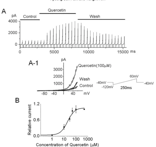
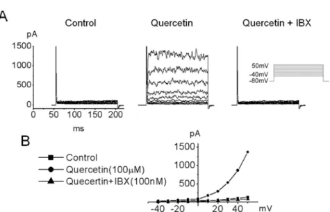
The Effect of Quercetin on the BK Ca in Umbilical Cord Vein-derived Mesenchymal Stem Cells

Kyoung Sun Park

1

and Yangmi Kim

2

*

1

Department of Physiology, College of Medicine, Seoul National University, Seoul, Korea

2

Department of Physiology, College of Medicine, Chungbuk National University, Cheongju, Korea Recently, we demonstrated that a large conductance Ca

2+

-activated K

+

channel (BK

Ca

) in human umbilical cord vein-derived mesenchymal cells (hUC-MSCs). In this study, we studied effects of quercetin, the flavonoid used for the modulator of cell cycle and the treatment of anti-tumor, on BK

ca

in hUC-MSCs using single channel and/or whole cell configuration. Single channel conductance was 277.8 ± 36.2 pS in hUC-MSCs and the value is similar to that of typical BK

Ca

channel reported in other cells. The single channel current was activated by increasing of intracellular Ca

2+

. Quercetin (30~100 µ M) increased BK

Ca

in whole cell patch configuration whereas EGCG was not affected on the channel. The activation effect of quercetin on BK

Ca

channel was also observed in inside-out configuration. The quercetin-induced BK

Ca

was increased a concentration-dependent manner with an IC

50

value of 19.9 µ M. Taken together, BK

Ca

in hUC- MSCs can be an important target for the action of quercetin and the channels are partly may contribute to maintaining homeostasis by regulation of K ion flux in hUC-MSCs.

Key words: Human umbilical cord vein-derived mesenchymal cells, quercetin, BK

Ca

Received 27 April 2010; Revised version received 21 June 2010; Accepted 23 June 2010

케르세틴 (quercetin) 플라보노이드 (flavonoid) 일종으로

폴리페놀 군에 속하며 우리가 먹는 과일이나 야채에 많 이 존재하는 항산화제로 알려져 있다 . 우리가 먹는 플라

노이드 중 60-75% 케르세틴이다 (Hertog and Hollman,

1996). 케르세틴은 심장질환 동물의 관상동맥에 작용하여

혈관확장효과를 나타내며 고혈압 동물 모델에서 혈압을 감소시키거나 , 심장비대를 방지하며 산화를 방지한다 (Duarte et al., 2001; Sanchez et al., 2006). 또한 케르세틴은

암 역할을 한다고도 알려져 있는데 췌장암세포 , 랫드의

전립선 암세포 , 그리고 전골수구성 백혈병 (promyeloleukemic)

세포인 HL-60 세포에서 케르세틴은 세포의 자살

포토시스 (apoptosis) 유발함이 보고 되었다 (Lee et al., 2002b; Wang et al., 2003). 또한 사람의 유방암세포에서

도 케르세틴에 의해 세포주기가 정지되고 아포토시스가 관찰되었다 (Choi et al., 2001). 이처럼 케르세틴은 항암 ,

항산화 작용 등을 나타내고 있으며 줄기세포에서도 이러 한 플라보노이드들이 분화를 조절한다고 보고되기 시작

하고 있다 . 돼지의 골수 배양 세포에서 케르세틴은 파골

세포형성 (osteoclastogenesis) 조절한다고 알려져 있으며 (Rassi et al., 2005), 다른 종류의 플라보노이드인 epigallocatechin gallate (EGCG) NF-kappaB 경유하여

파골세포분화를 억제하고 (Lin et al., 2009), hBM-MSCs

서는 지방세포 분화를 유발한다고 보고되었다 (Shin et al.,

2009). 케르세틴은 혈관 평활근의 이온통로에도 작용한

다 . 사람의 장간막동맥과 대동맥에서도 케르세틴에 의한 NO/cGMP 활성이 BK

Ca

를 활성화하여 과분극을 유도하고 이 과분극이 이온경사에 의해 세포내로의 칼슘 (Ca

2+

)

입이 촉진됨이 보고되었고 (Kuhlmann et al., 2005), 관상

동맥 (coronary arteries) 에서는 케르세틴이 과산화수소 (H

2

O

2

)

를 생산하여 BK

Ca

전류를 증가시킨다고 보고된 바 있다

(Cogolludo et al., 2007).

최근 우리는 제대정맥에서 유래된 중간엽 줄기세포

(human umbilical cord vein-derived mesenchymal stem cells, hUC-MSCs) 에서 BK

Ca

통로 (large conductance Ca

2+

- activated K channel), Na 통로 그리고 몇가지 종류의 K

통로가 기능적으로 발현됨을 보고하였다 (Park et al., 2007).

또한 우리는 최근 hUC-MSC 에 존재하는 BK

Ca

통로를 단 일 통로로를 기록하여 근위축성 측상경화증 (amyotrophic lateral sclerosis) 치료제인 riluzole 의해 활성화됨이

*Corresponding author: Yangmi Kim, Department of Physiology, College of Medicine, Chungbuk National University, 410 Seongbong-ro, Heungdeok-gu, Cheongju, Chungbuk 361-763 Tel: +82-43-261-2855

Fax: +82-43-272-1603

E-mail: [email protected]

(2)

204 Kyoung Sun Park and Yangmi Kim 인하였다 (Park et al , 2008). 연구에서는 hUC-MSC

분화 줄기세포에 존재하는 BK

Ca

에 플라보노이드가 어떤 영향을 주는지 조사하였다 . hUC-MSCs 존재하는 BK

Ca

에 대한 플라보노이드의 효과는 단일통로 수준 및 whole cell patch configuration 이용하여 확인하였다 .

재료 및 방법

제대정맥에서 유래된 사람의 중간엽 줄기세포의 배양

배양 과정은 제대 정맥을 모은 후 12-16 시간 사이에

시행 되었다 . 제대정맥을 모아 제대정맥을 phosphate buffered saline (GIBCO-BRL, Invitrogen Corporation, Grand Island, NY, USA) 씻어 혈관 내로 0.5%

collagenase (GIBCO-BRL, Invitrogen Corporation, Grand Island, NY, USA) 넣고 37

o

C 에서 5 시간 동안 방치하였

다 . 혈관을 부드럽게 마사지하여 떨어져 나온 세포를

집하였다 . 수집된 세포는 1,000 rpm 에서 10 동안 원심

분리하고 , Dulbecco s modified Eagle s medium-low glucose (GIBCO-BRL, Invitrogen Corporation, Grand Island, NY, USA), 100 U/mL penicillin, 100 µ g/mL streptomycin, and 10% fetal bovine serum (GIBCO- BRL, Invitrogen Corporation, Grand Island, NY, USA)

섞어 75 cm

2

culture flasks 넣어 37

o

C, 5% CO

2

배양 기에서 배양하였다 . 배양액은 2 일마다 바꾸어 주고 전기

생리학적인 실험에 사용하였다 .

전기생리학적 방법

실험에 사용한 용액 중 정상 세포 외 용액은 140 mM

NaCl, 5,4 mM KCl, 0.5 mM MgCl

2

, 1.8 mM CaCl

2

, 10 mM glucose, 5 mM HEPES 구성하였고 , pH 7.4

정하여 사용하였다 . 단일통로 기록 (single channel recording)

을 위해 inside-out patch 또는 cell-attached patch

때 유리전극 (pipette) 용액과 bath 용액은 150 mM KCl, 1 mM MgCl

2

, 10 mM HEPES, 5 mM EGTA 구성

하고 , pH 7.2 적정하였다 . Whole cell patch 고정

방법으로 전류를 기록할 경우는 pipette 용액을 140 mM KCl, 1 mM MgCl

2

, 5 mM Mg-ATP, 2 mM EGTA, 5 mM HEPES 구성하고 , pH 7.3 으로 적정하여 사용하였다 .

세포전체 전류 (whole-cell current) 단일통로 전류 (single channel current) gigaohm (G )-seal 형성하여 기록하

였다 . Whole-cell current 기록시에는 1~3 M 정도의

극저항을 가진 전극을 사용하고 , 단일통로 기록시에는

2~3 M 저항의 전극을 사용하였다 . 패치전극의 끝은

sylgard (Dow Corning, Midland, MI, USA) coating fire polish 하여 사용하였다 . 단일통로의 기록시 이온통로

의 활성도는 패치고정증폭기 (patch clamp amplifier, axopatch 200B, Union city, CA, USA) 기록하였다 .

록되는 신호는 8-pole bessel filter (-3 dB, Frequency Device) 를 사용하여 2 kHz 에서 filter 하였다 . Data 를

computer 옮길 때는 10 kHz sampling rate digidata 1322A interface (Axon instrument, Union city, CA, USA) 를 이용하여 computer 에 저장하였다 . 모든 data

는 pCLAMP program 9.0 (Axon instrument, Union city, CA, USA) 이용하여 분석하였다 . 모든 실험은 실온 (23-25

o

C) 에서 진행하였다 .

결 과

최근 우리는 사람의 제대정맥유래 중간엽 줄기세포

(human umbilical cord vein-derived mesenchymal stem cells, hUC-MSCs) 에서 whole cell patch configuration

사용하여 기능적으로 발현되는 이온통로를 규명하였다

(Park et al. , 2007). 연구에서는 이들 large- conductance Ca

2+

-activated K

+

(BK

Ca

) 통로가 플라보노이

드에 의해 어떻게 조절 되는지를 관찰하였다 . Figure 1A

는 hUC-MSC 에서 보이는 전형적인 전류의 양상이다 .

향 전류가 나타난 다음 외향 전류가 기록됨을 알 수 있 었다 . 내향 전류는 우리의 이전 보고에서 기록한

치의 약 30% 에서만 관찰되는 Na 전류임을 확인하였다 (Park et al. , 2007). 연구에서는 시도된 패치의 98%

에서 보이는 외향 전류에 대한 플라보노이드의 효과를 관

찰 하였다 . 외향 전류는 nosy 패턴을 가지고 있으며

BK

Ca

전류임이 확인 되었다 (Park et al., 2007; Park et

al ., 2008). EGCG (50 µ M) 외향 전류 내향 전류에

거의 영향을 주지 않았으며 이러한 현상을 전류 전압 곡 선으로 나타내었다 (Figure 1A and 1A-1). 반면 케르세틴

은 외향 전류를 활성화 시켰으며 이 활성은 케르세틴을 없애주면 천천히 정상 수준으로 회복되었고 (Figure 1B),

전류전압 곡선에서 케르세틴의 활성효과를 볼 수 있었다

(Figure 1B-1). 이러한 현상은 대부분의 패치에서 관찰

었다 (n=16). 이때 유지전압은 80 mV 하였으며 40 mV 에서 50 mV 까지 자극을 주어 기록하였다 .

케르세틴에 대한 전류의 활성을 시간대별로 보기 위해 램프 자극을 주어 10 간격으로 케르세틴의 효과를

록하였다 (Figure 2A and 2A-1). 유지전압을 80 mV

고 − 120 mV 에서 60 mV 까지 램프 자극을 주어 기록하였

다 . 케르세틴을 적용하기 전에는 전류가 증가하지 않았다

가 케르세틴을 적용하면 서서히 증가하기 시작하여 약 2

분 정도가 지나면 최대값에 도달함을 확인하였다 . Figure

2A-1 대표적인 전류를 전류 전압 곡선상에 나타낸

이다 . 전류의 크기는 +50 mV 에서 6.7 이상 증가하였

으며 (n=4), 케르세틴을 씻어내면 서서히 회복되나 완전히

회복되지는 않았다 . 케르세틴에 의해 외향 전류가 농도

의존적으로 증가되는지 보기 위해 각각의 다른 농도에서

(3)

전류를 기록하였다 . 케르세틴에 의해 조금씩 증가하기 시 작하여 50 µ M 에서 거의 최대 값에 이르렀고 , IC

50

는 약

19.9 µ M 정도였다 (Figure 2B, n=3).

케르세틴에 의해 증가된 전류가 어떤 전류인지 알아보 기 위해 단일통로 기록법을 이용하여 기록하였다 . Figure 3A 단일통로 수준에서 large-conductance Ca

2+

-activated K

+

channel (BK

Ca

) 을 hUC-MSCs 에서 기록한 통로는

40 mV 에서 277.8 ± 36.2 pS 정도의 단일통로 전도도를

였으며 (11.6 ± 1.5 pA), 통로는 세포 Ca

2+

막전압

에 의존적으로 활성이 증가하였다 (Figure 2A). 결과는

우리의 이전 결과와 유사하였다 (Park et al., 2008).

40 mV 전압에서 통로의 활성도 0.00117 였으며 평균

리는 시간 (mean open time) 은 0.09 ms 정도였다 (n=4).

40 mV 에서 기록되는 전류는 세포 안에서 세포 밖으로 나

가는 외향전류로 기록되었다 . 통로가 세포내 Ca

2+

어떻게 반응하는지 조사하기 위해 세포내 Ca

2+

농도를

변화시키면서 통로의 활성도를 비교하였다 . 세포내 Ca

2+

이 없는 상태에서는 활성을 보이는 통로가 하나였다가 세 포내 Ca

2+

농도가 높아지면 3 이상의 통로가 오랫동안

열리는 것이 관찰되었으며 , 세포내 Ca

2+

농도가 증가하

Figure 1. The effect EGCG (epigallocatechin 3-gallate) and quercetin on outward in hUC-MSCs. hUC-MSCs were voltage-

clamped under whole cell configuration at -80 mV. Voltage steps of 200 ms duration were then applied from -40 mV to 50 mV in 10-

mV increments every 10 s, showing outward currents a rapidly activating current with noisy oscillation, similar to large conductance

Ca

2+

activated K

+

current (BK

Ca

). The pipette solution consisted of 140 mM KCl, 1 mM MgCl

2

, 5 mM Mg-ATP and 2 mM EGTA; the

bath solution contained 143 mM NaCl, 5.4 mM KCl, 0.5 mM NaH

2

PO

4

, 0.5 mM MgCl

2

, 1.8 mM CaCl

2

, 5 mM HEPES and 10 mM

glucose. (A) Membrane currents were recorded in the presence of EGCG. (A-1) The current-voltage relationship (I-V relationship)

was plotted from A. Current-voltage relationship showed in the control ( ), 50 µ M EGCG ( ) and wash ( ). (B) Quercetin

increased outward current in hUC-MSCs. (B-1) The current-voltage relationship (I-V relationship) was plotted from B. I-V

relationships showed in the control ( ), 100 µ M quercetin ( ) and wash ( ).

(4)

206 Kyoung Sun Park and Yangmi Kim

면 평균 열리는 시간이 0.09 ms 에서 3.95 ms 로 증가하였 고 , 통로의 활성도 0.001 에서 0.31 증가 하였다 . 이러

한 결과는 세포내 Ca

2+

에 활성화 되는 BK

Ca

통로임을 증 명하며 우리의 이전 결과와 일치하였다 (Park et al., 2008).

이 통로에 inside-out patch 모드에서 bath( 세포내 ) 직접

케르세틴 (30 µ M) 적용하였을 반응을 관찰 하였다 .

케르세틴에 의해 단일 통로 활성화도의 변화는 +60 mV

에서 기록하였다 . 케르세틴을 적용하기 단일통로의

성도는 0.035 ± 0.001 이었으나 케르세틴 적용 후의 통로의

활성도는 0.056 ± 0.029 1.6 증가하였다 . 또한

로의 평균 열리는 시간은 1.43 ± 0.15 ms 에서 2.63 ± 0.42

ms 으로 약간 증가 하였다 (n=5). 이러한 결과는 케르세틴

이 채널에 직접 작용할 수도 있음을 제시한다 .

케르세틴에 의해 증가된 전류가 BK

Ca

임을 확인하기 위 해 케르세틴 처리 후 BK

Ca

특이적인 억제제의 효과를 관 찰하였다 (Figure 4). 유지전압은 80 mV 으로 whole cell patch 모드에서 막전압을 40 mV 에서 50 mV 까지

용하였을 경우 외향전류가 관찰 되었으며 , 외향전류는

케르세틴에 의해 활성화되고 활성화된 전류는 BK

Ca

특이

적인 억제제인 iberiotoxin 에 의해 완전히 억제되었으므로 케르세틴에 의해 증가된 전류는 BK

Ca

임을 확인하였다 .

이상의 결과들은 사람 제대정맥 유래 미분화 중간엽 줄

기세포 (hUC-MSC) 에 기능적으로 발현되는 이온통로 중

BK

Ca

가 케르세틴에 의해 활성화되며 , 이는 케르세틴에

해 이러한 이온 통로의 조절이 다른 세포에서처럼 줄기 세포의 신호전달체계에 영향을 주어 세포의 분화나 증식 과 같은 세포내 항상성의 변화에 기여할 수 있음을 제시 한다 .

고 찰

본 연구에서 사람 제대정맥 유래 미분화 줄기세포 (hUC-

MSCs) 에서 존재하는 외향 전류가 플라보노이드 케르

세틴에 의해 활성화됨을 확인하였고 , 케르세틴에 의해

성화된 전류가 BK

Ca

통로임을 확인하였다 .

BK

Ca

통로는 Ca

2+

통로를 통해 세포 밖에서 들어오는

Ca

2+

이나 세포내 소기관의 세포막 통로를 통해 방출되는

칼슘에 의해 세포내 Ca

2+

증가하면 열리는 통로로서

Figure 2. Effect of extracellular quercetin on the outward current in hUC-MSCs. (A) Ramp pulse was applied from -120 to 60 mV for

250 ms at a holding potential of -80 mV and the pulse was applied at every 10 s. The x-axis shows time and the y-axis shows

current. The outward current was activated by quercetin. Currents were reached a stable level in 2 min after the application of

quercetin (100 µ M) and recovered after washing with quercetin-free solution. (A-1) Quercetin -induced typical outward-rectifying

current vs. voltage. (B) Dose dependence of quercetin-induced outward current in hUC-MSC using the whole cell patch

configuration. Currents were measured at 50 mV and IC

50

was 19.9 µ M. (n=3 cells per concentration).

(5)

세포내의 K 이온들을 세포외로 방출함으로써 세포내를 안 정화 시키는 역할을 한다 . BK

Ca

통로는 혈관 평활근에서 혈관의 수축과 이완을 조절 하는데 결정적인 역할을 한 다고 알려져있고 혈관내피 세포의 증식이나 암세포의 증 식에 관여한다 (Wiecha et al. , 1998; Roger et al. ,

2004). BK

Ca

는 세포내의 전압을 안정적으로 음성값으로

유지하게 하며 BK

Ca

의 활성제는 세포를 더욱 안정화 시 키거나 세포내 Ca

2+

완충할 있는 역할을 하여 세포

의 항상성을 조절할 것으로 생각된다 .

케르세틴은 암세포에서 세포내의 신호전달에 기여하여 세포의 주기나 성장에 기여하며 혈관 평활근에서는 혈관 의 이완에 관여한다 . 케르세틴에 의한 BK

Ca

의 활성이 혈 관평활근에서는 NO 신호나 H

2

O

2

증가에 의해 일어난다 고 보고되었다 (Kuhlmann et al. , 2005; Cogolludo et al. , 2007). 연구에서 사용된 hUC-MSC 에서 케르세틴에

한 BK

Ca

활성은 NO 활성이나 H

2

O

2

에 의한 것인지는 결 론지을 수 없다 . 하지만 NO H

2

O

2

가 없는 상황인

inside-out patch configuration( 단일통로 기록법 중의 하나

임 ) 에서도 케르세틴만으로 통로의 활성이 나타났고 (Figure 3), Kuhlman et al. (2005) Cogolludo et al. (2007)

보고처럼 케르세틴에 의한 BK

Ca

의 활성양상이 NO 활성 이나 H

2

O

2

를 조성해 낼 수 있는 whole cell configuration ( 전체전류를 기록할 있는 방법 ) 양상보다는 낮게 나타

났기 때문에 세포내 케르세틴에 의한 H

2

O

2

의 활성이나

NO 의 증가에 의해 일어날 가능성도 배제 할 수 없다

(Figure 3 and Figure 4). 그러므로 세포의 종류에 따라

케르세틴에 의한 BK

Ca

의 활성경로는 다를 수 있다고 생 각된다 .

미분화 줄기세포의 분화시 세포안의 환경 변화 , 산화적

대사과정 (oxidative metabolism) 변화 다양한 생화학

적 및 병리학적 변화가 수반되므로 줄기세포의 분화 조 절에도 플라보노이드가 관여할 가능성이 있다 . 돼지의

수 배양 세포와 사람의 골수에서 유래된 중간엽 줄기세

포 (hBM-MSC) 에서 플라보노이드의 효과가 이러한 가능성

을 뒷받침한다 (Rassi et al. , 2005; Lin et al. , 2009;

Shin et al. , 2009). 또한 과정에서 일차적인 세포내

온 조절기인 이온통로가 기여 할 수 있을 것이라는 것은 자명하다 . 하지만 아직까지는 줄기세포에서 이온통로에

대한 연구는 아직 미흡하며 , 나아가 플라보노이드가

줄기세포에 존재하는 이온통로에 영향을 받는다는 보고 Figure 3. The effect of intracellular quercetin on large conductance Ca

2+

activated K

+

channels (BK

Ca

) using single channel recording in hUC-MSCs. Data were obtained from estimates of relative channel activity. (A) The trace shown the effect of intracellular Ca

2+

. Single channel currents recorded at +40mV from an inside-out patch configuration. Pipette solution contained 150 mM KCl, 1 mM MgCl

2

, 5 mM EGTA, and 10 mM HEPES. The bath solution consisted of 150 mM KCl 150 mM KCl, 1 mM MgCl

2

, 5 mM EGTA, and 10 mM HEPES and 0 µ M, 1 µ M CaCl

2

. The channel activity was increased by [Ca

2+

]

i

dependently. The letter “a” and “b” show an expanded scale of the A. Current amplitude of 1 main peak is about 12 pA. (B) The traces were showed the effect of quercetin on BK

Ca

channels. The channel activity was increased by intracellular application of quercetin (30 µ M). The letter “a” and “b” show an expanded scale of the A. The recording potential was +60mV. Current amplitude of 1 main peak is about 15 pA. The letter “close” represents the closed channel level and the letter “open” represents the open channel level. Abbreviations:

min, minutes; ms, milliseconds; pA, picoampere.

(6)

208 Kyoung Sun Park and Yangmi Kim

는 없다 . 연구에서 제대정맥 유래 미분화 줄기세포

(hUC-MSCs) 에서 플라보노이드가 줄기세포에 존재하는 이

온 통로에 대한 플라보노이드가 영향을 줄 수 있음을 증 명하였다 . 제대정맥 유래 미분화 줄기세포에서 BK

Ca

가 플 라보노이드 중 케르세틴에 의해 증가됨을 확인하였으므 로 이 통로가 플라보노이드에 의해 분화과정에 기여할 가 능성이 있을 것이라고 추측되므로 향후 계속적인 연구가 요구된다 .

수년 동안 케르세틴의 효과는 BK

Ca

뿐만 아니라 막전 압에 의해 활성화되는 전류인 K

V

전류와 Cl

전류 , Ca

2+

전류에서도 보고되었다 . 사람의 Kv1.5 통로가 케르세틴에

의해 활성화되고 (Yang et al. , 2009), 랫드의 결장에서는 Cl

분비를 증대시키기도 한다 (Cermak et al. , 1998).

드 꼬리동맥 평활근 (rat tail artery smooth muscle) 에서 L- type Ca

2+

통로를 활성화 시키며 (Saponara et al. , 2002),

골격근에서도 케르세틴이 단일 Ca

2+

방출 통로를 활성화

하여 세포내 Ca

2+

농도를 조절한다고 보고된 있다 (Lee

et al. , 2002a). 케르세틴에 의해 세포내 Ca

2+

방출통로를

통해 세포내 Ca

2+

증가할 있다는 보고는 hUC-MSC

에서도 BK

Ca

의 활성이 케르세틴에 의해 세포내 Ca

2+

가하여 BK

Ca

통로가 활성화 될 수 있을 가능성을 뒷받침 한다 . 하지만 우리의 연구는 세포내 Ca-free 상태 (10

−7

M

이하 ) 에서도 케르세틴에 의한 채널 활성이 나타났으므로

세포내 Ca

2+

증가에 의한 것이라기 보다는 케르세틴

자체가 직접 채널에 붙어서 작용할 수도 있음을 시사한 다 (Figure 3). 그리고 hUC-MSCs 존재하는 BK

Ca

통로는 플라보노이드에 의해 활성화되는 특성을 지녔으며 , whole

cell patch 모드뿐만 아니라 inside-out patch 모드에서도

플라보노이드에 의한 활성화 특성이 여전히 관찰되는 것 으로 보아 , 평활근에 존재하는 BK

Ca

가 케르세틴에 간접적 인 작용으로 활성이 증가된다는 결과와 상반된다 .

결론적으로 hUC-MSC 에서 플라보노이드에 의해 조절

되는 이온 통로의 특성을 규명함으로써 플라보노이드가 줄기 세포에서 세포의 분화 기전에 기여할 수 있을 가능 성을 제시하며 본 연구결과는 hUC-MSC 증식 조절

한 연구나 더 나아가서 UC-MSC 분화 조절을 연구하

는데 기초적인 자료로서 제공될 수 있을 것으로 생각된 다 . 또한 앞으로 줄기세포의 분화 증식시 플라보노이

드가 이온통로와 연관된 신호전달체계가 어떻게 연관되 어 있는지 연구하는 것이 필요하다 생각된다 .

감사의 글

이 논문은 2008 년도 충북대학교 학술연구지원사업의

연구비 지원에 의하여 연구되었습니다 . 참고문헌

Cermak, R., Follmer, U. and Wolffram, S. (1998) Dietary flavonol quercetin induces chloride secretion in rat colon. Am. J.

Physiol. 275(5 Pt 1), G1166-1172.

Choi, J. A., Kim, J. Y., Lee, J. Y., Kang, C. M., Kwon, H. J., Yoo, Y.

D., Kim, T. W., Lee, Y. S. and Lee, S. J. (2001) Induction of cell cycle arrest and apoptosis in human breast cancer cells by quercetin. Int. J. Oncol. 19(4), 837-844.

Cogolludo, A., Frazziano, G., Briones, A. M., Cobeno, L.,

Figure 4. Effect of quercetin and Iberiotoxin (IBX, a blocker of large conductance Ca

2+

-activated K

+

current) on BK

Ca

in hUC-MSCs.

(A) Current traces are shown for 200-ms depolarization pulses from -40 mV to +50 mV (in 10-mV increments) from a holding

potential of -80mV. Quercetin activates noisy outward currents in hUC-MSCs. Currents were recorded before (control), after the

exposure to quercetin (100 µ M) reached a stable level (3 min) and after application with quercetin and IBX solution. (B) The current-

voltage relationship (I-V relationship) was plotted from A. I-V relationships of currents showed in the control ( ), 100 µ M quercetin

( ) and 100 µ M quercetin with 100 nM iberiotoxin ( ).

(7)

Moreno, L., Lodi, F., Salaices, M., Tamargo, J. and Perez- Vizcaino, F. (2007) The dietary flavonoid quercetin activates BKCa currents in coronary arteries via production of H

2

O

2

. Role in vasodilatation. Cardiovasc. Res. 73(2), 424-431.

Duarte, J., Perez-Palencia, R., Vargas, F., Ocete, M. A., Perez- Vizcaino, F., Zarzuelo, A. and Tamargo, J. (2001) Antihypertensive effects of the flavonoid quercetin in spontaneously hypertensive rats. Br. J. Pharmacol. 133(1), 117-124.

Hertog, M. G. and Hollman, P. C. (1996) Potential health effects of the dietary flavonol quercetin. Eur. J. Clin. Nutr. 50(2), 63- Kuhlmann, C. R., Schaefer, C. A., Kosok, C., Abdallah, Y., 71.

Walther, S., Ludders, D. W., Neumann, T., Tillmanns, H., Schafer, C., Piper, H. M. and Erdogan, A. (2005) Quercetin- induced induction of the NO/cGMP pathway depends on Ca

2+

-activated K

+

channel-induced hyperpolarization- mediated Ca

2+

-entry into cultured human endothelial cells.

Planta Med. 71(6), 520-524.

Lee, E. H., Meissner, G. and Kim, D. H. (2002a) Effects of quercetin on single Ca

2+

release channel behavior of skeletal muscle. Biophys. J. 82(3), 1266-1277.

Lee, L. T., Huang, Y. T., Hwang, J. J., Lee, P. P., Ke, F. C., Nair, M.

P., Kanadaswam, C. and Lee, M. T. (2002b) Blockade of the epidermal growth factor receptor tyrosine kinase activity by quercetin and luteolin leads to growth inhibition and apoptosis of pancreatic tumor cells. Anticancer Res. 22(3), 1615-1627.

Lin, R. W., Chen, C. H., Wang, Y. H., Ho, M. L., Hung, S. H., Chen, I. S. and Wang, G. J. (2009) (-)-Epigallocatechin gallate inhibition of osteoclastic differentiation via NF-kappaB.

Biochem. Biophys. Res. Commun. 379(4), 1033-1037.

Park, K. S., Jung, K. H., Kim, S. H., Kim, K. S., Choi, M. R., Kim, Y. and Chai, Y. G. (2007) Functional expression of ion channels in mesenchymal stem cells derived from umbilical cord vein. Stem Cells 25(8), 2044-2052.

Park, K. S., Jung, K. H., Chai, Y. G., Kim, C. H., Shin, E. Y. and Kim. Y. (2008) Riluzole-induced activation of BKCa channels

and inhibition of voltage-gated Na channel in human umbilical cord vein-derived mesenchymal cells. Lab. Anim.

Res. 24(3), 287-292

Rassi, C. M., Lieberherr, M., Chaumaz, G., Pointillart, A. and Cournot, G. (2005) Modulation of osteoclastogenesis in porcine bone marrow cultures by quercetin and rutin. Cell Tissue Res. 319(3), 383-393.

Roger, S., Potier, M., Vandier, C., Le Guennec, J. Y. and Besson, P. (2004) Description and role in proliferation of iberiotoxin- sensitive currents in different human mammary epithelial normal and cancerous cells. Biochim. Biophys. Acta 1667(2), 190-199.

Sanchez, M., Galisteo, M., Vera, R., Villar, I. C., Zarzuelo, A., Tamargo, J., Perez-Vizcaino, F. and Duarte, J. (2006) Quercetin downregulates NADPH oxidase, increases eNOS activity and prevents endothelial dysfunction in spontaneously hypertensive rats. J. Hypertens. 24(1), 75-84.

Saponara, S., Sgaragli, G. and Fusi, F. (2002) Quercetin as a novel activator of L-type Ca

2+

channels in rat tail artery smooth muscle cells. Br. J. Pharmacol. 135(7), 1819-1827.

Shin, D. W., Kim, S. N., Lee, S. M., Lee, W., Song, M. J., Park, S.

M., Lee, T. R., Baik, J. H., Kim, H. K., Hong, J. H. and Noh, M. (2009) (-)-Catechin promotes adipocyte differentiation in human bone marrow mesenchymal stem cells through PPAR gamma transactivation. Biochem. Pharmacol. 77(1), 125-133.

Wang, S., Degroff, V. L. and Clinton, S. K. (2003) Tomato and soy polyphenols reduce insulin-like growth factor-I-stimulated rat prostate cancer cell proliferation and apoptotic resistance in vitro via inhibition of intracellular signaling pathways involving tyrosine kinase. J. Nutr. 133(7), 2367-2376.

Wiecha, J., Munz, B., Wu, Y., Noll, T., Tillmanns, H. and Waldecker, B. (1998) Blockade of Ca

2+

-activated K

+

channels inhibits proliferation of human endothelial cells induced by basic fibroblast growth factor. J. Vasc. Res. 35(5), 363-371.

Yang, L., Ma, J. H., Zhang, P. H., Zou, A. R. and Tu, D. N.

(2009) Quercetin activates human Kv1.5 channels by a residue I502 in the S6 segment. Clin. Exp. Pharmacol. Physiol.

36(2), 154-161.

수치

Figure 1.  The effect EGCG (epigallocatechin 3-gallate) and quercetin on outward in hUC-MSCs
Figure 2.  Effect of extracellular quercetin on the outward current in hUC-MSCs. (A) Ramp pulse was applied from -120 to 60 mV for 250 ms at a holding potential of -80 mV and the pulse was applied at every 10 s
Figure 4.  Effect of quercetin and Iberiotoxin (IBX, a blocker of large conductance Ca 2+ -activated K +  current) on BK Ca  in hUC-MSCs.

참조

관련 문서

This study was to determine the effects of allogenic umbilical cord blood (UCB)-derived mesenchymal stem cells (MSCs) and recombinant methionyl human granulocyte

MCT, monocrotaline; PAH, pulmonary arte- rial hypertension; hUCB-MSCs, human umbilical cord blood derived mesenchymal stem cells; C, control group; M, monocrotaline group; UD, early

Comparison of proliferative and multi lineage differentiation potential of human mesenchymal stem cells derived from umbilical cord and bone marrow.. Cells isolated

Human umbilical cord blood­derived mesenchymal stem cell transplantation attenuates severe brain injury by permanent middle cerebral artery occlusion in newborn

(2012) Human umbilical cord blood-derived mesenchymal stem cells improve neuropathology and cognitive impairment in an Alzheimer’s disease mouse model through modulation

Umbilical cord blood (UCB)-derived mesenchymal stem cells (MSCs) have been introduced as a possible therapy in acute lung injury and acute respiratory distress syndrome (ARDS)..

Candidate adult stem or progenitor cells include mononuclear cells (MNCs), endothelial progeni- tor cells (EPCs), or mesenchymal stem cells (MSCs) from cord blood (CB)-, bone

Human umbilical cord blood mesenchymal stem cell (MSC) engraftment.. The effects of MSC treatment on gross lesions of the radiation