http://dx.doi.org/10.4174/astr.2014.86.4.184 Annals of Surgical Treatment and Research
Outcomes of living donor liver transplantation using elderly donors
Jae Hyun Han, Young Kyoung You, Gun Hyung Na, Eun Young Kim, Soo Ho Lee, Tae Ho Hong, Dong Goo Kim
Department of Surgery, Seoul St. Mary’s Hospital, The Catholic University of Korea College of Medicine, Seoul, Korea
INTRODUCTION
Since the first report of human liver transplantation in 1963, patient and graft survival rates have continuously improved.
However, organ shortage has been considered as one of the major obstacles of this treatment option and has resulted in the need for donor pool expansion [1,2].
Living donor liver transplantation (LDLT), an alternative to classic deceased donor liver transplantation (DDLT), has become the mainstream treatment in East Asia because of the scarcity of cadaveric liver grafts. Furthermore, the proportion
of aged donors in LDLT has gradually increased to meet this requirement [3,4].
In cadaveric liver transplantation, commonly known donor risk factors include old age, hypotension duration, use of vasoactive agents, and the degree of steatosis [5,6], among which age is generally considered as one of the most significant risk factors that negatively impact graft and patient survival [7-9].
The morphological and functional problems surrounding elderly donor livers have been well described. The represen- tative morphological changes include a reduction in overall liver weight and size, as well as in the number of hepatocytes.
Purpose: Living donor liver transplantation (LDLT) using elderly donors is increasing in frequency in response to organ shortage. However, elderly donor graft has been reported to negatively affect graft patency and patient survival.
Methods: We retrospectively reviewed the medical records of 604 patients who underwent LDLT at Seoul St. Mary’s Hospital, The Catholic University of Korea between May 1999 and September 2012. Elderly donors were defined as those
≥55 years of age. Here, we evaluate the survival differences and causes of death of recipients of elderly donor grafts.
Results: The overall mortality rate of the recipients was significantly higher in the elderly donor group (group A) than in the younger donor group (group B: 46.2% vs. 18.1%, P = 0.004). The survival length of group A was significantly shorter than that of group B (31.2 ± 31.3 and 51.4 ± 40.8 months, P = 0.014). The significantly common causes of death in group A were biliary (41.7%) and arterial complication (16.7%), and it was higher than those in group B (P = 0.000 and P = 0.043, respectively).
Conclusion: LDLT using elderly donors could induce more serious complications and higher mortality rates than those at using younger donors. As such, careful donor selection is needed, especially with regard to assessing the condition of potential elderly donor livers. Furthermore, a large-volume and multicenter study of complications and outcomes of LDLT using elderly donor liver is required.
[Ann Surg Treat Res 2014;86(4):184-191]
Key Words: Liver transplantation, Living donor, Survival, Prognosis
Received September 23, 2013, Revised November 5, 2013, Accepted November 15, 2013
Corresponding Author: Young Kyoung You
Division of Hepatobiliary-Pancreas Surgery & Liver Transplantation, Department of Surgery, Seoul St. Mary’s Hospital, The Catholic University of Korea College of Medicine, 222 Banpo-daero, Seocho-gu, Seoul 137- 701, Korea
Tel: +82-2-2258-6102, Fax: +82-2-595-2992 E-mail: yky602@catholic.ac.kr
Copyright ⓒ 2014, the Korean Surgical Society
cc Annals of Surgical Treatment and Research is an Open Access Journal. All articles are distributed under the terms of the Creative Commons Attribution Non- Commercial License (http://creativecommons.org/licenses/by-nc/3.0/) which permits unrestricted non-commercial use, distribution, and reproduction in any medium, provided the original work is properly cited.
It is known that these changes are attributable to an annually decreased hepatic blood flow of 0.3%–0.5%; by age 65 years, the liver volume reaches 40%–45% of its peak volume. However, no significant decrease in synthetic function occurs [6,10].
Decreased clearance and increased sensitivity to certain drugs, especially warfarin, occurs because of decreasing micro- somal oxidation rates with increasing age along with an increa- sed incidence of cholelithiasis and related diseases [11]. More- over, some studies have stated that the prevalence of hepatic artery thrombosis after liver transplantation increases with the increase in donor ages [7,8].
Several studies have analyzed the impact of donor age on liver transplantation but have shown contradictory results [5,9,12-15]. Moreover, LDLT has been the sole focus of only a few studies.
The aims of this study were to retrospectively evaluate the differences in survival length of recipients of liver transplants from elderly living donors and review their complications and causes of death.
METHODS
We retrospectively reviewed the medical records of 604 LDLT recipients who underwent the procedure using a right liver graft between May 1999 and September 2012 in the Department of Surgery, Seoul St. Mary’s Hospital, The Catholic University of Korea. This study was approved by the Institutional Review Board of The Catholic University of Korea.
We defined elderly donors as those ≥55 years of age [16-18].
The recipients were divided into two groups: group A included those recipients whose donors were elderly (≥55 years of age), and group B included those recipients whose donors were not elderly (<55 years of age).
Preoperative evaluation
Donor age, sex, and the degree of hepatosteatosis were compared between the groups. Recipient age, sex, body mass index (BMI), and transplantation indications were also com- pared between the two groups. We measured the recipients’
Child-Turcotte-Pugh (CTP) and Model for End-stage Liver disease (MELD) scores in both groups to compare the preopera- tive disease severity. The patients of high-urgency were listed using United Network for Organ Sharing classification system, including status I and IIA.
The length of hospital stay, use of a T-tube, in-hospital mortality, and overall mortality were compared between the two groups to evaluate surgical outcomes. Postoperative compli- cations were compared according to arterial, biliary, and portal vein complications; hepatocellular carcinoma recurrence; and the development of infection.
The median follow-up period was 58.5 months (6–143
months).
Donor evaluation
Although the general donor age recommendation in our institution is 16–55 years of age, the donors in this study were 16–67 years old. None of the donors had substantial medical conditions, and almost all were family members of the recipients. After the surgery was scheduled, all the candidates reconfirmed their voluntary participation with social workers and transplantation coordinators. Furthermore, all the donor candidates (with the exception of those participating in emergency operations) underwent psychiatric evaluations by psychiatrists at the beginning of the donor evaluation.
The anatomical structure of the vasculature and biliary tree and liver consistency were evaluated using abdominal CT, ultrasonography, and magnetic resonance cholangio pancrea- tography (MRCP). In cases of suspicious fatty liver of more than a moderate degree on preoperative ultrasonography, a preoperative percutaneous liver biopsy was performed. A degree of hepatosteatosis up to 30% was considered optimal.
All the patients were evaluated by preoperative CT volumetry to calculate the remnant liver volume after an imaginary right hepatectomy. We considered an estimated remnant liver volume
>35% to be optimal. If the remnant left liver volume was estimated to be <35%, we shifted the procedure of a hepatic parenchymal cutting line 1 or 2 cm on the right lateral side to a conventional right hepatectomy procedure to ensure greater remnant liver volume in the donor. Donor candidates whose estimated remnant liver volume was <30% were excluded from the operation.
Technical aspects
A frozen section liver biopsy to evaluate the degree of hepatosteatosis was performed in all of the donors during the operation.
All of the hepatic grafts were perfused and preserved with iced Histidine-Triptophan-Ketoglutalate solution (Custodiol, JeniceParm, Seoul, Korea) through the portal vein and hepatic artery. The amount of perfusion solution used was at least three times that of the graft volume.
Until December 2004, T-tube insertion into the bile duct had been a routine procedure; however, since then, duct-to-duct biliary reconstruction without a stent became the standard technique, except in a few cases of expected stricture or leakage at the biliary anastomosis site such as multiple preoperative transarterial chemoembolization (attempted >10 times) for hepatocellular carcinoma treatment and retransplantation or previous Roux-en-Y hepaticodochojejunostomy.
Postoperative care of the recipient
All of the recipients were managed using a standardized
postoperative treatment protocol. Doppler ultrasonography was routinely performed to evaluate vascular structure patency and confirm bile duct dilatation on the first, third, and fifth postoperative days. On the 7th and 20th day after surgery, follow-up abdominal CT scans were performed to evaluate intra-abdominal statuses of vascular patency, liver regeneration status and the other intra-abdominal condition. On the 20th postoperative day, MRCP was performed to examine the status of the biliary system. CT scans with volumetry were performed regularly after discharge. If abnormal findings were suspected, then angiography, endoscopic retrograde cholangiopancreatography, or percutaneous transhepatic cholangiography was also performed, and an immediate interventional procedure was performed if indicated.
Immunosuppression
Immunosuppression regimens were composed of a calci- neurin inhibitor such as tacrolimus or cyclosporine, myco- phenolate mofetil (MMF), and prednisolone. The dose of tacrolimus was adjusted to maintain serum levels of 7–10 ng/
mL for the first month after surgery and 5–7 ng/mL thereafter.
The dose of cyclosporine was adjusted to maintain serum levels of 100–150 ng/mL for the first month after surgery and 50–100 ng/mL thereafter. Withdrawal of steroids was generally completed at the first month after surgery, and MMF was discontinued between 3 and 6 months after surgery. An interleukin-2 receptor blocker was administered on the day of surgery prior to the operation and on the fourth day after surgery.
Statistical analysis
Mean and standard deviation were used as numeric variables.
Continuous variables were compared using the Student t-test, whereas categorical variables were analyzed using the chi- square test.
Survival data were analyzed using the Kaplan-Meier method to analyze graft and patient survival rates. The survival time distribution between groups was analyzed using the log- rank test. A P-value less than 0.05 was considered statistically significant.
RESULTS
Donor
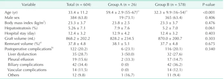
A total of 604 cases were included in this study. Of the patients, 26 were recipients whose donors were ≥55 years of age (group A), whereas the remaining 578 patients had donors who were <55 years of age (group B). The mean donor age was 59.4 ± 2.9 years (55–67 years) and 32.3 ± 9.9 years (16–54 years) in groups A and B, respectively. There was no statistically significant difference in the donor sex ratio between the groups (P = 0.406). The BMI score was 23.8 ± 2.5 kg/m2 in group A and 23.3 ± 3.7 kg/m2 in group B (P = 0.476). The degree of graft hepatosteatosis did not different statistically (7.9% ± 7.6% in group A vs. 5.2% ± 7.1% in group B, P = 0.061). The mean donor length of hospital stay was 12.9 ± 4.5 and 12.4 ± 3.2 days in groups A and B, respectively (P = 0.403). The mean graft liver volume was not significantly different between the groups (828.2 ± 234.5 mL vs. 870.0 ± 200.7 mL, respectively, P = 0.303), nor was the mean remnant liver volume of the donors (38.1% ± 5.1% vs. 37.7% ± 4.8%, respectively, P = 0.675). And
Table 1. Donor characteristics in the comparison study using elderly donors and younger donors in the living donor liver transplantation
Variable Total (n = 604) Group A (n = 26) Group B (n = 578) P-value
Age (yr) 33.4 ± 11.2 59.4 ± 2.9 (55–67)c) 32.3 ± 9.9 (16–54)c) <0.001
Male sex 384 (63.8) 19 (73.1) 365 (63.4) 0.406
Body mass index (kg/m2) 23.3 ± 3.7 23.8 ± 2.5 23.3 ± 3.7 0.476
Hepatosteatosis (%) 5.26 ± 7.1 7.9 ± 7.6 5.2 ± 7.0 0.061
Hospital stay (day) 12.4 ± 3.2 12.9 ± 4.2 12.4 ± 3.2 0.403
Graft volume (mL) 868.2 ± 202.2 828.2 ± 234.5 870.0 ± 200.7 0.303
Remnant volume (%)a) 37.8 ± 4.8 38.1 ± 5.1 37.7 ± 4.8 0.675
Postoperative complicationsb) 122 (20.2) 6 (23.1) 116 (20.1) 0.340
Liver dysfunction 35 (28.7) 3 (50.0) 32 (27.6)
Pleural effusion 19 (15.6) 2 (33.3) 17 (14.7)
Biliary complications 42 (34.4) 0 (0) 42 (36.2)
Vascular complications 14 (11.5) 0 (0) 14 (12.1)
Others 12 (9.8) 1 (16.7) 11 (9.4)
Values are presented as mean ± standard deviation or number (%) unless otherwise indicated.
Group A, recipients of livers from elderly donors (≥55 years of age); group B, recipients of livers from younger donors (<55 years of age).
a)Remnant donor liver volume, b)Postoperative complications. c)Mean ± standard deviation (range).
then, the postoperative complication rate of donors in each groups was compared. In group A, there were 3 cases of liver dysfunction (11.5%), 2 cases of pleural effusion (7.7%) and 1 case of postoperative ileus (3.8%). There was no case of biliary complication and vascular complication in elderly donor group.
All of the liver dysfunction was temporary, pleural effusion and postoperative ileus was spontaneously improved with conservative care. There was no statistically significantly difference in postoperative complication rate between two groups (P = 0.340) (Table 1).
Recipients
The recipients’ characteristics and posttransplantation out- comes are presented in Table 2. There were no statistically significant differences in age, BMI, CTP score, MELD score, or proportion of the high urgency patients. There was also no significant difference in anatomic variation such as portal vein thrombosis and biliary variation that could affect the postoperative surgical complication rate (P = 0.466). However, the sex ratio of the recipients between the groups did differ significantly as follows: in group B, 418 patients (72.7%) were male, whereas in group A, 13 patients (50%) were male (P = 0.023). A T-tube was used during bile duct anastomosis in 5 cases (19.2%) in group A and 138 cases (23.9%) in group B (P =
0.586). The overall lengths of hospital and intensive care unit stays did not differ between the groups (33.3 ± 16.9 days vs.
30.3 ± 21.7 days, respectively, P = 0.481; 7.1 ± 1.2 days vs.
7.3 ± 5.4 days, respectively, P = 0.821). The most common indication for liver transplantation in both groups was liver cirrhosis due to HBV infection (88.5% vs. 70.3%, respectively, P
= 0.048), followed by hepatocellular carcinoma (HCC: 53.8%
vs. 41.1%, respectively, P = 0.225). The mean AFP level in all the patients and that in the patients with HCC did not differ significantly between the groups (64.3 ± 135.8 ng/mL vs. 67.9
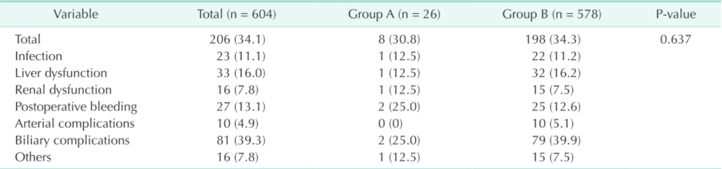
± 218.8 ng/mL, P = 0.933; 67.1 ± 133.6 ng/mL vs. 115.1 ± 07.0 ng/mL, P = 0.562, respectively). Among the patients with HCC, 2 patients (14.3%) in group A and 40 patients (16.8%) in group B experienced recurrence during the follow-up period. And then, the postoperative complication rate of recipient in each groups was compared. There were 8 cases of postoperative complication cases in group A. The most common postoperative complication was biliary complication (39.3%), there were 2 cases (25.0%) in group A and 79 cases (39.9%) in group B. However, there was no significant difference in overall complication rate between two groups (P = 0.637) (Table 3).
Although there was no significant difference in hospital mortality rate between the groups (7.7% vs. 7.4%, respectively, P = 0.955), the overall mortality rate was significantly higher
Table 2. Recipient characteristics and posttransplantation outcomes in the comparison study using elderly donors and younger donors in the living donor liver transplantation
Variable Total (n = 604) Group A (n = 26) Group B (n = 578) P-value
Age (yr) 49.8 ± 9.1 52.2 ± 11.8 49.7 ± 9.0 0.183
Male sex 431 (71.7) 13 (50.0) 418 (72.7) 0.023
Body mass index (kg/m2) 23.9 ± 3.6 23.8 ± 2.5 23.9 ± 3.7 0.880
CTP score 9.2 ± 2.6 10.2 ± 2.2 9.2 ± 2.6 0.055
MELD score 16.5 ± 9.8 19.4 ± 9.5 16.4 ± 9.8 0.130
High urgencya) 80 (13.2) 5 (19.2) 75 (13.0) 0.376
α-FP (ng/mL) 67.8 ± 215.1 64.3 ± 135.8 67.9 ± 218.1 0.933
α-FP of HCC (ng/mL) 112.4 ± 300.1 67.1 ± 133.6 115.1 ± 307.0 0.562
Anatomic variation 272 (45.0) 17 (65.4) 255 (44.1) 0.466
PV thrombosis 80 (29.4) 5 (29.4) 75 (29.4)
Bile duct variation 177 (65.1) 10 (58.8) 167 (65.5)
Others 15 (5.5) 2 (11.8) 13 (5.1)
Disease
HCC/HBV/HCV/Alc./others 251/428/27/65/80 14/23/0/2/1 237/405/27/63/79 0.219
T-tube 143 (23.7) 5 (19.2) 138 (24.0) 0.586
ICU stay (day) 7.3 ± 5.3 7.1 ± 1.2 7.3 ± 5.4 0.821
Hospital stay (day) 30.4 ± 21.5 33.3 ± 16.9 30.3 ± 21.7 0.481
Hospital mortality 45 (7.5) 2 (7.7) 43 (7.4) 0.955
Overall mortality 116 (19.2) 12 (46.2) 104 (18.1) 0.004
Survival (mo) 50.6 ± 40.6 31.2 ± 31.3 51.4 ± 40.8 0.013
Values are presented as mean ± standard deviation or number (%).
Group A, recipients of livers from elderly donors (≥55 years of age); group B, recipients of livers from younger donors (<55 years of age); BMI, body mass index; CTP, Child-Turcotte-Pugh; MELD, model for end stage liver disease; ICU, intensive care unit; HCC, hepatocellular carcinoma; PV, portal vein; Alc., alcohol.
a)High urgency included United Network for Organ Sharing status I and IIA.
in group A than in group B (46.2% vs. 18.1%, P = 0.004). A total of 12 patients in group A and 106 patients in group B died during the follow-up period. The causes of death in group A were biliary complications (n = 5, 41.7%), arterial complications (n = 2, 16.7%), HCC recurrence (n = 2, 16.7%), portal vein complications (n = 1, 8.3%), sepsis (n = 1, 8.3%), and other
causes (n = 1, 8.3%). In group B, the causes of death included HCC recurrence (n = 31, 29.2%), sepsis (n = 23, 21.7%), biliary complications (n = 10, 9.4%), portal vein complications (n = 9, 8.5%), arterial complications (n = 5, 5.7%), and other causes (n = 27, 25.5%) (Table 4). The overall survival length was 31.2 ± 31.3 months in group A and 51.4 ± 40.8 months in group B (P = 0.013). The 1-, 3-, and 5-year posttransplantation survival rates were respectively 63.4%, 58.5%, and 44.6% in group A and 86.9%, 84.3%, and 80.7% in group B (P < 0.001) (Fig. 1).
DISCUSSION
The use of elderly donor livers for LDLT remains contro- versial. Some studies reported favorable results [5,13,15], but others reported significantly increased risks of initial graft non function, and low graft and patient survival rates. However, almost all of the earlier studies were of DDLT [12,14,19,20], and only one report was of LDLT [4].
Liver transplantation for end-stage liver disease and HCC seems to provide better results than other treatment modalities, but the scarcity of graft donors remains its major obstacle.
Furthermore, there have been low numbers of brain death liver donors due to cultural customs, especially in Far East Asia. As Table 3. Comparison of the postoperative complications between two groups
Variable Total (n = 604) Group A (n = 26) Group B (n = 578) P-value
Total 206 (34.1) 8 (30.8) 198 (34.3) 0.637
Infection 23 (11.1) 1 (12.5) 22 (11.2)
Liver dysfunction 33 (16.0) 1 (12.5) 32 (16.2)
Renal dysfunction 16 (7.8) 1 (12.5) 15 (7.5)
Postoperative bleeding 27 (13.1) 2 (25.0) 25 (12.6)
Arterial complications 10 (4.9) 0 (0) 10 (5.1)
Biliary complications 81 (39.3) 2 (25.0) 79 (39.9)
Others 16 (7.8) 1 (12.5) 15 (7.5)
Values are presented as number (%).
Group A, recipients of livers from elderly donors (≥55 years of age); group B, recipients of livers from younger donors (<55 years of age).
Table 4. Cause of recipient death in the comparison study using elderly donors and younger donors in the living donor liver transplantation
Variable Total (n = 118) Group A (n = 12) Group B (n = 106) P-value
Arterial complications 8 (6.8) 2 (16.7) 6 (5.7) 0.043
Biliary complications 15 (12.7) 5 (41.7) 10 (9.4) <0.001
Portal complications 10 (8.5) 1 (8.3) 9 (8.5) 0.359
Recurrent HCC 33 (28.0) 2 (16.7) 31 (29.2) 0.647
Sepsis 24 (20.3) 1 (8.3) 23 (21.7) 0.722
Others 28 (23.7) 1 (8.3) 27 (25.5) 0.656
Values are presented as number (%).
Group A, recipients of livers from elderly donors (≥55 years of age); group B, recipients of livers from younger donors (<55 years of age); HCC, hepatocellular carcinoma.
Fig. 1. Kaplan-Meier survival curves of the recipients (P <
0.001) in living donor liver transplantation. Group A, recipients of livers from elderly donors (≥55 years of age); group B, recipients of livers from younger donors (<55 years of age).
such, family members would be the most promising source of liver grafts for LDLT in these areas. Therefore, we have no choice but to use the limited supplies of available liver grafts, meaning that we cannot deny elderly LDLT donor volunteers unless they are medically compromised. In this study, we tried to use of medically uncompromised donor graft and there was no significant difference in the post-operative complication rate of donors between usual and elderly group (P = 0.340).
Earlier studies stated that the cold ischemic time in DDLT negatively influences graft patency and is the major problem in grafts from elderly patients. However, this problem can be overcome in LDLT as demonstrated by the lack of cases of primary liver graft non function in the present study. Despite this, several complications other than primary liver graft non function occurred. Therefore, older donor age with or without other complications could negatively influence graft patency [21].
Our study showed that the outcomes including overall mortality rate and mean survival length in recipients of liver grafts from elderly living donors was significantly poorer than those of nonelderly donors (46.2% vs. 18.1%, P = 0.004).
Nevertheless, the 5 year overall survival rate was 79.5% in the elderly group and 81.5% in the nonelderly group. This is a promising result compared with those of earlier reports that revealed 60%–80% survival rates [22,23]. Moreover, there was no case of primary liver graft non function, a concern in DDLT using elderly donor grafts, and no other severe acute complications in our study. In addition, the hospital mortality rates did not differ significantly between the groups (7.7% vs.
7.4%, P = 0.955).
Some authors described that preoperative biliary and vascular anomaly was related with postoperative complication and eventually, could affect mortality rate [24,25]. In this study, there was no significant difference in anatomic variation between two groups (P = 0.466) and the postoperative complication rate was not so different between groups (P = 0.637). Furthermore, there was also no statistically significant difference in terms of age, CTP score, MELD score, proportion of the high urgency patients, or other preoperative medical conditions between the recipient groups. Therefore, donor age might be the sole risk factor for reduced recipient survival rate in our study.
Woodhouse and Wynne [10] described that liver age itself may be associated with a loss in proliferative response and regeneration. This causes increased susceptibility to irreversible damage such as viral infection, ischemic injury, and impaired hepatic blood flow despite the appearance of well-maintained liver graft function.
Mutimer et al. [20] and other several investigators [6,26]
described that the use of elderly donors for HCV-infected recipients promotes HCV recurrence and hepatic fibrosis or even
cirrhosis. This could explain the poor outcome of recipients of liver donations of elderly patients; however, it cannot explain our results because there were no patients with HCV in group A (recipients whose donors were ≥55 years of age). On the other hand, Lake [27] described that donor age was not related with disease recurrence or graft loss in patients with HBV.
Some investigators have suggested that the degree of hepa- tosteatosis of donor liver increases with age as a result of reduced protein synthesis, prolonged cholestasis, and delayed capacity for regeneration and described that with donor age, it was one of the most important risk factors for decreased graft and patient survival [10,28]. In our study, the degree of hepatosteatosis of group A seemed to be higher than that of group B (7.9% ± 7.6% vs. 5.2% ± 7.1%), but the difference was not statistically significant (P = 0.061).
In group A, the most common cause of death was biliary complications (41.7%), followed by hepatic artery thrombosis and recurrent HCC. Most of the biliary complications arose from biliary anastomotic strictures. Its prevalence is similar or slightly higher than those described by others [29,30]. Biliary complications are considered the most common problem after LDLT, but there have been no definite measurements for that without careful preoperative evaluation and the use of a meticulous surgical technique. Several studies have described that biliary complications, especially anastomotic and nonanastomotic strictures, are closely related to arterial complications, mainly hepatic artery thrombosis [7,8].
Due to increasing the disparity between donors and reci- pients, expansion of the donor pool has become a serious issue.
As the mean life expectancy continues to increase, increasing demands for elderly donors are emerging. However, our study shows a higher overall mortality rate in the group using elderly living donor livers (≥55 years of age) than in the group using younger living donor livers (<55 years of age).
Our study showed conflicting results compared with that of Li et al. [3] in which the use of older donors in LDLT had no negative influence on donor or recipient outcomes. However, that study had some limitation. First, there were fewer enrolled elderly donors (21 vs. 26) and much fewer younger donors (108 vs. 578) than in our study. Decisively, the criteria used by that study to distinguish elderly donors (≥50 years of age) was more generous than those used in our study (≥55 years of age), which followed those of earlier reports [16-18] and many other transplant centers. Furthermore, the mean age of the recipients in that study who received livers from the elderly donors was much younger than that in our study (41.10 ± 10.26 years vs.
55.2 ± 11.8 years), which would influence the result to be more favorable than ours.
The limitation of this study was that it was not controlled or prospective; rather, it was a retrospective study. It also recruited a relatively small number of patients, especially in the elderly
1. Nadig SN, Bratton CF, Karp SJ. Marginal donors in liver transplantation: expan- ding the donor pool. J Surg Educ 2007;64:
46-50.
2. Montalti R, Nardo B, Bertelli R, Beltempo P, Puviani L, Vivarelli M, et al. Donor pool expansion in liver transplantation.
Transplant Proc 2004;36:520-2.
3. Li C, Wen TF, Yan LN, Li B, Yang JY, Xu MQ, et al. Safety of living donor liver transplantation using older donors. J Surg Res 2012;178:982-7.
4. Yoshizumi T, Shirabe K, Soejima Y, Taketomi A, Yamashita N, Ikegami T, et al. Living donor liver transplantation in patients older than 60 years. Trans- plantation 2010;90:433-7.
5. Keswani RN, Ahmed A, Keeffe EB. Older age and liver transplantation: a review.
Liver Transpl 2004;10:957-67.
6. Serrano MT, Garcia-Gil A, Arenas J, Ber Y, Cortes L, Valiente C, et al. Outcome of liver transplantation using donors older than 60 years of age. Clin Transplant 2010;24:543-9.
7. Cescon M, Zanello M, Grazi GL, Cucchetti A, Ravaioli M, Ercolani G, et al. Impact of very advanced donor age on hepatic artery thrombosis after liver transplantation.
Transplantation 2011;92:439-45.
8. Stewart ZA, Locke JE, Segev DL, Dagher NN, Singer AL, Montgomery RA, et al.
Increased risk of graft loss from hepatic artery thrombosis after liver trans plan- tation with older donors. Liver Transpl 2009;15:1688-95.
9. Yersiz H, Shaked A, Olthoff K, Imagawa D, Shackleton C, Martin P, et al. Correlation
between donor age and the pattern of liver graft recovery after transplantation.
Transplantation 1995;60:790-4.
10. Woodhouse KW, Wynne HA. Age-related changes in liver size and hepatic blood flow. The influence on drug metabolism in the elderly. Clin Pharmacokinet 1988;
15:287-94.
11. Schmucker DL. Liver function and phase I drug metabolism in the elderly: a paradox. Drugs Aging 2001;18:837-51.
12. Deschênes M, Forbes C, Tchervenkov J, Barkun J, Metrakos P, Tector J, et al. Use of older donor livers is associated with more extensive ischemic damage on intra operative biopsies during liver trans- plantation. Liver Transpl Surg 1999;5:357- 61.
13. Ikegami T, Nishizaki T, Yanaga K, Shimada M, Kishikawa K, Nomoto K, et al. The impact of donor age on living donor liver transplantation. Transplantation 2000;70:
1703-7.
14. Grazi GL, Ravaioli M, Zanello M, Ercolani G, Cescon M, Varotti G, et al. Using elderly donors in liver transplantation. Trans- plant Proc 2005;37:2582-3.
15. Kuramitsu K, Egawa H, Keeffe EB, Kasahara M, Ito T, Sakamoto S, et al.
Impact of age older than 60 years in living donor liver transplantation. Transplan- tation 2007;84:166-72.
16. Santori G, Andorno E, Morelli N, Bottino G, Mondello R, Gianelli Castiglione A, et al. Impact of different cadaveric donor age cut-offs on adult recipient survival after liver transplantation: a single-center analysis. Transplant Proc 2005;37:2576-81.
17. Hesse UJ, Berrevoet F, Pattyn P, de Hemptinne B. Results of liver transplan- tation in elderly patients (> 55 years of age). Langenbecks Arch Chir Suppl Kongressbd 1996;113:419-21.
18. Adam R, Karam V, Delvart V, O'Grady J, Mirza D, Klempnauer J, et al. Evolution of indications and results of liver trans- plantation in Europe: a report from the European Liver Transplant Registry (ELTR). J Hepatol 2012;57:675-88.
19. Kim do Y, Choi MS, Lee JH, Koh KC, Paik SW, Yoo BC, et al. Older donor allografts are associated with poor patient survival after living donor liver transplantation for hepatitis B virus-related liver diseases.
Liver Int 2007;27:260-7.
20. Mutimer DJ, Gunson B, Chen J, Berenguer J, Neuhaus P, Castaing D, et al. Impact of donor age and year of transplantation on graft and patient survival following liver transplantation for hepatitis C virus.
Transplantation 2006;81:7-14.
21. Cassuto JR, Patel SA, Tsoulfas G, Orloff MS, Abt PL. The cumulative effects of cold ischemic time and older donor age on liver graft survival. J Surg Res 2008;148:38- 44.
22. Shah SA, Levy GA, Greig PD, Smith R, McGilvray ID, Lilly LB, et al. Reduced mortality with right-lobe living donor compared to deceased-donor liver trans- plantation when analyzed from the time of listing. Am J Transplant 2007;7:998- 1002.
23. Mizuno S, Yokoi H, Shiraki K, Usui M, Sakurai H, Tabata M, et al. Prospective study on the outcome of patients with
REFERENCES
group. Because of the small number of cases, a multivariate analysis of the cause of death was not possible. As such, we think that a large and multicenter analysis of LDLT using elderly donor livers is required.
In conclusion, based on our results, we believe that LDLT using elderly donors (≥55 years of age) could induce more serious complications such as biliary strictures or hepatic artery thrombosis and could cause higher morbidity and mortality
rates than LDLT using younger donors. As such, the selection of elderly donor livers should be made carefully.
CONFLICTS OF INTEREST
No potential conflict of interest relevant to this article was reported.
hepatocellular carcinoma registered for living donor liver transplantation: how long can they wait? Transplantation 2010;89:650-4.
24. Kasahara M, Egawa H, Tanaka K, Ogawa K, Uryuhara K, Fujimoto Y, et al. Varia- tions in biliary anatomy associated with trifurcated portal vein in right-lobe living- donor liver transplantation. Transplan- tation 2005;79:626-7.
25. Radtke A, Sgourakis G, Sotiropoulos GC, Molmenti EP, Nadalin S, Schroeder T, et al. Vascular and biliary anatomy of the right hilar window: its impact on recipient
morbidity and mortality for right graft live donor liver transplantation. World J Surg 2009;33:1941-51.
26. Boin IF, Ataide EC, Leonardi MI, Stucchi R, Seva-Pereira T, Pereira IW, et al. Elderly donors for HCV(+) versus non-HCV reci- pients: patient survival following liver transplantation. Transplant Proc 2008;40:
792-6.
27. Lake JR. Should liver transplantation be performed for patients with chronic hepa- titis B? Yes! Liver Transpl Surg 1995;1:260- 5.
28. Ono Y, Kawachi S, Hayashida T, Wakui M,
Tanabe M, Itano O, et al. The influence of donor age on liver regeneration and hepa- tic progenitor cell populations. Surgery 2011;150:154-61.
29. Duailibi DF, Ribeiro MA Jr. Biliary compli- cations following deceased and living donor liver transplantation: a review.
Transplant Proc 2010;42:517-20.
30. Takatsuki M, Eguchi S, Kawashita Y, Kanematsu T. Biliary complications in recipients of living-donor liver trans- plantation. J Hepatobiliary Pancreat Surg 2006;13:497-501.