Relationship Between Electromyographic Activity of the Abductor Hallucis and the Pressure of a Pinch Gauge During Short Foot Exercise
Kyung-mi Park, B.H.Sc., P.T.
Dept. of Physical Therapy, The Graduate School, Yonsei University Heon-seock Cynn, Ph.D., P.T.
Dept. of Physical Therapy, College of Health Science, Yonsei University
Dept. of Ergonomic Therapy, The Graduate School of Health and Environment, Yonsei University Houng-sik Choi, Ph.D., P.T.
Dept. of Physical Therapy, Hanseo University
Abstract
6)The aims of this study were to investigate the correlation between the electromyographic (EMG) activity of the abductor hallucis (AbdH) and the amount of pressure measured by a pinch gauge (PG), and to compare the EMG activity of AbdH and the pressure measured by the PG during short foot (SF) exercise in subjects with pes planus and in subjects with a neutral foot alignment. Fourteen subjects were recruited for this study (pes planus group=7; neutral foot alignment group=7). A surface EMG was used to collect AbdH activity, and a PG was positioned under the first metatarsophalangeal joint to measure the pressure produced by the first metatarsal head during the SF exercise. The AbdH activity and the pressure measured by the PG showed a positive good correlation (r=.80, p<.05). The EMG activity of the AbdH and the pressure measured by the PG were significantly lower for subjects with pes planus than for subjects with a neutral foot alignment (p<.05). Based on these findings, the PG can be recommended as an effective instrument for evaluating the performance of the AbdH. It may also be beneficial for monitoring how well the SF exercise is performed, and for providing visual feedback to patients with pes planus during SF exercise in a clinical setting.
Key Words: Medial longitudinal arch; Pes planus; Short foot exercise.
Introduction
The abductor hallucis (AbdH), which is the most medial muscle in the first intrinsic muscle layer of the plantar surface of the foot, has two important roles in maintaining stance, gait, and balance (Fiolkowski et al, 2003; Headlee et al, 2008). First, the AbdH sup- ports the medial longitudinal arch (MLA) of the foot.
Because the AbdH originates from the posteromedial calcaneus and inserts into the medial sesamoid of the hallux or proximal phalanx (Kendall et al, 2005), its functions include hallucis abduction, first meta- tarsophalangeal (MTP) flexion and supination, and the
inversion of the calcaneus in conjunction with the ele- vation of the MLA (Wong, 2007). Second, the AbdH controls pronation of the foot. Because the AbdH is almost perpendicular to the oblique midtarsal joint ax- is, this muscle stabilizes and supinates the midtarsal joint against the pronating force of ground reaction during the propulsive phase (Mann and Inman, 1964).
Several studies have found that the navicular drop is significantly increased after decreased muscle activity, or fatigue, of the AbdH (Fiolkowski et al, 2003;
Headlee et al, 2008).
Several different strengthening methods for intrinsic foot muscles, such as the AbdH, have been described in
Corresponding author: Heon-seock Cynn [email protected]
Parameters Mean±SD
Age (yrs) 22.6±2.7
Weight (㎏) 68.9±13.2
Height (㎝) 163.4±28.6
Table 1. General characteristics of subjects (N=14)
order to prevent overuse injuries in people who present with problems from excessive pronation (Fiolkowski et al, 2003; Headlee et al, 2008; Wong, 2007). These in- clude the ‘toe curl’ (TC), which is achieved by curling the toes on a towel, bunching the towel beneath the foot using interphalangeal and MTP flexion of the toes (Jung et al, 2011). Another exercise is the ‘short foot’
(SF), which is accomplished by shortening the foot in an antero-posterior direction and actively attempting to bring the head of the first metatarsal toward the heel without toe flexion (Prentice, 2009). Jung et al (2011) reported significantly greater electromyographic (EMG) activity in the AbdH during the SF exercise than dur- ing the TC exercise. However, it is difficult to monitor how well the SF exercise is performed because of the lack of appropriate measurement methods. We thought, based on our clinical experience, that the pinch gauge (PG), which is designed for objective measurement of finger pinch strength, can be a useful clinical instrument for evaluating the performance level of the SF exercise by determining the pressure loaded on a PG placed un- der the first metatarsal head. In addition, the PG would also provide visual feedback of AbdH activation by monitoring changes in pressure during the SF exercise.
Because the metatarsal bones and the navicular serve as the keystones of the MLA (Franco, 1987), the first metatarsal head may press the PG in an inferoposterior direction by bring the first metatarsal bone toward the heel during the SF exercise. Thus, the pressure de- termined by PG can indicate the force of the AbdH to flex the tarsometatarsal joint for elevating MLA.
Therefore, the aims of this study were to determine the association between the muscle activity of the AbdH and the pressure measured by the PG, and to compare the EMG activity of AbdH and the pressure loaded on the PG for the subjects with pes planus and for the subjects with a neutral foot alignment. We hy-
pothesized that there would be a positive correlation between the muscle activity of the AbdH and the amount of pressure determined by the PG. Moreover, we hypothesized that the EMG activity of AbdH and the measured pressure of subjects with a neutral foot alignment would be higher than those of the subjects with pes planus during the SF exercise.
Methods Subjects
Seven subjects with pes planus and 7 subjects with a neutral foot alignment participated in this study. Table 1 shows the demographic data of the subjects. We screened the foot type of the subjects by measuring the resting calcaneal stance position (RCSP) (neutral align- ment: between 2° of inversion and 2° of eversion) and their scores on the navicular drop (ND) test (neutral alignment: between 5 and 9 ㎜) (Cote et al, 2005;
Razeghi and Batt, 2002). To measure the RCSP, the ex- aminer drew a bisection line on the calcaneus in the prone position. After this, the subject was asked to stand in a relaxed position on a 20 ㎝ high wooden box.
The RCSP was measured using the posterior bisection line of the heel in relationship to the ground, using a gravity goniometer. In previous study, the intratester and intertester reliability of measurement of RCSP were investigated. ICC and SEM values for intratester were .85 and 1.2 and for intertester were .68 and 1.8, re- spectively (Sell et al, 1994). In addition, the intratester and intertester reliability of the ND test were evaluated in previous study. ICC and SEM values for intratester were .83 and 1.7 and for inttertester were .73 and 2.1, respectively (Sell et al, 1994). The exclusion criteria in- cluded past or current inflammatory arthritis, foot or an- kle surgery, diabetes, lower limb amputation, and foot
Figure 1. Hydraulic pinch gauge.
deformities such as hallux valgus, hammer toe, or claw toe. Prior to the study, the subjects had been explained about all of the procedures in detail, and they provided written, informed consent in order to participate.
Data collection and processing
Surface electromyography7)
EMG data were collected using a Noraxon TeleMyo 2400 system1) and analyzed using a Noraxon MyoResearch 1.06 XP software. To prepare the electrode sites, the skin was shaved and then cleaned with rubbing alcohol. Surface electrodes were positioned at an interelectrode distance of 2 ㎝. The reference electrode was placed on the lateral malleo- lus of the tested side. EMG data were collected for the AbdH, for which the electrode sites were set ap- proximately 1~2 ㎝ posterior to the navicular tuber- osity, which is just anterior to an imaginary line drawn through the anterior margin of the medial malleolus (Arinci Incel et al, 2003). The sampling rate was 1000 ㎐. A bandpass filter was used between 20 and 450 ㎐. The raw data were processed into the root mean square (RMS) with a window of 50 milliseconds. For normalization, maximum voluntary isometric contraction (MVIC) of the AbdH was measured in the manual muscle testing position, as recommended by Kendall et al (2005). The inves- tigator gripped the heel firmly, at a neutral ankle po- sition, and applied resistance to just the medial side
of the first metatarsal and proximal phalanx to create abduction of the MTP joint of the great toe. The subject was asked to adduct the hallux and forefoot against the pressure applied by the examiner. EMG data were collected for five seconds. The initial sec- ond and the last second of the EMG data were dis- carded, and the remaining three seconds of data were used to determine the mean amplitude of the MVIC.
The mean of three trials of MVIC measurement was calculated for data analysis. The means of three trials for each exercise were used for data analysis. The collected EMG amplitudes of the AbdH during each exercise were expressed as a percentage of mean MVIC (%MVIC).
The pressure determined by the PG (pinch gauge)8) The pressure was recorded by a hydraulic pinch gauge2). A PG was placed under the first metatarsal head to monitor how well the MTP joint of the great toe was kept on the ground during the SF exercise.
To keep the tested foot aligned in a straight line, the PG was embedded in a carpet (Figure 1). The PG was calibrated to be zeroed when the tested MTP joint of the great toe was placed on the PG and each trial was started. The subject was asked to maintain a maximum effort for five seconds. The examiner read the peak needle of the PG, which remained at maximum. Force was recorded in kilograms, and nor- malized by the participant’s weight to account for in- dividual differences. The mean of three trials was
1) Noraxon TeleMyo 2400T, Noraxon Inc., Scottsdale, AZ, U.S.A.
2) Hydraulic pinch gauge, Dynatronics Corporation, Salt Lake City, UT, U.S.A.
Subjects with pes planus Subjects with a neutral foot alignment p
Activity of AbdHa (%MVICb) 26.90±14.43* 69.29±16.30 <.05
Amount of Pressure (%Weight) 3.47±1.65 7.34±1.77 <.05
*Mean±SD, aabductor hallucis,bmaximum voluntary isometric contraction.
Table 2. EMG activity of the AbdH and the amount of pressure measured by the PG during the SF exercise, in subjects with pes planus and in subjects with a neutral foot alignment
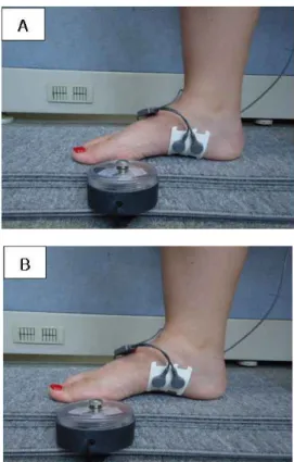
Figure 2. Short foot exercise with a PG. A:
Starting position, B: Maximum effort to elevate MLA.
calculated for data analysis.
Procedure
Before the testing session, the subjects were fami- liarized with the SF exercise and with the PG. The training session was approximately 20 minutes. The subject was asked to sit with hips and knees at ap- proximately 90° on a chair, and bring the head of the first metatarsal toward the measuring plate of the PG, and then to shorten the foot in an antero-posterior di- rection by maintaining contact between the head of the metatarsal and the measuring plate of the PG, without
toe flexion (Figure 2). The subject was instructed to perform the exercise with a maximum effort. The forefoot and heel were kept on the PG and ground, respectively, during the SF exercise. Three trials of five seconds were performed to measure the EMG ac- tivity of the AbdH and the pressure on the PG during the SF exercise. A five minute rest was allowed be- tween trials to minimize the testing effect and fatigue.
Statistical Analysis
Pearson’s correlation was used to determined the as- sociation between the muscle activity of the AbdH and the pressure measured by the PG. Kolmogorov-Smirnov Z-tests were performed to assess whether continuous data approximated a normal distribution. An independent t-test was used to compare the activity of the AbdH and the pressure measured by the PG in subjects with pes planus and subjects with a neutral foot alignment.
The statistical significance level was set at .05. All statistical analyses were performed using PASW Statistics version 18.0 software.
Results
There were no differences in age, weight, and height between the subjects with pes planus and those with a neutral foot alignment (p>.05). The muscle ac- tivity of the AbdH and the pressure measured by the PG showed a strong positive correlation (r=.80, p=.001).
Correlation coefficient >.75 was considered good to excellent correlation (Portney and Watkins, 2009). All of the continuous variables were found to approximate a normal distribution (Kolmogorov-Smirnov Z-test, p>.05). The EMG activity of the AbdH, and the pres-
sure measured by the PG, were significantly lower for subjects with pes planus than for those with a neutral foot alignment (p<.05) (Table 2).
Discussion
Clinically, the SF exercise, which emphasizes MTP joint flexion for enhancing the longitudinal and transverse arches, has been advocated as a means of improving neuromuscular control and the strength of the foot’s in- trinsic muscles (Greenman, 2003; Rothermel et al, 2004).
The SF exercise is performed with the foot on the ground (in a partial or full weight-bearing position), with the patient elevating their MLA while keeping the fore- foot and rearfoot on the ground (Greenman, 2003;
Rothermel et al, 2004). Therefore, it is important to mon- itor how well the SF exercise is performed, and how much the AbdH is activated, during the SF exercise.
In this study, the AbdH activity and the pressure measured by the PG showed a strong positive correla- tion, which supports our research hypothesis. We as- sumed that the pressure measured by a PG positioned under the first MTP joint would indicate the activity of the AbdH, the intrinsic muscle activated to hold and improve the MLA. To our knowledge, this is the first SF exercise study to simultaneously measure the per- formance of intrinsic muscle through PG pressure and AbdH activity. Moreover, because the current study showed a relationship between the pressures measured by the PG and the AbdH activity in subjects with pes planus and subjects with a neutral foot alignment, the PG can be used for patients with pes planus, in a clin- ical setting, as a visual feedback instrument for mon- itoring and motivating AbdH activity and performance.
For SF exercise in the sitting position, the EMG activity of the AbdH was significantly higher for sub- jects with a neutral foot alignment than for subjects with pes planus, thus our research hypothesis was supported. We chose to use surface EMG to assess the AbdH activity. The AbdH is the most superficial and largest of the plantar intrinsic muscles, and Kura
et al (1997) assumed that the EMG activity of this muscle was representative of all the underlying plantar intrinsics muscles as well; it acts during flexion of the first MTP joint. Our findings agree with the results from previous studies that compared the activity of the intrinsic musculature, during ambulation, in individuals with flat-foot and normal-foot structures (Banks et al, 2001). Although the task performed by the subjects was different, the flatfooted patients showed decreased EMG activity in the AbdH. Decreased AbdH activity may cause accelerated and excessive pronation of the subtalar joint after initial contact, and into midstance, placing an increased load on the plantar fascia, liga- ments, and foot musculature that are attempting to slow down and limit pronation (Banks et al, 2001; Cole et al, 2005; May et al, 2002; Wearing et al, 2004).
Our results showed that the pressure measured by the PG in subjects with pes planus was significantly lower than that of subjects with a neutral foot alignment. We postulated that the pressure measured by the PG indicates the force produced by pulling the forefoot toward the heel during the SF exercise. This pressure can thus be implied as the level of perform- ance for elevating and maintaining the MLA during the SF exercise. Previous studies hypothesized that the primary biomechanical cause of certain foot and ankle syndromes that lead to pes planus-including plantar fasciitis, achilles tendinitis, hallux valgus, and tibialis posterior and tibialis anterior overuse syndromes-is not excessive pronation, but rather a lack of pronation control (Hintermann and Nigg, 1998; Kaufman et al, 1999; Van Boerum and Sangeorzan, 2003). Therefore, it is important to make efforts to address the dynamic control of pronation during static stance and ambula- tion by strengthening the intrinsic muscles. It may help to increase muscular support for the arch, thus increasing control of pronation (Fiolkowski et al, 2003;
Franco, 1987).
This study has several limitations. The first limitation is that we did not measure the EMG activity of other foot intrinsic muscles which could have contributed to bring the first metatarsal head in inferoposterior direc-
tion and elevate the MLA during the SF exercise.
However, a previous study postulated that the AbdH is representative of all the underlying intrinsic foot muscles (Kura et al, 1997). The second limitation is that we did not measure the angle of the MLA during the SF exercise. However, we asked subjects to elevate the MLA by pulling the forefoot to heel, and we observed how the height of the MLA changed during the SF exercise. Moreover, Jung et al (2011) reported that the SF exercise elevated the MLA during the exercise. The third limitation is that we did not measure the extrinsic foot muscles, such as the tibialis posterior, the tibialis anterior, and the flexor hallucis longus, which contribute to foot supination and lead to compensation. Further study is needed to determine the activity of the extrinsic foot muscles during SF exercise.
Conclusion
This study showed a strong positive correlation between the muscle activity of the AbdH and the pressure measured by the PG during the SF exercise.
In addition, the EMG activity of AbdH and the pres- sure measured by the PG during the SF exercise were significantly higher for the subjects with a neu- tral foot alignment than for the subjects with pes planus. Therefore, the PG can be recommended as an effective instrument for evaluating the performance of AbdH activation in the elevation and maintenance of the MLA. It may also be beneficial for monitoring how well the SF exercise is performed, as well as providing visual feedback for patients with pes planus doing the SF exercise in a clinical setting.
References
Arinci Incel N, Genç H, Erdem HR, et al. Muscle im- balance in halluxvalgus: An electromyographic study. Am J Phys Med Rehabil. 2003;82(5):345-349.
Banks AS, Downey MS, Martin DE, et al. McGlamry’s
Comprehensive Textbook of Foot and Ankle Surgery, 3rd ed. Philadelphia PA, Williams and Wilkins, 2001.
Cole C, Seto C, Gazewood J. Plantar fasciitis:
Evidence-based review of diagnosis and therapy.
Am Fam Physician. 2005;72(11):2237-2242.
Cote KP, Brunet ME, Gansneder BM, et al. Effects of pro- nated and supinated foot postures on static and dy- namic postural stability. J Athl Train. 2005;40(1):41-46.
Fiolkowski P, Brunt D, Bishop M, et al. Intrinsic pedal musculature support of the medial longi- tudinal arch: An electromyography study. J Foot Ankle Surg. 2003;42(6):327-333.
Franco AH. Pes cavus and pes planus. Analyses and treatment. Phys Ther. 1987;67(5):688-694.
Greenman P. Principles of Manual Medicine. 2nd ed.
Baltimore: Lippincott Williams & Wilkin, 2003.
Headlee DL, Leonard JL, Hart JM, et al. Fatigue of the plantar intrinsic foot muscles increases navicular drop. J Electromyogr Kinesiol. 2008;18(3):420-425.
Hintermann B, Nigg BM. Pronation in runners. Implications for injuries. Sports Med. 1998;26(3):169-176.
Jung DY, Kim MH, Koh EK, et al. A comparison in the muscle activity of the abductor hallucis and the medial longitudinal arch angle during toe curl and short foot exercises. Phys Ther Sport.
2011;12(1):30-35.
Kaufman KR, Brodine SK, Shaffer RA, et al. The effect of foot structure and range of motion on musculoskeletal overuse injuries. Am J Sports Med. 1999;27(5):585-593.
Kendall FP, McCreary EK, Provance PG, et al. Muscles:
Testing and function, with posture and pain. 5th ed. Baltimore, Lippincott Williams & Wilkins, 2005.
Kura H, Luo ZP, Kitaoka HB, et al. Quantitative analysis of the intrinsic muscles of the foot.
Anat Rec. 1997;249(1):143-151.
Mann R, Inman VT. Phasic activity of intrinsic muscles of the foot. J Bone Joint Surg Am. 1964;46:469-481.
May TJ, Judy TA, Conti M, et al. Current treatment of plantar fasciitis. Curr Sports Med Rep.
2002;1(5):278-284.
This article was received October 5, 2011, was reviewed October 14, 2011, and was accepted November 2, 2011.
Portney LG, Watkins MP. Foundations of Clinical Research: Applications to Practice. 3rd ed.
Pearson/Prentice Hall, Upper Saddle River, N.J., 2009.
Prentice WE. Rehabilitation Techniques in Sports Medicine. 4th ed. New York, McGraw Hill Higher Education, 2009.
Razeghi M, Batt ME. Foot type classification: A critical review of current methods. Gait Posture.
2002;15(3):282-291.
Rothermel SA, Hale SA, Hertel J, et al. Effect of active foot positioning on the outcome of a balance train- ing program. Phys Ther Sport 2004;5(2):98-103 Sell KE, Verity TM, Worrell TW, et al. Two meas-
urement techniques for assessing subtalar joint position: A reliability study. J Orthop Sports Phys Ther. 1994;19(3):162-7.
Van Boerum DH, Sangeorzan BJ. Biomechanics and pathophysiology of flat foot. Foot Ankle Clin.
2003;8(3):419-430.
Wearing SC, Smeathers JE, Yates B, et al. Sagittal movement of the medial longitudinal arch is un- changed in plantarfasciitis. Med Sci Sports Exerc. 2004;36(10):1761-1767.
Wong YS. Influence of the abductor hallucis muscle on the medial arch of the foot: A kinematic and anatomical cadaver study. Foot Ankle Int.
2007;28(5):617-620.