Abstract
The succulent leaves of Aloe have been widely used for medicinal and cosmetic industries. In this study, a comparative foliar anatomy in six cultivated Aloe species in Korea was carried out using light microscopy. These taxa show characteristics similar to those found in other Aloe species within the genus. The leaves of six cultivated species in this study are amphistomatic, tetracytic, and sunken stomata. The size range of the stomata and stomata index (SI) are 26.8-60.9 × 18.0-48.5 μm (length
× width), 1.0-11.9, respectively. Epidermal cells varied from rectangular to hexagon, with distinctive striate anticlinal wall. Druse, raphides, and styloids oxalate crystals are also found in the parenchyma cells. Moreover, xerophytic features which adapted to survive in water stressed condition are briefly discussed. These results of the anatomical characteristics of studied taxa could provide essential information for identifying species as herbal medicinal resources in Aloe family.
국내 재배산 알로에 6 분류군 잎의 비교해부학적 연구
송준호, 양선규, 문병철, 최고야 한국한의학연구원 한약연구부
A comparative foliar anatomical study of
six cultivated Aloe species (Asphodelaceae) in Korea
Jun-Ho Song, Sungyu Yang, Byeong Cheol Moon, Goya Choi
Herbal Medicine Research Division, Korea Institute of Oriental Medicine
Correspondence: Goya Choi
Herbal Medicine Research Division, Korea Institute of Oriental Medicine, 1672 Yuseong-daero, Yuseong-gu, Daejeon, 34054, Rep. of Korea
Tel: +82-42-868-9348, E-mail: serparas@kiom.re.kr
Received 2018-03-02, revised 2018-03-14, accepted 2018-03-17, available online 2018-03-19 doi:10.22674/KHMI-6-1-3
Keywords: Aloe, Comparative foliar anatomy, Stomata, Epidermal cells, Crystals, Aloe barbadensis L.
서론
알로에속(Aloe-Aloineae-Alooideae-Asphodelaceae)은 전 세계적으로 400~530 여종이 속해 있는 과 내 가장 큰 속으로, 남아프리카, 마다가스카르가 주요 다양성의 중심으로 보고되어 있고, 주로 아프 리카, 아라비아를 비롯한 열대 지역에 광범위하게 분포한다1-5).
한의학적으로는 노회(蘆薈), 라틴 생약명은 Aloe 로, 그 기원을 A. barbadensis L. (=A. vera L. ex Webb), A. ferox Mill., A. africana Mill. 또는 A. spicata Baker 및 이들 잡종의 잎에서 얻은 액즙(液 汁)을 건조한 것으로 규정하고 있다6,7). 노회는 사하약(瀉下藥) 중 공하약(攻下藥)으로 분류되며, 습 관성 변비나 열결변비(熱結便秘)를 사하시켜 치료하고, 간경실열(肝經實熱)로 인한 두훈두통(頭暈頭 痛)과 번조이노(煩燥易怒), 경간(驚癎) 등에 유효한 것으로 알려져 있다6). 한편, 약리학적으로는 본 속에 속하는 일부 분류군에서 항염증, 항균, 항암, 항산화, 간보호 등의 효과가 보고되는 등 꾸준한 연구가 진행되고 있다8-11).
잎에서 확인할 수 있는 형태학적, 해부학적 형질은 식별형질로서 매우 중요한 역할을 해왔으며, 분류 학적, 계통학적으로 그 유용성이 검증된 바 있다12,13). 알로에속 분류군의 잎 표피 형질 및 해부학적 연구는 대부분 아프리카 자생 분류군에 한해서 형질 기재 및 분류학적 유용성 검증을 위해 수행되었고, 건생식물(xerophytes)로서 갖는 형태학적 형질에 대해 일부 보고된 바 있다14-19). 우리나라에서는 속 내 일부 분류군이 제주도, 거제도를 비롯한 남부지역에서 재배되고 있으나, 재배식물로서 알로에 잎에 대한 형태학적, 해부학적 연구는 전무한 실정이다.
따라서, 본 연구에서는 우리나라에서 주로 재배되고 있는 알로에 6 분류군에 대해 잎 표피 형질과 해부학적 형질을 관찰 및 기재하며, 이들 형질에 대한 비교 형태학적 접근을 시도하였다. 이를 통해 한약재 노회 기원식물의 내부형태 기초 자료 축적과 분류학적 식별형질 및 형태감별 형질로서 잎 표피 및 해부학적 형질에 대한 유용성을 검증하고자 하였다. 또한 건생식물로서 수분스트레스에 반응하기 위해 적응한 형질에 대해 고찰하고자 하였다.
본론
1. 재료 및 방법
1) 식물 및 표본 재료
알로에속에 속한 식물은 교목 또는 관목이거나 줄기가 없는 다년생 초본으로, 잎은 로제트상으로 두껍고, 다육성이며, 엽선은 예두이거나 매우 뾰족하고, 엽연에는 대부분 단단한 치아상 거치가 있거나 가시가 있다. 화서는 아정생이며, 비스듬히 서거나 직립하는 총상화서이고, 화피는 원주형으로, 대개 적색, 주황색, 황색이며, 드물게 녹색 또는 백색이고, 열매는 포배개열하는 삭과이다1,20,21).
연구에 사용한 재료는 국내에서 재배되고 있는 알로에 6 분류군으로 경상남도 거제시 거제면 읍내로 소재의 알로에농장(34°51′57″ N, 128°34′57″ E)에서 생체를 수집하여 관찰하였으며, 실험에 사용된 재료는 건조표본으로 제작한 후에 표본번호를 부여하여 한국한의학연구원 한약표준표본관(KIOM)에 증거표본으로 보관하였다(Appendix). 실험에 사용한 모든 분류군의 잎 시료는 모두 성숙한 개화기의
잎으로 선별하였고, 일관성 있게 정중앙 부위를 대상으로 비교하였으며, 가능한 분류군 당 2 개체의 재료를 이용하여 관찰하였다.
2) 잎 표피 및 해부학적 실험 방법
잎 표피 형질: 잎의 향축면(상면; adaxial surface)과 배축면(하면; abaxial surface)을 구분하였고, 표피에서 깊이 5 mm 정도로 절편을 내어 Jeffrey's solution22)에 60℃에서 5~10 분간 담가두었다. 이후 Safranin O 로 염색하여 봉입제(Permount™ SP15-100, Fisher Scientific, Fair Lawn, USA)를 이용, 영구표본으로 제작하였다. Stomatal index (SI)는 0.5 mm2 당 기공의 수 / (0.5 mm2 당 기공의 수 + 0.5 mm2 당 표피세포의 수) × 100 으로 측정 및 계산하였다.
해부학적 형질: 잎의 해부학적인 구조를 밝히기 위해서 파라핀 매몰법을 사용하였다23). 70% 알코올 에 보관된 시료는 알코올시리즈(alcohol series: 80%, 90%, 95%, 98%, 100% Ⅰ, 100% Ⅱ에서 각각 약 30 분 정도 보관)을 거쳐 탈수한 후, 녹는점이 56~58℃인 파라핀(Paraplast, Leica Microsystems, Wetzlar, Germany)과 치환시켜 조직포매기(modular tissue embedding center; Leica EG1150H, Leica Microsystems, Wetzlar, Germany)로 embedding cassettes 에 매몰한 후 조직절편기(sliding microtome; Leica SM 2010R, Leica Microsystems, Wetzlar, Germany)를 이용하여 5~10 μm 두께로 절단하였다. 절단된 절편은 슬라이드 글라스(slide glass) 위에 부착시켜 신전기(slide warmer; C-SL, Chang Shin Scientific Co, Korea)에 올려 신장시켜 완전 건조시켰다. 준비된 슬라이드는 xylene 을 이용한 탈파라핀(deparaffin)과정과 Safranin O 와 Fastgreen FCF 를 이용한 이중염색(double stain- ing)을 거친 후, 봉입제로 봉입하였다. 잎 표피형질과 해부학적 형질 관찰을 위해 영구표본은 광학현미 경(light microscopy; Olympus BX-53 Microscope, Olympus, Tokyo, Japan)을 이용하였으며, 현미 경용 디지털카메라(digital camera for microscopes; Olympus DP21, Olympus, Tokyo, Japan)로 촬 영하였다.
해부학적 형질에 관한 기재는 Anatomy of the Dicotyledons24)와 Esau’s Plant Anatomy25)에서 제시 된 용어를 따랐다.
2. 결과 및 고찰
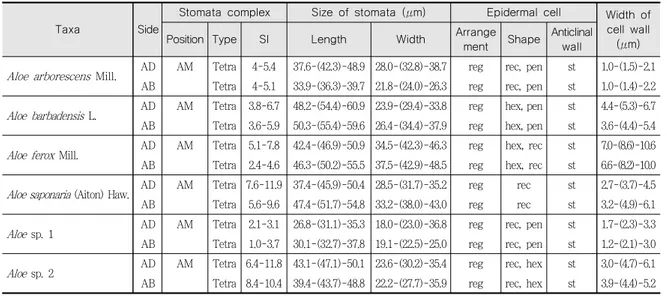
국내에서 재배되고 있는 알로에속 6 분류군[알로에(A. arborescens Mill.), 알로에베라(A. barba- densis), 희망봉알로에(A. ferox), 알로에사포나리아(A. saponaria (Aiton) Haw.), 미동정 2 분류군 (Aloe sp. 1, sp. 2)]의 잎 표피 형질(기공복합체의 유형, 기공의 크기, stomata index, 표피세포의 배열, 형태, 수층벽의 구조, 세포벽의 두께)과 해부학적 구조(기공, 공변세포의 위치, 큐티클, 결정체의 종류, 유관속)를 관찰하고, 기재하였다(Table 1).
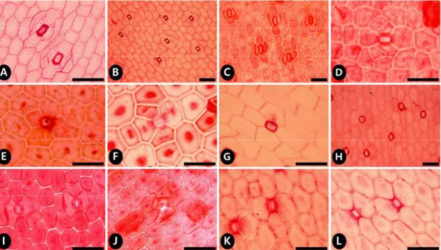
연구된 알로에속 6 분류군의 기공복합체는 모든 분류군에서 향축면과 배축면 양면 모두에 기공이 존재하는 양면기공엽(amphistomatic type)으로 나타나(Fig. 1; Table 1), 기존의 연구와 동일한 것으 로 확인되었다18,19,26). 공변세포의 크기는 26.8~60.9 × 18.0~48.5 μm (길이 × 너비)로 분류군마다 차이 를 보이는데, 알로에베라가 평균 54.9 × 31.9 μm 로 가장 크게 나타났으며, Aloe sp. 1 이 평균 31.9
× 22.8 μm 로 가장 작게 관찰되었다(Table 1). 또한, 대부분의 분류군에서 배축면에 분포하는 기공의
크기가 향축면에 비해 큰 것으로 나타났고, stomatal index 는 대부분 향축면이 배축면보다 높게 나타 났다(Table 1). 이는 배축면의 기공의 크기가 비교적 큰 것에 반해 일정 면적 내 표피세포 당 기공의 분포는 적게 나타나는 것으로 판단된다. 한편, 기존에 보고된 남아프리카 자생 희망봉알로에의 경우 양면 기공의 빈도가 유사하게 관찰된 반면19), 본 연구에서 관찰한 국내 재배 희망봉알로에의 경우에는 향축면 기공의 빈도가 높게 나타나 상이한 차이를 보였다. 기공의 빈도는 식물체의 성장과정 및 환경 요건에 따라 직접적인 영향을 받는다는 보고가 있어27), 본 형질이 분류군의 종 간 식별형질 및 형태감 별형질로는 유용하지 않은 것으로 판단되었다. 연구된 모든 분류군에서 기공복합체의 형태는 두 개의 공변세포가 4 개의 부세포(subsidiary cell)로 둘러싸이는 4 세포형(tetracytic)으로 관찰되어, 기존 연 구와 동일하게 나타났다18,19,26).
표피세포는 관찰한 모든 분류군에서 규칙적(regular)으로 배열되어 있었으며, 부세포의 수층벽 (anticlinal wall)은 직선형(straight)으로 관찰되었다(Fig. 1). 표피세포의 형태는 직사각형 (rectangular), 오각형(pentagon), 육각형(hexagon)으로 다양하게 나타났고, 동일 분류군 내에서는 배 축면, 향축면이 거의 동일한 형태로 관찰되었다.
세포벽의 두께는 희망봉알로에에서 평균 8.4 μm 로 가장 두껍게 나타나 본 연구에서 관찰한 6 분류군 내에서 뚜렷하게 구분되었으며, Aloe sp. 1 에서 평균 2.2 μm 로 가장 얇게 나타났다. 비후된 세포벽은 매우 건조한 환경에 저항하기 위한 기본적인 구조로 알려져 있다18). 관찰한 6 분류군의 세포벽 두께는 1.0~10.6 μm 로 다양하였고, 이중 희망봉알로에는 6.6~10.6 μm 로 다른 분류군과 구별되어 주요 식별 형질로 유용하게 나타났다.
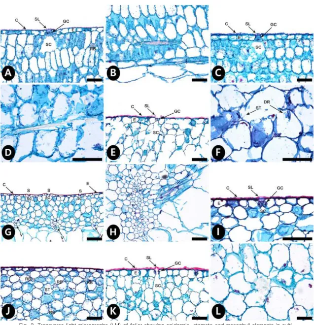
잎의 횡단면을 관찰한 결과, 모든 분류군에서 큐티클층은 두껍게 확인되었으며, 공변세포는 표피세포 보다 함몰(sunken)된 구조를 나타냈고, stomatal ledges 와 기공 밑 세포 간극(stomatal chamber)이 관찰되었다(Fig. 2). 특히, 본 연구에서 관찰한 모든 분류군에서 나타난 함몰된 공변세포는 건생식물의 일반적인 형질 중 하나로 기존의 연구와 동일한 결과14-19)로 확인되었다. 함몰된 공변세포의 입구는 stomatal ledges 또는 stomatal pits 로 구성되는데, 이들의 형태가 분류학적으로 유용한 것으로 보고
Taxa Side
Stomata complex Size of stomata (μm) Epidermal cell Width of cell wall
Position Type SI Length Width Arrange (μm)
ment Shape Anticlinal wall
Aloe arborescens Mill. AD AM Tetra 4-5.4 37.6-(42.3)-48.9 28.0-(32.8)-38.7 reg rec, pen st 1.0-(1.5)-2.1 AB Tetra 4-5.1 33.9-(36.3)-39.7 21.8-(24.0)-26.3 reg rec, pen st 1.0-(1.4)-2.2
Aloe barbadensis L. AD AM Tetra 3.8-6.7 48.2-(54.4)-60.9 23.9-(29.4)-33.8 reg hex, pen st 4.4-(5.3)-6.7 AB Tetra 3.6-5.9 50.3-(55.4)-59.6 26.4-(34.4)-37.9 reg hex, pen st 3.6-(4.4)-5.4
Aloe ferox Mill. AD AM Tetra 5.1-7.8 42.4-(46.9)-50.9 34.5-(42.3)-46.3 reg hex, rec st 7.0-(8.6)-10.6 AB Tetra 2.4-4.6 46.3-(50.2)-55.5 37.5-(42.9)-48.5 reg hex, rec st 6.6-(8.2)-10.0
Aloe saponaria (Aiton) Haw. AD AM Tetra 7.6-11.9 37.4-(45.9)-50.4 28.5-(31.7)-35.2 reg rec st 2.7-(3.7)-4.5 AB Tetra 5.6-9.6 47.4-(51.7)-54.8 33.2-(38.0)-43.0 reg rec st 3.2-(4.9)-6.1
Aloe sp. 1 AD AM Tetra 2.1-3.1 26.8-(31.1)-35.3 18.0-(23.0)-36.8 reg rec, pen st 1.7-(2.3)-3.3 AB Tetra 1.0-3.7 30.1-(32.7)-37.8 19.1-(22.5)-25.0 reg rec, pen st 1.2-(2.1)-3.0
Aloe sp. 2 AD AM Tetra 6.4-11.8 43.1-(47.1)-50.1 23.6-(30.2)-35.4 reg rec, hex st 3.0-(4.7)-6.1 AB Tetra 8.4-10.4 39.4-(43.7)-48.8 22.2-(27.7)-35.9 reg rec, hex st 3.9-(4.4)-5.2 Abbreviations - AB, abaxial side; AD, adaxial side. Position: AM, amphistomatic. Type: Tetra, tetracytic. SI, stomatal index.
Arrangement: reg, regular. Shape: hex, hexagon; pen, pentagon; rec, rectangular. Anticlinal wall: st, striate.
Table 1. Overview of representative stomatal and epidermal characters on leaves of taxa studied in the genus Aloe.
된 바 있으며14), 표면에 나타나는 표피상납질(epicuticular waxes)의 유무 및 발달 정도 역시 식별형질 로서 유용한 것으로 알려져 있다15). 추후 주사전자현미경(scanning electron microscope; SEM)을 이용한 미세형태학적 연구를 통해 국내 재배 알로에 분류군 내 형질의 분류학적 유용성을 확인할 필요 가 있을 것으로 사료된다.
식물체 내 결정화 현상(crystallization)은 식물체의 물리적인 보호, 옥살산염의 방출, 칼슘의 저장 및 광합성 중 빛의 조절 등 다양한 기능과 연관이 있는 것으로 보고된 바 있다28). 결정체(crystal)는 본 연구에서 관찰한 6 분류군의 유세포(parenchymatous cell) 내에서 침상 결정체(raphide)가 확인되 었고, 알로에베라에서만 침상 결정체, 선정체(druse)와 막대형 결정체(styloid)가 동시에 관찰되어 다른 분류군과 구별되었다(Fig. 2). 추후 줄기와 뿌리 등의 다른 영양기관에 분포하는 결정체의 종류 및 성장적 변이에 따른 결정체의 양 등에 대한 연구가 필요할 것으로 판단하였다.
알로에는 열대지방에 서식하는 건생식물로 알려져 있다. 일반적으로 건생식물의 잎은 수분손실을 막기 위해 두꺼운 큐티클층을 갖고 있으며, 함몰된 공변세포를 지니고29,30), CO2 전도계수(leaf con- ductance)를 최대로 하기 위해 양면기공엽을 갖는 등31,32), 건조한 환경에 적응하기 위한 형질을 나타 낸다고 보고된 바 있다. 본 연구를 통해 기존에 보고된 열대 지방의 알로에 형질과 국내 재배 알로에 6 분류군의 잎 형질이 유사함을 확인할 수 있었으며, 건생식물로서 나타나는 형질 역시 동일하게 관찰 되었다.
Fig. 1. Light micrographs (LM) of foliar clearings of cultivated Aloe species in Korea. A-B. A. arborescens. C-D. A. barbadensis (=A. vera). E-F. A. ferox. G-H. A. saponaria. I-J. Aloe sp. 1. K-L. Aloe sp. 2. A, C, E, G, I, K. Adaxial surface.
B, D, F, H, J, L. Abaxial surface. All scale bars = 100 ㎛.
Fig. 2. Transverse light micrographs (LM) of foliar showing epidermis, stomata and mesophyll elements in culti- vated Aloe species in Korea. A-D. A. arborescens. A. Part of the adaxial surface. B. Raphide bundle (acicular crystals) in the mesophyll. C. Part of the abaxial surface. D. Raphides (acicular crystals) in the mesophyll. E-G. A. barbadensis (=A. vera). E. Part of the adaxial surface. F. Druse and styloid crystals in the mesophyll. G. Part of the abaxial surface. H. Vascular bundle of A. saponaria. I-J. Part of the adaxial surface of Aloe sp. 1. K-L. Aloe sp. 2. K. Part of the adaxial surface. L. Group of druse in the mesophyll. All scale bars = 100 ㎛ except figure G (200 ㎛). C, cuticle; DR, druse; E, epidermis;
GC, guard cell; RB, raphide bundle; S, stomata; SC, stomata cavity; SL, stomatal ledge; ST, styloid;
V, vein.
결론
본 연구에서는 한약 자원 노회의 기원식물인 알로에와 동속 이종을 포함한 재배식물 총 6 분류군 잎의 해부학적 연구를 수행하였으며, 이를 통해 한약재 내부형태 기초 자료를 축적하였다. 본 연구 결과, 기원식물인 알로에베라는 가장 큰 기공을 갖고, 침상 결정체, 선정체와 막대형 결정체를 모두 지니고 있어 다른 분류군과 구별되었으며, 희망봉알로에는 표피세포의 세포벽이 가장 두꺼워 다른 분류군과 뚜렷하게 구분되었다. 이처럼, 표피세포의 형태, 세포벽의 두께, 공변세포의 크기, 결정체의 종류 및 분포 등의 형질로 알로에속 내 기원종을 포함한 일부 분류군을 구별할 수 있었고, 동정, 감별을 위한 식별형질로의 가능성을 확인하였다. 또한 건생식물로서 자생지 분류군과 동일하게 나타나는 두꺼 운 큐티클층, 양면기공엽, 함몰된 기공의 형질을 확인하였다. 추후 표피상납질, 병층벽(periclinal wall) 의 형태 등 주사전자현미경을 이용한 미세형태학적 형질 추가를 통해 미세형태학적 형질의 분류학적 유용성 및 감별을 위한 형질 검증이 필요할 것으로 사료된다.
감사의 글
본 연구는 한국한의학연구원 K-herb 과제의 일환으로 진행 중인 ‘한약자원의 발굴 및 보전’ 과제 (K18401)의 지원에 의해 수행되었습니다. 본 연구 수행에 필요한 표본을 제공해주신 한국한의학연구 원 한약표준표본관(index herbariorum code KIOM)께 감사드립니다.
참고문헌
1. Reynolds GW. The aloes of tropical Africa and Madagascar. Mbabane: The Aloes Book Fund.
1966.
2. Viljoen AM. A chemotaxonomic study of phenolic leaf compounds in the genus Aloe. Ph.D.
dissertation. Johannesburg: Rand Afrikaans University. 1999.
3. Glen HF and Hardy DS. Aloaceae: Aloe. In Germishuizen G (ed.), Flora of Southern Africa.
Vol. 1. Pt. 1. Pretoria: National Botanical Institute. 2000.
4. Klopper RR and Smith GF. The genus Aloe L. (Apshodelaceae: Alooideae) in Namaqualand, South Africa. Haseltonia. 2007;13:1-13.
5. Klopper RR, Van Wyk AE and Smith GF. Phylogenetic relationships in the family Asphodelace ae (Asparagales). Biodiversity & Ecology. 2010;3:9-36.
6. 주영승. 증보운곡본초학. 전주:도서출판 우석. 2013:585-7.
7. Korea Institute of Oriental Medicine. Defining Dictionary for Medicinal Herbs[Korean, 'Hanya k Giwon Sajeon'] (2018). Published on the Internet; http://boncho.kiom.re.kr/codex/ (accesse d 2018-02-21).
8. Reynolds T and Dweck A. Aloe Vera Leaf Gel: A Review Update. Journal of Ethnopharmacolog y. 1999;68:3-37.
9. Norikura T, Kennedy DO, Nyarko AK, Kojima A and Matsui-Yuasa I. Protective effect of Aloe
extract against the cytotoxicity of 1,4-napththoquinone in isolated rat hepatocytes involves modulations in cellular Thiol levels. Pharmacology and Toxicology. 2002;90(5):278-84.
10. Subramaniam S, Sathish Kumar D and Arulselvan P. Wound healing potential of Aloe vera leaf gel studied in experimental rabbits. Asian Journal of Biochemistry. 2006;1(2):178-85.
11. Kametani S et al. Chemical constituents of Cape Aloe and their synergistic growth-inhibition effect on Ehrlich ascites tumor cells. Bioscience, Biotechnology, and Biochemistry. 2007;71 (5):1220-9.
12. 송준호, 홍석표. 쉬땅나무족(Sorbarieae Rydb., 장미과) 잎의 해부학적 형질 및 분류학적 유용성.
한국식물분류학회지. 2014;44(2):119-31.
13. 송준호, 홍석표. 쉬땅나무족(조팝나무아과: 장미과) 잎표피 미세형태학적 형질의 분류학적 유용성.
한국식물분류학회지. 2016;46(2):119-212.
14. Newton LE. Taxonomic use of the cuticular surface features in the genus Aloe (Liliaceae).
Botanical Journal of the Linnean Society. 1972;65(3):335-9.
15. Cutler DF, Brandham PE, Carter S and Harris SJ. Morphological, anatomical, cytological and biochemical aspects of evolution in East African shrubby species of Aloë L. (Liliaceae). Botan ical Journal of the Linnean Society. 1980;80(4):293-317.
16. Beaumont J, Cutler DF, Reynolds T and Vaughan JG. Secretory tissues in the East African shrubby aloes. Botanical Journal of the Linnean Society. 1986;92(4):399-402.
17. Smith GF and Van Wyk AE. Systematic leaf anatomy of selected genera of southern African Alooideae (Asphodelaceae). South African Journal of Botany. 1992;58(5):349-57.
18. Coopoosamy RM and Naidoo KK. Anatomical features of the medicinal importance of Aloe excelsa (Berger). African Journal of Biotechnology. 2011;10(39):7622-32.
19. Wintola OA and Afolayan AJ. The foliar anatomy and micromorphology of Aloe ferox Mill.
(Asphodelaceae). African Journal of Traditional, Complementary and Alternative medicines.
2014;11(2):350-7.
20. Chen X and Gilbert MG. Aloe. In Wu ZY and Raven PH (eds.), Flora of China. Vol. 24. Beijing
& St. Louis: Science Press and Missouri Botanical Garden Press. 2010:160-1.
21. Daru BH et al. Molecular and morphological analysis of subfamily Alooideae (Asphodelaceae) and the inclusion of Chortolirion in Aloe. Taxon. 2013;62(1):62-76.
22. Johansen DA. Plant microtechnique. New York: McGraw-Hill Book Co. 1940.
23. Ruzin SE. Plant Microtechnique and Microscopy. New York & Oxford: Oxford University Press. 1999.
24. Wilkinson HP. The plant surface (mainly leaf). In Metcalfe CR and Chalk L (eds.), Anatomy of the Dicotyledons. 2nd ed. Vol. I. Oxford: Clarendon Press. 1979:97-165.
25. Evert RF. Esau’s Plant Anatomy: Meristems, Cells, and Tissues of the Plant Body: Their Structure, Function, and Development. Hoboken, New Jersey: John Wiley & Sons, Inc. 2006.
26. Nazli A and Kumar K. Studies on the stomata of two medicinally important plants Aloe abyss inica and Haworthia limifolia. Annals of Plant Sciences. 2015;4(12):1239-42.
27. Santos LDT, Thadeo ML, Iarema RM, Meira SA and Ferreira FA. Foliar anatomy and histoche mistry in seven species of Eucalyptus. Revista Árvore. 2008;32(4):769-79.
28. Franceschi VR and Nakata PA. Calcium oxalate in plants: Formation and function. Annual Review of Plant Biology. 2005;56(1):41-71.
29. Wickens GE. Anatomical and morphological adaptations. In Wickens GE (ed.), Ecophysiology of Economic Plants in Arid and Semi-Arid Lands. Heidelberg: Springer-Verlag 1998:145-60.
30. Cutler DF, Botha CEJ and Stevenson DW. Adaptive features. In Cutler DF, Botha CEJ and Stevenson DW (eds.), Plant Anatomy: an applied approach. Oxford: Blackwell. 2008:135-53.
31. Wood JG. The physiology of xerophytism in Australian plants: the stomatal frequencies, tran spiration and osmotic pressures of sclerophyll and tomentose-succulent leaved plants. The Journal of Ecology. 1934;22(1):69-87.
32. Fahn A and Cutler DF. Xerophytes. Gebrüder Borntraeger, Berlin, Stuttgart. 1992.
Appendix
Materials and voucher specimen data used in this study:
Aloe arborescens Mill.: Geoje-si, Gyeongsangnam-do, 15, Jan, 2018 (KIOM201801020712~16);
Aloe barbadensis L. [=Aloe vera (L.) Burm.f.]: Geoje-si, Gyeongsangnam-do, 15, Jan, 2018 (KIOM201801020702~6); Aloe ferox Mill.: Geoje-si, Gyeongsangnam-do, 15, Jan, 2018 (KIOM201801020707); Aloe saponaria (Aiton) Haw. [=Aloe maculata All.]: Geoje-si, Gyeongsangnam-do, 15, Jan, 2018 (KIOM201801020717~19); Aloe sp. 1: Geoje-si, Gyeongsangnam-do, 15, Jan, 2018 (KIOM201801020708~9); Aloe sp. 2: Geoje-si, Gyeongsangnam-do, 15, Jan, 2018 (KIOM201801020710~11).
ⓒ The Author(s) 2018, khmi.or.kr