고지혈증이 동반되지 않은 양측성 종골건 황색종
- 1례 보고 -
인제대학교 의과대학 정형외과학교실
정병현・정형진・김동수・성열보・안종국・권칠수・심성실・김진호
- Abstract -
Bilateral Achilles Tendon Xathoma without Hypercholesterolemia
- A Case Report -
Byung Hyon Jung, M.D., Hyung Jin Chung, M.D., Dong Soo Kim, M.D., Yeol Bo Sung, M.D., Jong Guk Ahn, M.D., Chil Soo Kwon, M.D.,
Sung Sil Sim, M.D., Jin Ho Kim, M.D.
Department of Orthopedic Surgery, Paik Hospital, Inje University, Korea
Xanthoma is a localized collection of tissue histocytes containing lipid. The majority of tendi- nous xanthomas probably occurs in the setting of hypercholesterolemia especially in bilateral Achilles tendon xanthomas. Xanthoma of the Achilles tendon is a rather rare, interesting orthopaedic condition that has important ramifications in internal medicine and dermatology because the lesion is associated with a specific disturbance of lipid metabolism. We experienced one case of normolipidemic and symptomatic Achilles tendon xanthoma. Surgical intervention was carried out for cosmetic and symptomatic reasons, the patient undergoing total resection and a reconstruction of the Achilles tendon by the combinedV-Y muscle flap and modified Lindholm technique.
Key Words : Xanthoma, Achilles Tendon
─ 194 ─
※통신저자 : 정 병 현
경기도 고양시 일산구 대화동 2 2 7 0번지 인제대학교 의과대학 정형외과학교실 Tel : 0344) 910-7310
대 한 골 관 절 종 양 학 회 지
J. of Korean Bone & Joint Tumor Soc.
Volume 5, Number 3, September, 1999
서 론
황색종은 지방질을 포함한 조직구의 국소 침착으로 알려져 있다1 ). 황색종은 피부, 피하조직 혹은 심부연 부조직 어디에도 발생할 수 있으며 고지혈증과 관련 될 수도 있고 아닐 수도 있다. 발진성, 편평성, 건, 결절성 황색종으로 분류하며 건 황색종의 대부분, 특 히 양측성으로 오는 경우는 고지혈종이 동반되는 경 우가 대부분이다1 ). 저자들은 정상 지혈을 보이면서 양측 아킬레스건에 발생한 약 7㎝의 황색종 1례를 경 험하고 전절제와 재건술 및 물리치료로 비교적 좋은
결과를 얻었기에 문헌고찰과 함께 보고하는 바이다.
증례보고
2 2세 남자환자로 1년 전부터 양측 아킬레스건에 점차 커지는 비후성 종괴로 인한 외관상의문제 및 보행시 자극으로 인한 동통으로 내원하였다. 과거력 및 가족력상 특이사항은 없었다. 이학적 소견상 전 신상태는 양호하였으며 양측 아킬레스건에 약 3×7
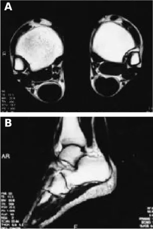
㎝ 정도의 종괴가 촉지되었다. 압통이 있었으나 열 감은 없었으며 족관절의 운동범위는 정상이었다. 방 사선 소견 : 단순방사선 소견상 양측 아킬레스건의 음영이 넓어진 소견을 보였고, 연속성이 유지되고 있으며 석회침착이나 골조직의 변화등은 없었다. 자 기공명영상 소견상 양측 모두 시상면에서 아킬레스 건이 저강도의 비후된 소견을 보이고 있으며 고강도 의 수직선이 보이고 있다. 측상면에서 앞쪽으로 convexity margin이 보이고 있으며 저강도의 원형 구조를 고강도 신호가 둘러싼 형태를 취하고 있다 (Fig. 1-A,B). 검사소견 : 혈액침강속도(ESR) 5
㎜/hr, 콜레스테롤(Cholesterol) 156㎎/ d L ( 1 3 0 - 2 3 0㎎/dL), 고밀도지단백(HDL) 61㎎/ d L ( 3 5 - 6 5㎎ /dL), 중성지방(TG) 92㎎/ d L ( 3 0 - 1 9 0㎎/dL), 저 밀도지단백(LDL) 76.6㎎/ d L으로 정상소견 보였다.
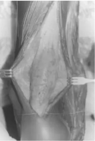
수술소견 : 아킬레스건의 원위부 약 3×7㎝에 걸쳐 황백색의 덩어리가 보였으며 전 제거후 V-Y 술식으 로 재건술 시행하였다(Fig. 2). 조직학적 소견 : 절 개면은 황백색이었으며 다수의 다핵성 거대세포 다 각형 포말세포, 방추성 기질세포 및 콜레스테롤 등 이 보였다(Fig. 3).
토 론
Fabey 등2 )은 1 7 3례를 통한 분석에서 황색종은 주로 중수지 관절의 신전건에 발생하며, 아킬레스건 이나 슬개건에도 발생할 수 있으며 드물게는 삼두박 근건이나 발가락의 신전건에도 발생할 수 있다고 하 였다. 건에 발생하는 황색종의 9 0 %는 신전건에 발 생하여, 약 반수에서 아킬레스건에 발생한다. 황색 종은 대부분 가족성 고지혈증과 관련되어 있으며 그 수와 크기는 콜레스테롤치와 나이등과 관련되어 있
다7 , 9 ). 따라서 황색종은 그 자체 보다도 고지혈증으
─ 195 ─
— 정병현 외 : 고지혈증이 동반되지 않은 양측성 종골건 황색종 —
Fig. 1-A. Sagittal T1-weighted MR image shows dif- fuse increase in size of Achilles tendon with parallel vertical striations of increased signal intensity in tendon substance.
Fig. 1-B. Axial T1-weighted MR image shows enlarged tendon with convex anterior margin. Diffuse stippled pattern presumably caused by colla- gen is surrounded by high-signal foamy histo- cytes and inflammatory reaction.
A
B
로 인한 심혈관질환을 예방하고 치료하는데 더 큰 의의를 가진다5 ). 정상 지혈증에서 발생한 황색종의 발생기전에 대한 기술은 많지 않으며4 ) Parker 등8 ) 은 원인에 따라 1) 지질단백의 용량 및 모양의 변화 2) 림프구증식증 3) 지질단백이상 없이 발생하는 국 소적 원인으로 분류하였다. Mancuso 등6 )은 지질 폐측 사이드의 존재와 높은 농도의 L p ( a ) *가 황색종 의 유발요인이라고 하였다. 수술적 방법으로는 부분 적 절제술 그리고 아전절제술 혹은 전절제술후의 근 막, 건전이술 또는 건재건술 등의 방법이 있다3 ). 아 킬레스근에 발생한 황색종은 통증이 있을 때 혹은 미용목적으로 수술적으로 제거할 수도 있다. 수술적 제거후 재발율은 6 0 %로 보고하고 있으나3 ) 대부분은 재수술적 필요는 없었다고 하였다. 저자들은 정상지 혈증을 보이면서 양측 아킬레스건에 발생한 황색종 에 대해 전제거( 7㎝) 및 V-Y 요법과 Lindholm 요 법으로 제건술 시행후 장하지 석고붕대를 통한 점진 적인 발목운동의 교정을 시행하였으며 1 6개월간 관
찰한바 양호한 결과를 얻었다고 사료되므로 발표하 는 바이다.
REFERENCES
01) Enzinger FM and Weiss SW : Xanthoma. S o f t Tissue Tomor, 3: 310-323, 1995.
02) Fabey JJ, Stark HH and Donovan WF : Xanthoma of the Achilles tendon. Seven cases with familial hyperbetalipoproteinemia. J Bone Joint Surg, 1197-1211, 1973.
03) Gang CJ, Ha SH and Lee SH : Tendinous and tuberous xanthomas without hypercholesterolemia.
J Korean Orthop Assoc, 24:1512-1516, 1985.
04) Harlan WR Jr, Graham JB and Estes EH : Familial hypercholesterolemia. A genetic and meta- bolic study. Am J Med, 45:77-110, 1966.
05) Jensen J, Blankenhorn DH and Kornerup V : Coronary disesase in familial hypercholesterolemia.
Circulation, 36:77-82, 1967.
06) Mancuso G, La Regina G and Bagnoli M : Normolipidemic tendinoces and tuberous xan- thomatosis. Arch Dermatol, 193:27-32. 1996.
07) Mishkel MA : The diagnosis and management of the patient with xanthomaosis. An experience with thirth-five cases. Quart J Med, 36:107-134, 1967.
08) Parker F : Normocholesterolemic xanthomatosis.
Arch Dermatol, 122:1253-1257,1986.
09) Piper J and Orrild L : Essential familial hypercho- lesterolemia and xanthomatosis. Am J Med, 21:34- 46, 1956.
─ 196 ─
— 대한골관절종양학회지 : 제 5 권 제 3 호 1999년 —
Fig. 2. The intraoperative photograph showed egg sized, yellowish mass of the Achilles tendon.
Fig. 3. The photomicrograph of the excised mass (H &
E, ×450) showed scattered xantomas cells with multinucleated giant cells.