146
Copyrights © 2014 The Korean Society of RadiologyINTRODUCTION
Tendinous xanthoma is commonly seen in patients with fa- milial hypercholesterolemia (FH) and produces nodular masses in the Achilles tendon and other tendons, and earlier clinical di- agnosis of FH depends on the detection of xanthomas (1). Early detection of FH is important in order to alter its clinical course (2). To our knowledge, the sonographic findings of tendinous xanthoma in the multiple tendons have been rarely reported.
We present a case of multiple xanthomas of ankles and elbows in a patient with FH.
CASE REPORT
A 52-year-old woman was referred for evaluation of multiple soft tissue lesions. She first noticed lesions of the bilateral Achil- les tendons at the age of 30. Subsequently, similar lesions that were slightly painful gradually developed in the elbows and feet.
The laboratory studies showed slightly increased levels of total cholesterol (230 mg/dL, upper normal value 200 mg/dL) and
low-density lipoprotein (LDL) (146 mg/dL, upper normal value 130 mg/dL). She also had family history of similar lesions. Ra- diographs, sonography, and MR studies were performed for the selected lesions.
Radiographs of the ankles and elbows revealed normal osse- ous architecture with increased soft tissue density posterior to
Case Report
pISSN 1738-2637 / eISSN 2288-2928 J Korean Soc Radiol 2014;71(3):146-149 http://dx.doi.org/10.3348/jksr.2014.71.3.146
Received April 23, 2014; Accepted July 7, 2014 Corresponding author: Ik Yang, MD
Department of Radiology, Kangnam Sacred Heart Hospital, Hallym University College of Medicine, 1 Singil-ro, Yeongdeungpo-gu, Seoul 150-950, Korea.
Tel. 82-2-829-5241 Fax. 82-2-832-1845 E-mail: [email protected]
This is an Open Access article distributed under the terms of the Creative Commons Attribution Non-Commercial License (http://creativecommons.org/licenses/by-nc/3.0) which permits unrestricted non-commercial use, distri- bution, and reproduction in any medium, provided the original work is properly cited.
Tendinous xanthomas are often associated with type 2 hyperlipoproteinemia, in which low-density lipoprotein derived from the circulation accumulates in the ten- dons. Therefore, the demonstration of tendinous xanthomas is helpful in diagnosing familial hypercholesterolemia (FH). In this study, we describe the sonographic and MR features of the case of bilateral multiple xanthomas of the ankles and elbows in a 52-year-old female patient with FH.
Index terms Xanthoma
Familial Hypercholesterolemia Tendon
Ultrasonography MRI
Multiple Xanthomas in a Patient with Familial Hypercholesterolemia: A Case Report
가족성 고콜레스테롤혈증 환자에서의 다발성 황색종: 증례 보고
Meehyun Park, MD, Ik Yang, MD, Hyun Suk Cho, MD
Department of Radiology, Kangnam Sacred Heart Hospital, Hallym University College of Medicine, Seoul, Korea
Fig. 1. Lateral radiograph of right ankle (A) and left elbow (B) show soft tissue mass at the posterior aspect (arrows). No bone or joint ab- normality is shown. Left ankle and right elbow demonstrated same ra- diographic finding.
A B
Meehyun Park, et al
147
jksronline.org J Korean Soc Radiol 2014;71(3):146-149
the metabolism and elimination of LDL are impaired and cho- lesterol is subsequently accumulated in soft tissues. This leads to the development of xanthoma in the Achilles and other tendons and accelerated development of coronary artery disease (1).
Coronary heart disease rarely occurs before the development of tendon xanthoma (2). So, early detection of tendinous xantho- ma can protect patients with FH from cardiovascular athero- sclerosis and early death.
Tendinous xanthomas are grayish-yellow to yellow masses composed of lipid-filled xanthoma cells, extracellular cholester- ol, and inflammatory cells. On clinical examination, these de- posits cannot be separated from the underlying tendon, and they move with the tendon on flexion and extension. The en- largement is fusiform or nodular and is usually non-tender and bilateral (3, 4). The most common locations are the extensor tendons of the hands, the feet, and the Achilles tendons. The av- erage age of onset of Achilles tendon xanthoma is 22, but onset during childhood occurs in homozygous patients and is associ- the ankle and elbow joints along the expected course of the
Achilles and triceps tendon (Fig. 1). Sonography demonstrated marked enlargement and mixed echoic pattern in the bilateral Achilles tendons, and similar findings were also shown in the triceps, anterior tibialis, posterior tibialis, peroneus longus, and flexor hallucis longus tendons (Figs. 2, 3). MR imaging of the ankles showed marked heterogeneous fusiform enlargement of the above multiple tendons with regions of increased signal in- tensity and numerous interspersed trabeculated linear areas with diminished signal intensity (Fig. 4).
The patient underwent open biopsy of the triceps tendon le- sion. Histopathology showed numerous lipid-filled foamy cells consistent with tendinous xanthoma (Fig. 5).
DISCUSSION
FH is characterized by an elevated serum LDL concentration.
Due to decreased numbers of functionally active LDL receptors,
Fig. 2. A 52-year-old woman who presented with multiple soft tissue lesions in both ankles.
A-C. Longitudinal (A) and transverse (B, C) ultrasound images show slightly heterogeneous stippled appearance of soft tissue masses along the anterior tibialis (A), peroneal (B), and Achilles (C) tendons. Posterior tibialis revealed similar sonographic finding.
B
A C
Fig. 3. A 52-year-old woman demonstrated soft tissue lesions also in both elbows.
A, B. Gray scale (A) and color Doppler ultrasound image at triceps tendon (B) show soft tissue mass with mild degree increased vascularity.
A B
Multiple Xanthomas in a Patient with Familial Hypercholesterolemia
148
J Korean Soc Radiol 2014;71(3):146-149 jksronline.orgment and mixed echoic pattern of the affected tendons. Des- camps et al. (8) reported that Achilles tendon of thickness above 5.8 mm was the most useful threshold for the diagnosis of FH, procuring sensitivity of 75% and specificity of 85%. In our case, Achilles tendon thickness was measured to be 30 mm on US, and this was notable not only for the pronounced enlargement of the Achilles tendon, but also for its similar findings at the tri- ceps, anterior tibialis, posterior tibialis, peroneus longus, and flexor hallucis longus tendons.
ated with more severe coronary atherosclerosis (5).
Radiographs of tendinous xanthomas show soft tissue enlarge- ment of uniformly increased density and absence of calcifica- tions. At the ankle, symmetric bilateral fusiform soft tissue en- largement of the Achilles tendon suggest the diagnosis of tendon xanthomas (5, 6).
Ultrasonography (US) with high resolution linear-array equip- ment has been rated as superior to MRI, for the evaluation of xanthomatous lesions (7). Sonography shows marked enlarge-
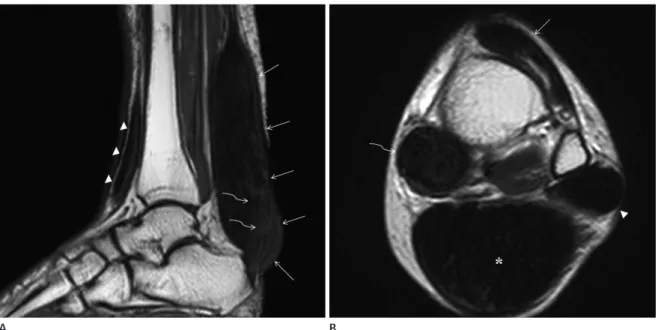
Fig. 4. A 52-year-old woman who presented with multiple soft tissue lesions in both ankles.
A. Sagittal T1-weighted MR image shows fusiform enlargement of the Achilles tendon (arrows) and of the tibialis anterior ten- don (arrowheads). Note numerous trabeculated areas of low signal representing remnant collagen fascicles (curved arrows).
B. Axial T2-weighted MR image of the ankle reveals enlargement of the tibialis anterior (arrow), peroneus longus (arrowhead), flexor hallucis longus (curved arrow), and Achilles tendon (*) with abnormal heterogeneous signal intensity.
Fig. 5. A 52-year-old woman who underwent open biopsy of triceps tendon lesion.
A, B. Gross image (A) and photomicrograph (hematoxylin and eosin, × 10) (B) of the tendinous xanhoma in triceps tendon. The cut surface is homogenously brightly yellow. On histopathology, it is characterized by lipid laden foam cells with large areas of cholesterol clefts, consistent with tendinous xanthoma.
A
A
B
B
Meehyun Park, et al
149
jksronline.org J Korean Soc Radiol 2014;71(3):146-149
milial hypercholesterolemia. Arteriosclerosis 1989;9(1 Suppl):I75-I80
3. Fahey JJ, Stark HH, Donovan WF, Drennan DB. Xanthoma of the achilles tendon. Seven cases with familial hyper- betalipoproteinemia. J Bone Joint Surg Am 1973;55:1197- 1211
4. Doyle JR. Tendon xanthoma: a physical manifestation of hyperlipidemia. J Hand Surg Am 1988;13:238-241
5. Glueck CJ, Levy RI, Frederickson DS. Acute tendinitis and arthritis. A presenting symptom of familial type II hyperli- poproteinemia. JAMA 1968;206:2895-2897
6. Gilbert PD, Kain TM, March HC. Hypercholesteremic xan- thomata of the tendons. Am J Roentgenol Radium Ther Nucl Med 1957;77:109-114
7. Bude RO, Adler RS, Bassett DR. Diagnosis of Achilles ten- don xanthoma in patients with heterozygous familial hy- percholesterolemia: MR vs sonography. AJR Am J Roent- genol 1994;162:913-917
8. Descamps OS, Leysen X, Van Leuven F, Heller FR. The use of Achilles tendon ultrasonography for the diagnosis of famil- ial hypercholesterolemia. Atherosclerosis 2001;157:514-518 9. Kelman CG, Disler DG, Kremer JM, Jennings TA. Xantho-
matous infiltration of ankle tendons. Skeletal Radiol 1997;26:256-259
Our case shows the typical findings of fusiform tendon en- largement, consisting of globular heterogeneous signal intensity on T1- and T2-weighted sequences and interspersed trabeculat- ed areas of diminished signal intensity coursing throughout the affected areas. These characteristic of low-signal trabeculations represent areas of residual normal collagen fascicles; the globu- lar areas of mixed signal intensity represent the xanthomatous deposits (9). Not only bilateral Achilles tendons, but also other multifocal tendons were involved in our case.
In conclusion, multiple tendon xanthomas are pathognomic for the familial hypercholesterolemia, so the role of radiologist is important for early diagnosis that can alter its clinical course.
Imaging findings are typical; symmetric bilateral fusiform soft tissue enlargement of the Achilles tendon and similar findings of other multifocal tendons should suggest the diagnosis of ten- don xanthoma in familial hypercholesterolemia.
REFERENCES
1. Kwiterovich PO Jr. Pediatric implications of heterozygous familial hypercholesterolemia. Screening and dietary treatment. Arteriosclerosis 1989;9(1 Suppl):I111-I120 2. Thompson GR, Seed M, Niththyananthan S, McCarthy S,
Thorogood M. Genotypic and phenotypic variation in fa-
가족성 고콜레스테롤혈증 환자에서의 다발성 황색종: 증례 보고
박미현 · 양 익 · 조현숙
건황색종은 보통 2형 고지질단백혈증에서 동반되며, 체내 순환 중인 저밀도 지단백이 건에 축적되면서 발생한다. 그러므 로, 건황색종의 입증은 가족성 고콜레스테롤혈증의 진단에 도움이 된다. 건황색종의 초음파, 자기공명영상(MRI) 소견을 고찰하는 몇몇 보고들이 있으나 대부분이 양측 아킬레스건(Achilles tendon)에 생긴 경우를 보고하고 있으며 양측 발목과 팔꿈치에 생긴 다발성 건황색종의 영상소견을 보고한 사례는 없었다. 이에 우리는 가족성 고콜레스테롤혈증을 가진 52세 여성에서, 양측 발목과 팔꿈치에 보인 다발성 건황색종의 초음파와 자기공명영상 소견을 보고한다.
한림대학교 의과대학 강남성심병원 영상의학과