www.jkfas.org pISSN 1738-3757 eISSN 2288-8551 J Korean Foot Ankle Soc 2016;20(1):39-42 http://dx.doi.org/10.14193/jkfas.2016.20.1.39
a smoker and had a body mass index (BMI) of 27.14 kg/m2. No hereditary prothrombotic conditions were known to afflict the pa- tient himself or his relatives.
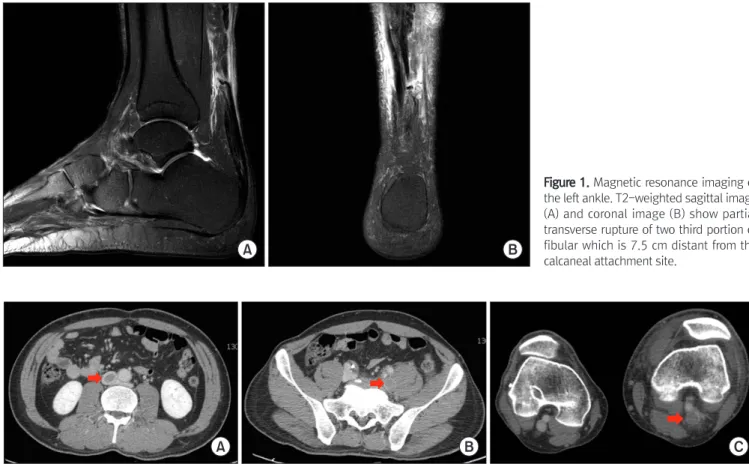
On physical examination, Thompson squeeze test was positive but palpable gap between the ruptured proximal end and distal end of left Achilles tendon was not clear. Magnetic resonance im- aging revealed partial transverse rupture of distal two third of lat- eral portion which is 7.5 cm distant from the calcaneal attachment site (Fig. 1). The patient agreed to undergo surgical repair and on the following day, operation was done. He underwent surgery under spinal anesthesia, and a tourniquet was inflated for seventy- four minutes. Skin incision along medial border of Achilles tendon was made and distal one third of medial portion of the Achilles tendon was ruptured at mid calf. We tried to pass the medial por- tion of the Achilles tendon proximally through the paratenon after island incision was made on ruptured site. However, it failed thus we prolonged incision to repair the tendon (Fig. 2). The tendon was sutured using a modified Krackow technique and additional side-to-side interrupted suture. The patieint was subsequently im- mobilized in a short leg cast in a plantar flexed position and was sent home 4 days after the operation on crutches.
Two weeks postoperatively, the patient visited the outpatient clinic to remove the stitch and change the cast into a ninety degree position of ankle. Surgical wound was clean and no clinical prob- lem was found. However, approximately 3 weeks postoperatively, Achilles tendon ruptures are common and the incidence of
Achilles tendon rupture is reported variably from 18 to 37.3 per 100,000.1,2) Both operative and non-operative treatments of Achil- les tendon rupture include a period of immobilization which is a risk factor for deep vein thrombosis (DVT).3) DVT is an important source of morbidity in orthopaedic surgery and can progress to pulmonary embolism (PE) which is a significant source of mortal- ity. The rates of reported DVT in patients with Achilles tendon rupture range from less than 1% to 34%.4) However, DVT after the Achilles tendon repair have not yet been reported. Here, we re- port a case of DVT following Achilles tendon repair that required angiointervention to remove the thrombus.
CASE REPORT
A 49-year-old male presented to our institution with a painful left heel after playing soccer. He complained about difficulty in walking. He had been taking amlodipine 5 mg per day for 2 years to control his hypertension and had a history of Achilles tendon repair in the right ankle 7 years prior to current injury. He was not
Case Report
This is an Open Access article distributed under the terms of the Creative Commons Attribution Non-Commercial License (http://creativecommons.org/licenses/CC
by-nc/4.0) which permits unrestricted non-commercial use, distribution, and reproduction in any medium, provided the original work is properly cited.
Copyright 2016 Korean Foot and Ankle Society. All rights reserved.ⓒ
Achilles tendon rupture is thought to be increasing with participation in sports activities. Both operative and non-operative treatments of Achilles tendon rupture include a period of immobilization. Complications following treatment of the Achilles tendon rupture include recurrence of rupture, flexor weakness, infection, and wound problems. However, deep vein thrombosis (DVT) after operative treatment of the Achilles tendon has not been reported. We report on a case of DVT after Achilles tendon repair.
Key Words: Achilles tendon, Deep vein thrombosis, Complications, Repair, Intervention
Deep Vein Thrombosis after Achilles Tendon Repair:
A Case Report
Hoseong Jang, Yong Eun Shin, Sung Hyun Kim, Hyun-Woo Park
Department of Orthopedic Surgery, Dankook University College of Medicine, Cheonan, Korea
Received January 3, 2016 Revised February 4, 2016 Accepted February 5, 2016 Corresponding Author: Hyun-Woo Park
Department of Orthopedic Surgery, Dankook University College of Medicine, 119 Dandae-ro, Dongnam-gu, Cheonan 31116, Korea
Tel: 82-41-550-3296, Fax: 82-41-556-3238, E-mail: [email protected] Financial support: None.
Conflict of interest: None.
40 Vol. 20 No. 1, March 2016
DISCUSSION
DVT and PE are serious and feared complications after ortho- paedic surgery. It is well known that DVT and PE could develop the patient visited the outpatient clinic because of painful swell-
ing and purple skin color of left lower extremity. The diameter of the left thigh was 62 cm and that of right thigh was 53 cm. After removal of cast, the symptom persisted and he was referred to the cardiothoracic surgeon. Doppler ultrasonography of the left leg showed the presence of the thrombus from popliteal vein to femoral vein. Computed tomographic venography showed exten- sive thrombosis in the infrarenal inferior vena cava (IVC) and left lower extremity (Fig. 3). Laboratory findings showed negative an- tinuclear antibody, antineutrophil cytoplasmic antibody and lupus anticoagulant. The results of antithrombin 3, protein C antigen, and protein S antigen were normal. Angiointervention was done for thrombolysis and to remove the thrombus. IVC filter had been placed for 10 days (Fig. 4). Swelling decreased after angiointer- vention and the diameter of the left thigh decreased from 62 cm to 54.3 cm. The heparin sodium (20,000 IU) was used for 5 days and rivaroxaban for 6 months. The patient could perform activities of daily living and american orthopaedic foot and ankle society hind- foot score was 85 at postoperative 6 months.
Figure 1. Magnetic resonance imaging of the left ankle. T2-weighted sagittal image (A) and coronal image (B) show partial transverse rupture of two third portion of fibular which is 7.5 cm distant from the calcaneal attachment site.
A B
A B
Figure 2. The intraoperative photo of left ruptured Achilles tendon. (A) Medial portion of the Achilles tendon was ruptured at mid calf. (B) Af- ter prolonged skin incision made.
A B C
Figure 3. Computed tomography venography shows thrombus (arrows). (A) Infrarenal inferior vena cava. (B) Left iliac vein. (C) Left popliteal vein.
www.jkfas.org 41 Hoseong Jang, et al. Deep Vein Thrombosis after Achilles Tendon Repair
is reported as a risk factor for DVT and PE after foot surgery.6) In ad- dition, congestive heart failure (CHF) is an independent risk factor for DVT in one study.7) On the other hand, in a retrospective study of 1,172 patients who had Achilles tendon ruptures, age older than 40 years, CHF, history of DVT or PE and obesity did not predict occurrence of DVT or PE.2) Although age older than 40 years did not predict occurrence of DVT or PE, all patients with DVT and PE were older than 40 years and aging which often is associated with obesity and decline in muscle strength, might lead to functional and metabolic changes. These changes might predispose a patient to DVT or PE.8) In the present case, patient’s BMI was 27.14 kg/m2 and he had no history of CHF and DVT or PE. Reviewing our case in retrospect, immobilization and age were possible risk factors for our patient.
The best treatment for thromboembolic disease is prevention.
However, the use of thromboprophylaxis after an Achilles ten- don rupture is controversial and recent antithrombotic guidelines published by the American College of chest Physicians did not specifically mention the need for DVT prophylaxis with regard to Achilles tendon rupture.9) In a randomized, controlled study, Lapidus et al.1) demonstrated a total DVT rate of 34% to 36% in pa- tients treated with either dalteparin (5,000 IU) or placebo, conclud- ing that thromboprophylaxis did not affect the incidence of DVT during immobilization after Achilles tendon rupture. On the other hand, in a randomized, controlled trial, Lassen et al.10) reported the DVT rate of 9% treated with low-molecular-weight heparin (reviparin 1,750 anti-Xa units) and 19% in the placebo group after ankle fracture or Achilles tendon rupture.
In this report, we presented a rare case of DVT following Achil- after total knee or hip arthroplasty. Although a few studies report-
ed the risk of developing thromboembolism after acute Achilles tendon rupture, DVT and PE after Achilles tendon rupture are not widely known. Because most DVT are asymptomatic and many clots resolve spontaneously. There isn’t a sufficient report about the angiointerventive treatment for DVT after surgery of Achilles tendon rupture and a few case reports were only about PE after Achilles tendon rupture.
The incidence of DVT after Achilles tendon rupture was highly variable in the literature and reported DVT ranges from less than 1% to 34%.4) Some reported the rate of only symptomatic DVT and others reported the rate of both asymptomatic and symptomatic DVT. This is the reason for discrepancy of incidence which is re- lated to the different study designs.
Lapidus et al.1) classified DVT into 3 groups: distal and proximal DVT or PE where the DVT located. Proximal DVT was defined when a thrombosis is located in the popliteal vein or any more proximal veins with or without involvement of the calf veins and most DVT were located in distal veins and asymptomatic. How- ever, proximal extension of thromboses from deep calf veins to more proximal veins can be as high as 28% and asymptomatic proximal DVT was closely related to the risk of PE. In the present case, symptomatic proximal DVT was found.
Many possible factors are believed to contribute to the risk of developing DVT or PE with cast immobilization of the lower limb following injury.4) These include trauma, immobilization, surgery.
However, in prospective and retrospective studies, there was no difference in DVT or PE incidence between surgically and nonsurgi- cally treated Achilles tendon ruptures.2,5) Obesity (BMI >30 kg/m2)
A B C
Figure 4. Angiointervention. (A) Venous flow is blocked at inferior vena cava (IVC) (arrow). (B) IVC filter placement and filling of thrombus is found (arrow). (C) Venous flow is recovered after thrombus removal (arrow).
42 Vol. 20 No. 1, March 2016
tomatic deep venous thrombosis after Achilles tendon rupture1 J Foot Ankle Surg1 2013;52:584-71
51 Nilsson-Helander K, Thurin A, Karlsson J, Eriksson BI. High incidence of deep venous thrombosis after Achilles tendon rup- ture: a prospective study1 Knee Surg Sports Traumatol Arthrosc1 2009;17:1234-81
61 Felcher AH, Mularski RA, Mosen DM, Kimes TM, DeLoughery TG, Laxson SE. Incidence and risk factors for venous thrombo- embolic disease in podiatric surgery1 Chest1 2009;135:917-221 71 Howell MD, Geraci JM, Knowlton AA. Congestive heart failure
and outpatient risk of venous thromboembolism: a retrospec- tive, case-control study1 J Clin Epidemiol1 2001;54:810-61 81 Di Nisio M, Di Iorio A, Porreca E, Abate M, Ferrante N, Bandinelli
S, et al. Obesity, poor muscle strength, and venous thromboem- bolism in older persons: the InCHIANTI study1 J Gerontol A Biol Sci Med Sci1 2011;66:320-51
91 Falck-Ytter Y, Francis CW, Johanson NA, Curley C, Dahl OE, Schulman S, et al; American College of Chest Physicians. Pre- vention of VTE in orthopedic surgery patients: antithrombotic therapy and prevention of thrombosis, 9th ed: American College of Chest Physicians Evidence-Based Clinical Practice Guidelines1 Chest1 2012;141 2 Suppl:e278S-325S1
101 Lassen MR, Borris LC, Nakov RL. Use of the low-molecular- weight heparin reviparin to prevent deep-vein thrombosis after leg injury requiring immobilization1 N Engl J Med1 2002;347:726- 301
les tendon repair who required angiointervention to remove the thrombus. The patient had a possibility of DVT progressing to PE which is a significant source of mortality.
Therefore, we believe that patients should be warned of the risk of postoperative DVT and PE before Achilles tendon repair which include a period of postoperative immobilization and thrombopro- phylaxis for high risk patient could be considered. We believe that this study may assist clinicians to make a proper diagnosis and to administer correct treatment.
REFERENCES
11 Lapidus LJ, Rosfors S, Ponzer S, Levander C, Elvin A, Lärfars G, et al. Prolonged thromboprophylaxis with dalteparin after surgical treatment of achilles tendon rupture: a randomized, placebo- controlled study1 J Orthop Trauma1 2007;21:52-71
21 Patel A, Ogawa B, Charlton T, Thordarson D. Incidence of deep vein thrombosis and pulmonary embolism after Achilles tendon rupture1 Clin Orthop Relat Res1 2012;470:270-41
31 Coon WW. Venous thromboembolism1 Prevalence, risk factors, and prevention1 Clin Chest Med1 1984;5:391-4011
41 Makhdom AM, Cota A, Saran N, Chaytor R. Incidence of symp-