169
Simultaneous Bilateral Quadriceps Tendon Rupture in Patient with Secondary Hyperparathyroidism due to Chronic Renal Failure: A Case Report
Jong Joon Shim1, Jae Chan Shim1, Jeong Ju Ha2, Kyoung Eun Lee1, Ghi Jai Lee1, Ho Kyun Kim1, Jung Ho Suh1
1Department of Radiology, Seoul Paik Hospital, InJe University College of Medicine
2Department of Orthopedic Surgery, Seoul Paik Hospital, InJe University College of Medicine
Simultaneous bilateral spontaneous rupture of the quadriceps tendon is a very rare condition and only a few cases have been reported in the literature. The etiology is not clear yet. But it occurs infrequently in patients with chronic metabolic disorders, such as secondary hyperparathyroidism due to chronic renal failure. We describe a case of simultaneous sponta- neous bilateral quadriceps tendon tupture in a 36-year-old male patient with secondary hyperaprathyroidism due to chronic renal failure.
Index words : Quadriceps∙Hyperparathyroidism∙Magnetic resonance imaging (MRI)
Simultaneous bilateral rupture of the quadriceps tendon is very rare, usually occurring in the elderly or in patients suffering from a chronic illness such as gout, collagen vascular disease, diabetes mellitus, hyperthyroidism, or chronic renal failure (1, 2).
Quadriceps tendon rupture is diagnosed clinically, but where there is no history of trauma or a hematoma present at physical examination masks the defect, spontaneous rupture may be difficult to diagnose. Magnetic resonance imaging(MRI) is the most accurate imaging modality for assessing tendon rupture and for preoperative planning (3). We present a case of simultaneous bilateral quadriceps tendon rupture in a patient with chronic renal failure and
secondary hyperparathyroidism, and describe the MRI findings.
A 36-year-old man presented to our hospital for acute onset of pain in both knees with an inability to extend the knees after a trivial fall following a minor slip. On his past medical history, he had been on hemodialysis for the past 5years due to chronic renal failure. On the physical examination, swelling of anterior aspect of the distal thigh, bruised skin dimpling at upper pole area and inferior displacement of the patella were identified. Evaluation of range of motion revealed complete loss of active knee extension.
The abnormal laboratory findings were as follows:
serum creatinine 10.1 mg/dL, serum alkaline phosphatase 341 IU/L, serum calcium 9.3 mg/dL, parathyroid hormone 1066 pg/mL. So hyperparathy- roidism with chronic renal disease was revealed.
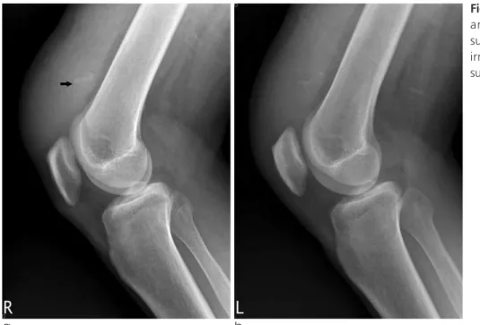
Plain radiographs of both knees revealed irregularly- shaped calcified densities in the supra patella soft
CASE REPORT INTRODUCTION
www.ksmrm.org JKSMRM 16(2) : 169-172, 2012
Print ISSN 1226-9751
�Received; June 7, 2012�Revised; July 30, 2012
�Accepted; July 30, 2012
Corresponding author : Jae Chan Shim, M.D., Department of Radiology, Inje University College of Medicine, Seoul Paik Hospital, 85 Jeo-dong 2nd street, Jung-gu, Seoul 100-032, Korea.
Tel. 82-2-2270-0139, Fax. 82-2-2266-2799 E-mail : [email protected]
Case Report
tissue(Fig. 1A, B). To further evaluate these finding, both knees MRI were performed. Sagittal T1- weighted and proton density MR images of the knee joints revealed complete detachment of the quadriceps tendon from the superior pole of the patella and proximal retraction of the ruptured tendon.
Hypointense T1 and hyperintense T2 signal intensities were noted between the patella and detached tendon in both knees (Fig. 2A-D). Surgical repair was undertaken and the MRI findings were confirmed.
Both quadriceps tendons were completely disrupted just proximal to the superior pole of the patella without bony fragment. The avulsed tendons’ ends were ragged with some dystrophic clacifications and the upper border of patellae was softened due to some degree of bone resorption.
A normal quadriceps tendon is one of the strongest tendins in the body, being able to withstand a load of 15 to 30 kg/mm (4). Simulataneous bilateral sponta- neous rupture of the quadriceps tendon is uncommon, and have been described as a complication of various diseases, such as, secondary hyperparathyroidism, systemic lupus erythematosus, diabetes mellitus, gout, rheumatoid arthritis, obesity, and end stage chronic renal failure (5). In patients with underlying disease,
spontaneous bilateral rupture of the quadriceps tendon may occur during ordinary daily activities such walking or even steeping from a car (4). The microscopic findings include fatty or myxoid degener- ation, calcification within the tendon, cystic softening, and decreased collagen with marked loss of nuclei (4, 6). The pathogenesis of bilateral quadriceps tendon rupture in chronic renal failure is unclear, and more than one factor may be included. Changes in collagen and the ground substance in patients with renal failure include ischemia and dystrophic calcification. Systemic acidosis, as well as the direct effects of parathyroid hormone and subperiosteal bone resorptionm with subsquent weakening of the tendon-bone intervace, have been implicated in the pathogenesis (4, 7, 8).
Renal failure as an underlying cause of bilateral quadriceps tendon rupture is directly related to the duration of renal failure and the period of dialysis (4, 7). In this 36-year-old man, chronic renal insufficiency and secondary hyperparathyroidism were precursors and causative conditions for the attenuation that precipitated spontaneous rupture.
Primary and secondary hyperparathyroidism has contributed to quadriceps tendon rupture by causing subperiosteal bone resorption, which weakens the osteotendinous junction. The result of this may be repeated minor avulsion fracture of the bone cortex, ultimately leading to scarring and weakening of the tendon attatchment site, and total rupture (9, 10).
DISCUSSION
JKSMRM 16(2) : 169-172, 2012
170
a b
Fig. 1. Lateral radiographs of right (a) and left (b) knees demonstrate suprapatellar soft tissue swelling and irregular shaped calcifications in the suprapatellar soft tissue (arrow in a).
Plain radiography is useful for demonstrating indirect signs of tendon rupture, such as poorly defined suprapatellar swelling and forward tilting of patella. Small fragments of avulsed bone or dystrophic calcification may be observed in the suprapatellar region. The patella is often, but not always, low lying (1).
In summary, a case of simultaneous, spontaneous, bilateral rupture of the quadriceps tendon in sceondary hyperparathyroidism is presented.
Although quadriceps tendon injuries are easily diagnosed by clinical findings, physical examination, plain radiography, and especially MRI are necessary for determining the site, extension and pattern of tendon rupture as well as for preoperative planning of repair or tendon reconstruction.
References
1. Dunnick NR. Image interpretation session: 1999. Bilateral Simultaneous Bilateral Quadriceps Tendon Rupture in Patient with Secondary Hyperparathyroidism due to Chronic Renal Failure � Jong Joon Shim, et al.
171
a b
Fig. 2. Sagittal T1 (TR 316.7, TE 11.2) and T2-weighted (TR 4083.3, TE 90.0) MR images of the right (a, b) and left (c, d) knees reveal disruption of the quadriceps tendons from the superior pole of the patella and proximal retraction of the torn (arrows in b and d). Joint effusion fluid and extensive deep and superficial soft tissue edema is seen around quadriceps tendon on T2-weighted image.
c d
quadriceps tendon rupture and multiple brown tumors in a patient with secondary hyperparathyroidism. Radiographics 2000;20:262-263
2. Calvo E, Ferrer A, Robledo AG, Alvarez L, Castillo F, Vallejo C. Bilateral simultaneous spontaneous quadriceps tendons rupture. A case report studied by magnetic resonance imaging.
Clin Imaging 1997;21:73-76
3. Lee YS, Son SB, Han CW, Kang SW. Bilateral simultaneous quadriceps tendon rupture in a patient with secondary hyperparathyroidism: a case report. J Korean Soc Radiol 2001;
45:507-511
4. Lombardi LJ, Cleri DJ, Epstein E. Bilateral spontaneous quadri- ceps tendon rupture in a patient with renal failure. Orthopedics 1995;18:187-191
5. Shah MK. Simultaneous bilateral rupture of quadriceps tendons:
analysis of risk factors and associations. South Med J 2002;95:
860-866
6. Anderson WE 3rd, Habermann ET. Spontaneous bilateral quadriceps tendon rupture in a patient on hemodialysis. Otrho Rev 1988;17:411-414
7. Bhole R, Flynn JC, Marbury TC. Quadriceps tendon ruptures in uremia. Clin Orthop 1985;195:200-206
8. Newberg A, Wales L. Radiographic diagnosis of quadriceps tendon rupture. Radiology 1977;125:367-371
9. De Franco P, Varghese J, Brown WW, Bastani B. Secondary hyperparathyroidism, and not beta 2-microglobulin amyloid, as a cause of spontaneous tendon rupture in patients on chronic homodialysis. Am J kidney Dis 1994;24:951-955
10. Meneghello A, Bertoli M. Tendon disease and adjacent bone erosion in dialysis patients. Br J Radiol 1983;56:915-920 JKSMRM 16(2) : 169-172, 2012
172
통신저자 : 심재찬, (100-032) 서울시 중구 저동2가 85번지, 인제의대 서울백병원 영상의학과
Tel. (02) 2270-0139 Fax. (02) 2266-6799 E-mail: [email protected]
만성신부전증에 의한 이차성 부갑상선기능항진증 환자에서 양쪽 대퇴사두건 동시 파열: 증례 보고
1인제의대 서울백병원 영상의학과
2인제의대 서울백병원 정형외과
심종준1∙심재찬1∙하정구2∙이경은1∙이기재1∙김호균1∙서정호1
양측 대퇴사두건의 동시성 파열은 매우 드물며 몇 개의 증례만이 문헌에 보고되어 있다. 정확한 병인은 아직까지 알 려지지 않았다. 그러나 만성신부전에 의한 이차성 부갑상선기능항진증과 같은 만성 대사성 질환을 앓고 있는 환자들 에게 드물게 발생한다. 저자들은 만성신부전에 의한 이차성부갑상선기능항진증 환자에서 대퇴사두건이 양측 동시 파 열된 1예를 경험하였기에 문헌고찰과 함께 보고하고자 한다.
대한자기공명의과학회지 16:169-172(2012)