Simultaneous bilateral rupture of the quadriceps ten- don is very rare, usually occurring in the elderly or in patients suffering from a chronic illness such as gout, collagen vascular disease, diabetes mellitus, hyper- parathyroidism, or chronic renal failure (1, 2).
Quadriceps tendon rupture is diagnosed clinically, but where there is no history of trauma or a hematoma pre- sent at physical examination masks the defect, sponta- neous rupture may be difficult to diagnose. Bilateral rupture is even more difficult to detect, since its appear- ance at physical examination may be symmetric.
Magnetic resonance imaging (MRI) is the most accurate imaging modality for assessing tendon rupture and for preoperative planning, though its findings have not been reported in the Korean medical literature. We re- port a case of simultaneous bilateral quadriceps tendon rupture in a patient with chronic renal failure and sec- ondary hyperparathyroidism, and describe the radiolog-
ic findings, including those of plain film and MRI.
Case Report
A 36-year-old woman presented with bilateral knee pain and swelling immediately after falling over on a sidewalk. She was unable to ambulate without assis- tance. According to her past medical history, she had ex- perienced chronic renal failure secondary to chronic glomerulonephritis caused by IgA nephropathy diag- nosed 12 years ago, and had been on maintenance he- modialysis for five years. Physical examination revealed bilateral, painful knee distension. After needle aspira- tion of approximately 60 cc of bloody fluid from both knee joints, physical examination revealed an indistinct palpable defect at the suprapatellar region of the knees.
The abnormal laboratory findings were as follows:
serum creatinine, 10.5 mg/dL; serum alkaline phos- phatase, 2992 IU/L; serum calcium, 8.3 mg/dL; BUN, 55.8 mg/dL; total protein, 5.6 g/dL; albumin, 3.3 g/dL;
hemoglobin, 8.2 g/dL; hematocrit, 25.9%. Electromyo- graphic studies showed no electrodiagnostic findings of peripheral neuropathy or lower motor neuron denerva- tion. A radiographic survey of the entire skeleton demonstrated characteristic findings of secondary hy- perparathyrodism including the salt-and -pepper ap-
Bilateral Simultaneous Quadriceps Tendon Rupture in a Patient with Secondary
Hyperparathyroidism: A Case Report1
Yeon Soo Lee, M.D., Sang Beom Son, M.D., Chang Whan Han, M.D.2, Si Won Kang, M.D.
Simultaneous bilateral rupture of the quadriceps tendon without a significant histo- ry of trauma may occur in association with chronic metabolic disorders such as chron- ic renal failure and secondary hyperparathyroidism, though has rarely been reported.
We describe a case of spontaneous bilateral quadriceps tendon rupture in a 36-year-old female patient with secondary hyperparathyroidism.
Index words : Knee, MR Tendons, injuries Tendons, MR
1Department of Radiology, Taejon St Mary’s Hospital, The Catholic University of Korea
2Department of Orthopedic Surgery, Taejon St Mary’s Hospital, The Catholic University of Korea
Received February 12, 2001; Accepted August 20, 2001
Address reprint requests to : Yeon Soo Lee, M.D., Department of Radiology, Taejon St Mary’s Hospital, The Catholic University of Korea, 520-2 Taehung-dong, Chung-ku, Taejon 301-723, Korea.
Tel. 82-42-220-9625 Fax. 82-42-257-0511 E-mail: [email protected]
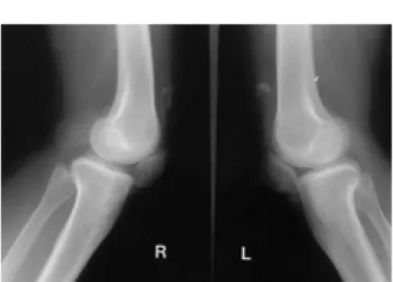
pearance of the skull, a rugger-jersey spine, and sub- chondral bone resorption of the hand and acromioclav- icular joints (Fig. 1). Plain radiographs of both knees re- vealed low-lying patellae bilaterally, and irregularly- shaped calcified densities in the suprapatellar soft tissue (Fig. 2). To further evaluate these findings, MR T1- and T2- weighted imaging of both knees was performed.
Sagittal T1- and T2- weighted MR images of the knee joints revealed complete detachment of the quadriceps tendon from the superior pole of the patella and proxi- mal retraction of the ruptured tendon. Hypointense T1 and hyperintense T2 signal intensities were noted be- tween the patella and detached tendon in both knees, where multiple, focal, bony fragments of high T1 and intermediate T2 signal intensity were present in the re- tracted quadriceps tendons (Fig. 3). Surgical repair was undertaken and the MRI findings were confirmed. Both quadriceps tendons were completely disrupted just proximal to the superior pole of the patella and old hem- orrhage with scarred fibrous tissues was found between the ruptured tendon and patella. In both knees, some os- sified fragments and focal subperiosteal erosion of the patella were also noted.
Discussion
A normal quadriceps tendon is one of the strongest tendons in the body, being able to withstand a load of 15 to 30 kg/mm (3). In patients with underlying disease,
─ 508 ─
A B
Fig. 1. Plain radiographic findings rep- resenting secondary hyperparathy- roidism
A. Bandlike sclerosis in the superior and inferior margins of the vertebral bodies is seen.
B. Subperiosteal resorptions (arrows) including brown tumor (arrowhead) are seen in the phalanges.
C. Subchondral resoprtion (arrow) is noted in the distal clavicle.
C
Fig. 2. Lateral radiographs of the both knees demonstrate low lying patellae with irregular shaped calcifications in the supra- patellar soft tissues. The soft tissue planes demarcating the dis- tal quadriceps tendons are indistinct.
spontaneous bilateral rupture of the quadriceps tendon may occur during ordinary daily activities such as walk- ing or even stepping from a car (3). The microscopic findings include fatty or myxoid degeneration, calcifica- tion within the tendon, cystic softening, and decreased collagen with marked loss of nuclei (3, 4). A similar pathology is seen in old age, when obesity, diabetes, ath- erosclerosis, chronic renal failure and inflammatory re- actions such as those accompaning gout, tuberculosis,
syphilis, and acute bacterial infections may occur (3).
The pathogenesis of bilateral quadriceps tendon rup- ture in chronic renal failure is uncelar, and more than one factor may be involved. Changes in collagen and the ground substance in patients with renal failure include ischemia and dystrophic calcification. Systemic acidosis, as well as the direct effects of parathyroid hormone and subperiosteal bone resorption, with subsequent weak- ening of the tendon-bone interface, have been implicat-
A B
Fig. 3. Sagittal T1- and T2-weighted MR images of the right (A, B) and left (C, D) knees reveal disruption of the quadri- ceps tendons from the superior pole of the patella and proximal retraction of the torn quadriceps tendon (open ar- rows) including focal ossified frag- ments (arrows). Suprapatellar fibrous tissue and hemorrhage are also noted.
C D
ed in the pathogenesis (3, 5, 6). It has been suggested that a chronic acidotic state leads to elastin deposition in the tendons, predisposing them to spontaneous rupture.
A reduced turnover of collagen, with replacement by elastic tissue, has been postulated as a cause of weaken- ing of the tendon (5, 7). In contrast to the ruptures seen in elderly or obese patients with fatty degeneration, ten- don ruptures in dialysis patients tend to occur at a younger age, usually under 40 years (5).
Renal failure as an underlying cause of bilateral quadriceps tendon rupture is directly related to the du- ration of renal failure and the period of dialysis (3, 5). In this 36-year-old-female, chronic renal insufficiency and secondary hyperparathyroidism were precursors and causative conditions for the attenuation that precipitated spontaneous rupture. The osteotendinous junction is thought to be weakened by bone resorption secondary to hyperparathyroidism, and it may be for this reason that it is the most frequently involved site (3). Primary or secondary hyperparathyroidism has contributed to quadriceps tendon rupture by causing dystrophic calci- fication and subperiosteal bone resorption, which weak- ens the osteotendinous junction. The result of this may be repeated minor avulsion fracture of the bone cortex, ultimately leading to scarring and weakening of the ten- don attachment site, and total rupture (7, 8). Thus, the most important causes of tendon rupture appear to be primary tendon disease and bone erosion at the site of tendon insertion. In our case, avulsion fractures of the patellar cortex were not present, but there were focal subperiosteal erosions in both patellae and ossified frag- ments with combined dystrophic calcification in the re- tracted quadriceps tendons.
To prevent residual deformity and functional loss, ear- ly diagnosis and repair are necessary. The clinical find- ings are not always clear, and after hematoma forma- tion, the tendon defect may not be readily palpable (9).
Plain radiography is useful for demonstrating indirect signs of tendon rupture, such as poorly defined suprap-
atellar swelling and forward tilting of the patella. Small fragments of avulsed bone or dystrophic calcification may be observed in the suprapatellar region. The patella is often, but not always, low lying (1). Sonography can demonstrate effusion and a ruptured tendon, though tendon separation and proximal retraction are most clearly defined by MRI. In summary, a case of simulta- neous, spontaneous, bilateral rupture of the quadriceps tendon in secondary hyperparathyroidism is presented.
Although the diagnosis is clinical, MRI can accurately determine the site and extent of tendon rupture, find- ings which can be very helpful for confirming the clini- cal diagnosis and for preoperative planning.
References
1. Dunnick NR. Image interpretation session: 1999. Bilateral quadri- ceps tendon rupture and multiple brown tumors in a patient with secondary hyperparathyroidism. Radiographics 2000;20:262-263 2. Calvo E, Ferrer A, Robledo AG, Alvarez L, Castillo F, Vallejo C.
Bilateral simultaneous spontaneous quadriceps tendons rupture. A case report studied by magnetic resonance imaging. Clin Imaging 1997;21:73-76
3. Lombaridi LJ, Cleri DJ, Epstein E. Bilateral spontaneous quadri- ceps tendon rupture in a patient with renal failure. Orthopedics 1995;18:187-191
4. Anderson WE 3rd, Habermann ET. Spontaneous bilateral quadri- ceps tendon rupture in a patient on hemodialysis. Orthop Rev 1988;17:411-414
5. Bhole R, Flynn JC, Marbury TC. Quadriceps tendon ruptures in uremia. Clin Orthop 1985;195:200-206
6. Newberg A, Wales L. Radiographic diagnosis of quadriceps tendon rupture. Radiology 1977;125:367-371
7. De Franco P, Varghese J, Brown WW, Bastani B. Secondary hy- perparathyroidism, and not beta 2-microglobulin amyloid, as a cause of spontaneous tendon rupture in patients on chronic he- modialysis. Am J Kidney Dis 1994;24:951-955
8. Meneghello A, Bertoli M. Tendon disease and adjacent bone ero- sion in dialysis patients. Br J Radiol 1983;56:915-920
9. Barasch E, Lombardi LJ, Arena L, Epstein E. MRI visualization of bilateral quadriceps tendon rupture in a patient with secondary hyperparathyroidism: implications for diagnosis and therapy.
Comput Med Imaging Graph 1989;13:407-410
─ 510 ─
대한방사선의학회지 2001;45:507-511
이차 부갑상선기능항진증 환자에서 대퇴사두건 양측 동시 파열: 1예 보고1
1가톨릭대학교 의과대학 대전성모병원 진단방사선과
2가톨릭대학교 의과대학 대전성모병원 정형외과
이연수・손상범・한창환2・강시원
특별한 외상의 병력없이 양측 대퇴사두건이 동시 파열되는 경우는 매우 드물며, 만성 신장질환이나 이차 부갑상 선기능항진증과 같은 만성 대사질환에서 발생할 수 있다. 저자들은 만성 신부전에 의한 이차 부갑상선기능항진증 환자에서 대퇴사두건이 양측 동시 파열된 1예를 경험하였기에 자기공명영상과 단순 촬영 소견을 문헌고찰과 함께 보고하고자 한다.