401
Copyrights © 2015 The Korean Society of Radiology
INTRODUCTION
Unilateral lower limb edema can be caused by several condi- tions such as deep vein thrombosis (DVT), lymphedema, and chronic venous insufficiency (1). It is necessary to also be aware of the uncommon causes of this condition. We report an unusu- al case of a patient with unilateral limb edema, which occurred due to a synovial cyst that was treated successfully with surgical intervention.
CASE REPORT
A 52-year-old woman, with diabetes mellitus and a 10-year history of hypertension, presented with a gradually worsening right leg edema without pain which persisted for a month.
There was no history of trauma, arthritis, or immobilization.
During physical examination, no abnormality was found except for the presence of pitting edema in the right leg. Ultrasonogra- phy (US), contrast-enhanced computed tomography (CT), and
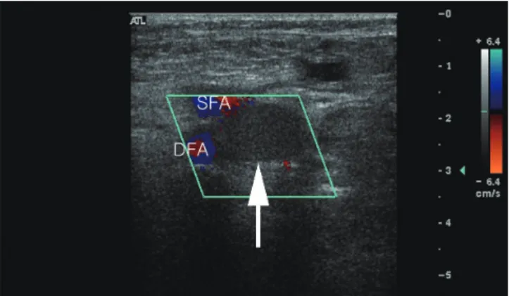
ascending venography were performed to rule out the DVT. US showed 2.1 cm sized hypoechoic mass compressing the right common femoral vein (Fig. 1). There was no blood flow within the mass on Doppler US. The CT scan demonstrated a 2.3 cm sized homogenous hypodense, well-defined, cystic mass com- pressing the right common femoral vein in front of the right hip joint (Fig. 2). No deep venous thrombosis was detected in the right lower extremity. Ascending venography was performed to demonstrate extrinsic compression and the absence of clots. It showed filling defect of the right common femoral vein, without the development of collateral venous flow (Fig. 3). On the basis of these findings, differential diagnosis included synovial cyst, ganglion cyst, paralabral cyst, and noninflammatory pseudotu- mor. During the surgery, a cystic mass of 2.5 cm in size was found protruding from the right hip joint and compressing the posterior wall of the left common femoral vein. The cystic mass was easily separated from the right common femoral vein and was excised successfully. No communication was found be- tween the hip joint space and the cyst. Histological examination
Case Report
pISSN 1738-2637 / eISSN 2288-2928 J Korean Soc Radiol 2015;72(6):401-404 http://dx.doi.org/10.3348/jksr.2015.72.6.401
Received November 3, 2014 Accepted January 29, 2015
Corresponding author: Il Soo Chang, MD
Department of Radiology, Konkuk University School of Medicine, 120-1 Neungdong-ro, Gwangjin-gu, Seoul 143-729, Korea.
Tel. 82-2-2030-5487 Fax. 82-2-2030-5549 E-mail: [email protected]
This is an Open Access article distributed under the terms of the Creative Commons Attribution Non-Commercial License (http://creativecommons.org/licenses/by-nc/3.0) which permits unrestricted non-commercial use, distri- bution, and reproduction in any medium, provided the original work is properly cited.
A synovial cyst of the hip joint is a rare cause of unilateral leg edema, and it is usu- ally associated with arthropathies such as rheumatoid arthritis and osteoarthritis.
An asymptomatic synovial cyst of the hip joint that is not associated with an ar- thritic condition occurs infrequently. In this paper, we described the case of a 52-year-old woman who presented with unilateral right leg edema caused by a sy- novial cyst of the hip joint.
Index terms Leg
Edema Hip Joint Synovial Cyst Veins
Synovial Cyst of the Hip Joint as a Rare Cause of Unilateral Leg Edema: A Case Report
1고관절 활액막낭종에 의한 편측 하지부종: 증례 보고1
Jihun Kang, MD
1, Il Soo Chang, MD
1, Sang Woo Park, MD
1, Ik Jin Yun, MD
2, Hyung Kyu Park, MD
3, Wan Seop Kim, MD
3, Hui Jin Lee, MD
1, Na Ra Kim, MD
1, Sung Gyu Moon, MD
1Departments of 1Radiology, 2Surgery, 3Pathology, Konkuk University School of Medicine, Seoul, Korea
Synovial Cyst of the Hip Joint
402
J Korean Soc Radiol 2015;72(6):401-404 jksronline.orgtant. Moreover, distinguishing between synovial and ganglion cyst using imaging is not always possible (5). It is known that sy- novial cyst is usually accompanied by the conditions affecting the hip, such as rheumatoid arthritis and osteoarthritis (6). A sy- novial cyst is a herniation of the synovial membrane caused by inflammation with an overproduction of synovial fluid (7). Such a cyst may cause unilateral edema in the lower extremity, when it compresses the adjacent venous structure (2). The most com- mon clinical manifestation of a synovial cyst of the hip joint is a palpable mass or the pain in the inguinal area or in the anterior thigh, due to compression of the femoral nerve (8). Iliopsoas bursitis presents with a triad of findings, which includes a mass in the inguinal region, pressure effect on the nearby structures, and roentgenologic arthritic changes (9). However, Iwata et al.
(4) insisted that the presence of an inguinal mass and groin pain is not mandatory for the diagnosis of iliopsoas bursitis. In addi- tion, one report has shown that in only 55% of the cases with a synovial cyst of the hip joint, compression of the adjacent vein confirmed the diagnosis of a synovial cyst (Fig. 4). Surgical re-
moval of the synovial cyst restored normal venous return and promptly relieved the patient’s symptoms. The patient was dis- charged on the fifth postoperative day.
DISCUSSION
Benign cystic lesion, associated with compression of the femo- ral vein and lower leg edema around the hip joint is a rare condi- tion. Until now, 33 cases have been reported in literature, includ- ing this case (2-4). Histologically, there are two types of cysts:
synovial and ganglion cysts. While synovial cyst is a juxta-artic- ular fluid collection lined by synovial cells, ganglion cyst is thought to be a myxomatous degeneration of certain fibrous tis- sue structures without the lining of synovial cell (3). Since the treatment for these two para-articular cysts is similar, clinical differentiation of ganglion and synovial cysts may not be impor- Fig. 1. Doppler ultrasonography shows a hypoechoic (not anechoic) mass compressing the right common femoral vein (white arrow). Note that there is no color signal that would suggest the presence of blood flow. The two color signals by the mass represent superficial (upper) and deep femoral artery (lower).
DFA = deep femoral artery, SFA = superficial femoral artery
Fig. 3. Ascending venography (prone position) shows extrinsic com- pression of the right common femoral vein (white arrow). There is no evidence of deep vein thrombosis.
Fig. 2. The CT scan shows a cystic mass compressing the right com- mon femoral vein (white arrow).
Fig. 4. The excised lesion showed a cyst with thin fibrous cyst wall.
The cyst wall was lined by a layer of flat, elongated synovial cells (ar- rows) (hematoxylin & eosin, × 200).
Jihun Kang, et al
403
jksronline.org J Korean Soc Radiol 2015;72(6):401-404
such as pain and a mass in the inguinal region. In the presence of compression of the adjacent vessel or nerve, surgical excision is the treatment of choice for a synovial cyst of the hip joint.
REFERENCES
1. Ely JW, Osheroff JA, Chambliss ML, Ebell MH. Approach to leg edema of unclear etiology. J Am Board Fam Med 2006;
19:148-160
2. Colasanti M, Sapienza P, Moroni E, Mosiello G, Postacchini F, di Marzo L. An unusual case of synovial cyst of the hip joint presenting as femoral vein compression and severe lower limb edema. Eur J Vasc Endovasc Surg 2006;32:468-470 3. Matsumoto H, Yamamoto E, Kamiya C, Miura E, Kitaoka T,
Suzuki J, et al. Femoral vein compression resulting from a ganglion of the hip joint: a case report. Ann Vasc Dis 2012;5:233-236
4. Iwata T, Nozawa S, Ohashi M, Sakai H, Shimizu K. Giant il- iopectineal bursitis presenting as neuropathy and severe edema of the lower limb: case illustration and review of the literature. Clin Rheumatol 2013;32:721-725
5. Bhan C, Corfield L. A case of unilateral lower limb swelling secondary to a ganglion cyst. Eur J Vasc Endovasc Surg 2007;33:371-372
6. Sartoris DJ, Danzig L, Gilula L, Greenway G, Resnick D. Syno- vial cysts of the hip joint and iliopsoas bursitis: a spectrum of imaging abnormalities. Skeletal Radiol 1985;14:85-94 7. Williams RA, Marks LJ. Synovial cyst causing an inguinal
mass. Br Med J 1978;2:91-92
8. Sugiura M, Komiyama T, Akagi D, Miyata T, Shigematsu H.
Compression of the iliac vein by a synovial cyst. Ann Vasc Surg 2004;18:369-371
9. Melamed A, Bauer CA, Johnson JH. Iliopsoas bursal extension of arthritic disease of the hip. Radiology 1967;89:54-58 10. Wunderbaldinger P, Bremer C, Schellenberger E, Cejna M,
Turetschek K, Kainberger F. Imaging features of iliopsoas bursitis. Eur Radiol 2002;12:409-415
was accompanied by a hip joint condition such as rheumatoid arthritis and osteoarthritis (2). Our patient also presented with unilateral edema in the lower leg, without a history of hip joint disease and other clinical manifestations such as pain or the presence of a palpable mass. The mass may have been difficult to palpate and it may not have caused compression of the femo- ral nerve, because of its small size and location under the femo- ral vein. Therefore, because a synovial cyst of the hip joint can only cause the lower limb edema without any other symptoms, it should be considered in the differential diagnosis of other conditions such as DVT and lymphedema.
Of the various imaging tools, US may be the most cost-effec- tive, simple, and quick assessment of a synovial cyst in the ingui- nal area. However, it is difficult to differentiate between a cystic and solid mass, when the cyst contains debris and hyperplastic synovium (10). Although contrast-enhanced CT scan is a suit- able imaging modality for the evaluation of the cyst size and its effect on the surrounding structures, magnetic resonance imag- ing (MRI) is the most accurate imaging technique for assessing the characteristics of soft tissue abnormalities and those associ- ated joint disorders (3). All communications between the ilio- psoas bursa and hip joint were demonstrated on MRI, whereas US demonstrated these in only 56% and CT in 60% of the cases (10). Ascending venography is useful for differentially diagnos- ing the synovial cyst of the hip joint and DVT, and it also helps to avoid anticoagulation therapy. However, recent developments in imaging techniques have decreased the need for venography.
A synovial cyst of the hip joint can be treated by surgical exci- sion, needle aspiration, total hip arthroplasty, or instillation of a steroid or sclerosing agent (2, 4). In asymptomatic cases, it may be left untreated. However, when the vessels or nerves around the inguinal region are compressed by the cyst, it should be sur- gically excised because of the associated high rate of recurrence (37%) (2).
In conclusion, this case demonstrates that unilateral edema in the lower extremity may be caused by a synovial cyst of the hip joint, without any accompanying arthropathies or symptoms
Synovial Cyst of the Hip Joint
404
J Korean Soc Radiol 2015;72(6):401-404 jksronline.org고관절 활액막낭종에 의한 편측 하지부종: 증례 보고1
강지훈
1· 장일수
1· 박상우
1· 윤익진
2· 박형규
3· 김완섭
3· 이희진
1· 김나라
1· 문승규
1고관절에 발생하는 활액막낭종은 편측 하지부종의 드문 원인이며, 대개는 류마티스 관절염이나 퇴행성 관절염과 같은 관 절질환과 연관되어 있다. 관절질환이나 증상을 동반하지 않는 고관절의 활액막낭종은 더욱 드물다. 이에 고관절 활액막낭 종에 의해 발생한 편측 하지부종을 보인 52세 여자 환자에 대해서 보고하고자 한다.
건국대학교 의학전문대학원 1영상의학교실, 2외과학교실, 3병리과학교실