779 서 론
최근, 인간 뇌의 기능적 이미지를 얻기 위한 방법으로 양 성자방출단층촬영 (positron emission tomography; PET)과 기 능적 자기공명영상술 (functional magnetic resonance imaging;
fMRI)이 있다. 양성자방출단층촬영과 기능적 자기공명영상 술은 신경활성의 분포를 알 수 있는 국부 대뇌 혈류 (re- gional cerebral blood flow)와 뇌의 혈액 산소포화도를 측정 한다. 이 두 방법은 3차원에서 밀리미터 이하를 구분할 수 있는 높은 공간 해상도라는 장점이 있으나 신경 활성을 간 접적으로 측정한다는 단점이 있다. Electroencephalography
Resolution Electromagnetic Tomography (LORETA)를 이용한 Cross Spectrum 분석
Location of Brain Electrical Source Activation according to Visually Stimulated Sexual Arousal: A Cross Spectral Analysis using Low Resolution Brain Electromagnetic Tomography (LORETA)
Sung Chul Kam, See Min Choi, Sung Uk Jeh, Jeong Seok Hwa, Ky Hyun Jung, Suk Won Jeong1, Oh-Young Kwon1, Jae Seog Hyun From the Departments of Urology and 1Neurology, School of Medicine, Gyeongsang National University, Jinju, Korea
Purpose: Low resolution brain electromagnetic tomography (LORETA) is a kind of functional imaging technique and it is also an up-to-date tech- nique for conducting electroencephalography (EEG) analysis. We tried to investigate the locations on the cerebral cortex that are activated by visually stimulated sexual arousal.
Materials and Methods: Thirty-three male volunteers (age range: 24.7±1.7 years) among all the right-handed medical students at our university were enrolled in this study. The EEGs included the segments recorded during resting, watching a music-video, intermission and watching a porno-video.
The LORETA images of the cross-spectral analysis were obtained with using segments of LORETA-KEY (KEY Institute for Brain-Mind Research, Switzerland) software.
Results: In the statistical nonparam etric m aps (SnPM) of each spectrum and the delta, theta and alpha waves did not show the increased current density. The beta 1, 2 and 3 activity showed the point of maximal current densities in the anterior parahippocampal gyrus of the left limbic lobe and the superior temporal gyrus of both temporal lobes, the superior tem- poral gyrus of the right temporal lobe, the precuneus of the right parietal lobe, the medial frontal gyrus of the left frontal lobe, the middle occipital gyrus of the right occipital lobe, the superior temporal gyrus of both temporal lobes and the superior frontal gyrus of the right frontal lobe.
Conclusions: The sexual arousal by visual stimulation may activate the anterior parahippocampal gyrus of the left limbic lobe, the superior temporal gyrus of both temporal lobes, the precuneus of the right parietal lobe, the medial frontal gyrus of the left frontal gyrus, and the middle occipital gyrus of the right occipital lobe. (Korean J Urol 2006;47:779-785) ꠏꠏꠏꠏꠏꠏꠏꠏꠏꠏꠏꠏꠏꠏꠏꠏꠏꠏꠏꠏ
Key Words: Tomography, Arousal, Visual stimulation
대한비뇨기과학회지 제 47 권 제 7 호 2006
경상대학교 의과대학 비뇨기과학교실,
1신경과학교실
감성철․최세민․제성욱․화정석
정기현․정석원1․권오영1․현재석
접수일자:2006년 3월 3일 채택일자:2006년 4월 4일
교신저자: 현재석
경상대학교병원 비뇨기과 경남 진주시 칠암동 90번지 ꂕ 660-702
TEL: 055-750-8196 FAX: 055-757-4503 E-mail: hyunjs@gshp.
gsnu.ac.kr
(EEG)는 신경 활성을 직접적으로 측정할 수 있지만 두개골 밖에서 EEG 측정은 기본적인 제한이 있기 때문에 전기적 신경 활성의 3차원적 재구성에 필요한 정보를 얻을 수 없었 다. 이러한 단점을 보완하기 위해 1994년에 Pascual-Marqui1 가 low-resolution brain electromagnetic tomography (LORETA) 를 소개하였다. LORETA는 기준전극의 영향을 받지 않고, 3차원적으로 뇌 활성화 부위를 찾는 역 문제 (Inverse prob- lem)풀기의 한 방법으로, 동일한 성상의 신경 세포들의 집단 은 동시에 활성화되고 동기화된다는 이론에 기초하여, 많은 역 문제 풀기의 해 중에서 가장 부드러운 (smoothest)분포를 보이는 3차원적 전기소스의 분포를 계산하여 나타낸다.2 EEG를 분석하여 3차원적 기능 영상을 얻는 LORETA는 신경활성을 직접적으로 측정하는 최신 기술이다. 저자들은 시각적 성적 자극을 주었을 때 대뇌 피질에서 활성화되는 부분을 LORETA를 이용하여 찾아보았다.
대상 및 방법
오른손잡이인 40명의 건강한 남자 대학생에게 시각적 성
적 자극을 주고 EEG를 측정하였다. 40명 중 7명은 EEG상 잡파 (artifact)가 많아 평가할 수 없어 제외하였다. 오른손잡 이인 남자 대학생 중 33명 (평균나이; 24.7±1.7)이 실험에 등록되었다. 모든 참가자에서 실험 모델을 자세히 설명하 였으며 그들의 동의를 얻었다. 연구에 앞서 모든 참가자에 게 실험에 참가한 동기에 대해 인터뷰하였다. 그리고 중대 한 내과적 질환, 약물 남용의 과거력, 정신 질환, 성기능 장 애의 과거력이 있는 경우에는 실험에서 제외하였다.
모든 EEG는 국제 10-20 체계에 맞추어 23 채널 (Fp1, Fp2, F3, F4, C3, C4, P3, P4, O1, O2, F7, F8, T3, T4,T5, T6, Fz, Cz, Pz, A1, A2, T1, T2)을 이용하여 기록하였다. 표본율은 200Hz이다. 각각 5분식 휴식, 뮤직 비디오시청, 휴식, 포르 노 비디오 시청 순으로 EEG가 기록되었다. EEG 기록이 끝 난 후 시각적 자극을 받았을 때 성적 흥분을 얼마나 느꼈는 지에 대한 설문지를 작성하였다. 성적 흥분은 발기의 정도 로 평가하였는데 발기가 없으면 0, 약간의 발기는 1, 중간 정도의 발기는 2, 완전 발기는 3으로 설문지를 작성하였다.
1을 기록한 사람이 19명, 2를 기록한 사람이 9명, 3을 기록 한 사람이 5명이었다. 뮤직 비디오와 포르노 비디오를 보면
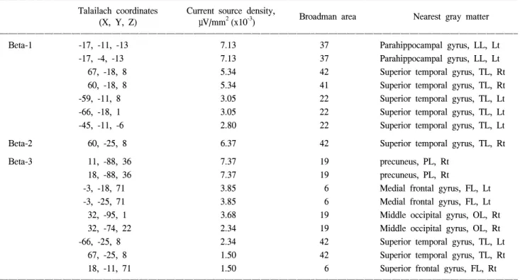
Table 1. The maximal points of current density of the beta
ꠚꠚꠚꠚꠚꠚꠚꠚꠚꠚꠚꠚꠚꠚꠚꠚꠚꠚꠚꠚꠚꠚꠚꠚꠚꠚꠚꠚꠚꠚꠚꠚꠚꠚꠚꠚꠚꠚꠚꠚꠚꠚꠚꠚꠚꠚꠚꠚꠚꠚꠚꠚꠚꠚꠚꠚꠚꠚꠚꠚꠚꠚꠚꠚꠚꠚꠚꠚꠚꠚꠚꠚꠚꠚꠚꠚꠚꠚꠚꠚꠚꠚꠚꠚꠚꠚꠚꠚꠚꠚꠚꠚꠚꠚꠚꠚꠚꠚꠚꠚꠚꠚꠚꠚꠚꠚꠚꠚꠚꠚꠚꠚꠚꠚꠚ Talailach coordinates Current source density,
Broadman area Nearest gray matter (X, Y, Z) μV/mm2(x10-3)
ꠏꠏꠏꠏꠏꠏꠏꠏꠏꠏꠏꠏꠏꠏꠏꠏꠏꠏꠏꠏꠏꠏꠏꠏꠏꠏꠏꠏꠏꠏꠏꠏꠏꠏꠏꠏꠏꠏꠏꠏꠏꠏꠏꠏꠏꠏꠏꠏꠏꠏꠏꠏꠏꠏꠏꠏꠏꠏꠏꠏꠏꠏꠏꠏꠏꠏꠏꠏꠏꠏꠏꠏꠏꠏꠏꠏꠏꠏꠏꠏꠏꠏꠏꠏꠏꠏꠏꠏꠏꠏꠏꠏꠏꠏꠏꠏꠏꠏꠏꠏꠏꠏꠏꠏꠏꠏꠏꠏꠏꠏꠏꠏꠏꠏꠏ
Beta-1 -17, -11, -13 7.13 37 Parahippocampal gyrus, LL, Lt
-17, -4, -13 7.13 37 Parahippocampal gyrus, LL, Lt
67, -18, 8 5.34 42 Superior temporal gyrus, TL, Rt
60, -18, 8 5.34 41 Superior temporal gyrus, TL, Rt
-59, -11, 8 3.05 22 Superior temporal gyrus, TL, Lt
-66, -18, 1 3.05 22 Superior temporal gyrus, TL, Lt
-45, -11, -6 2.80 22 Superior temporal gyrus, TL, Lt
Beta-2 60, -25, 8 6.37 42 Superior temporal gyrus, TL, Rt
Beta-3 11, -88, 36 7.37 19 precuneus, PL, Rt
18, -88, 36 7.37 19 precuneus, PL, Rt
-3, -18, 71 3.85 6 Medial frontal gyrus, FL, Lt
-3, -25, 71 3.85 6 Medial frontal gyrus, FL, Lt
32, -95, 1 3.68 19 Middle occipital gyrus, OL, Rt
32, -74, 22 2.34 19 Middle occipital gyrus, OL, Rt
-66, -25, 8 2.34 42 Superior temporal gyrus, TL, Lt
67, -25, 8 1.50 42 Superior temporal gyrus, TL, Rt
18, -11, 71 1.50 6 Superior frontal gyrus, FL, Rt
ꠏꠏꠏꠏꠏꠏꠏꠏꠏꠏꠏꠏꠏꠏꠏꠏꠏꠏꠏꠏꠏꠏꠏꠏꠏꠏꠏꠏꠏꠏꠏꠏꠏꠏꠏꠏꠏꠏꠏꠏꠏꠏꠏꠏꠏꠏꠏꠏꠏꠏꠏꠏꠏꠏꠏꠏꠏꠏꠏꠏꠏꠏꠏꠏꠏꠏꠏꠏꠏꠏꠏꠏꠏꠏꠏꠏꠏꠏꠏꠏꠏꠏꠏꠏꠏꠏꠏꠏꠏꠏꠏꠏꠏꠏꠏꠏꠏꠏꠏꠏꠏꠏꠏꠏꠏꠏꠏꠏꠏꠏꠏꠏꠏꠏꠏ LL: limbic lobe, TL: temporal lobe, PL: parietal lobe, FL: frontal lobe, OL: occipital lobe, Lt: left, Rt: right. The beta 1, 2 and 3 activity showed the point of maximal current densities in the anterior parahippocampal gyrus of the left limbic lobe and the superior temporal gyrus of both temporal lobes, the superior temporal gyrus of the right temporal lobe, the precuneus of the right parietal lobe, the medial frontal gyrus of the left frontal lobe, the middle occipital gyrus of the right occipital lobe, the superior temporal gyrus of both temporal lobes and the superior frontal gyrus of the right frontal lobe.
서 측정한 각각의 EEG를 5초 동안 잡파가 없는 부분 5군데 를 선택하였다 (fileter: 1.6-60Hz). 자료를 재분석하여 얻은 데이터를 brain electrical source analysis (BESA)에 의해 처리 하여 공간적 분석을 시행하였다. 데이터는 디지털화되어 6 개 [delta (1-3Hz), theta (4-7Hz), alpha (8-12Hz), beta1 (13-18Hz), beta2 (19-21Hz), beta3 (22-30Hz)]의 주파수 영역으로 나누어 졌다.
LORETA-KEY (KEY Institute for Brain-Mind Research, Switzerland) software는 비교 공간 (cross-spectral)분석에 의 해 대뇌속의 전류밀도를 얻을 수 있다. LORETA의 현재 사 용 중인 버전은 Montreal Neurologic Institute의 Brain Imaging Center에서 디지털화된 자기공명영상으로 만들어진 Talairach human brain atlas3에 등록된 Three-shell spherical head model 을 사용하고 있다. Three-shell spherical head model과 Talairach head geometry 사이의 등록은 Towle4에 의해 발표된 실질적 EEG전기 좌표를 사용하였다. LORETA의해 영역 (solution space)은 Montreal Neurologic Institute의 Brain Imaging Center
에서 만들어진 유사한 Digitized Probability Atlas에 의해 정 의된 Talairach Atlas에서 대뇌 회백질과 해마부위에 국한된 다. 따라서 7mm 범위의 공간 해상도에서 총 2394개의 격자
(voxel)가 이 신경해부학적 범위에서 산출된다.1,2
모든 부분을 교차 스펙트럼 분석 (cross spectral analysis) 하여 LORETA 영상을 얻은 후에, 뮤직 비디오를 보았을 때 와 포르노 비디오를 보았을 때의 통계적 비모수적 지도 [statistical non parametric maps (SnPM)]를 각 주파수 영역에 서 측정하였다 (delta 1-3/sec, theta 4-7/sec, alpha 8-12/sec, beta- 1 13-18/sec, beta-2 19-21/sec, and beta-3 22-30/sec). 각 주파수 영역에서 가장 전류 밀도가 높은 부분을 LORETA-KEY software로 계산하여 Brodmann영역 및 대뇌 피질의 해부학 적인 위치를 구하였다.
결 과
시각적 성적 자극을 주었을 때 뇌의 전류 밀도는 기저 상
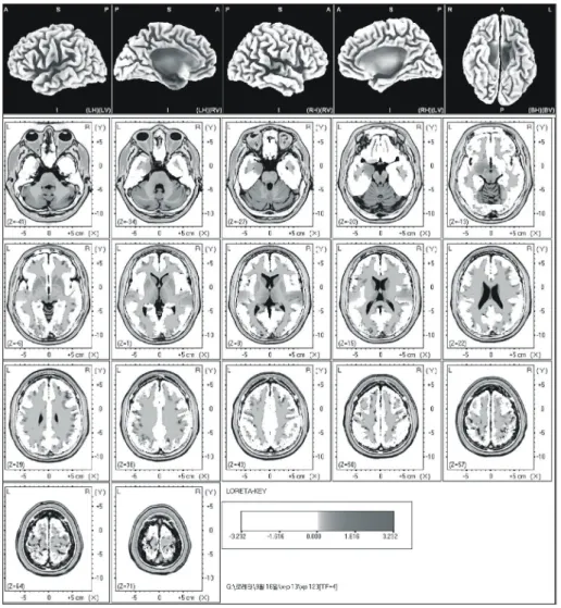
Fig. 1. The anatomical areas of current density increment of the beta 1 (13- 18Hz) band induced by visual-sexual arousal. The current density is increased in the parahippocampal gyrus, the su- perior temporal gyrus of the left hemis- phere and the superior temporal gyrus of the right hemisphere.
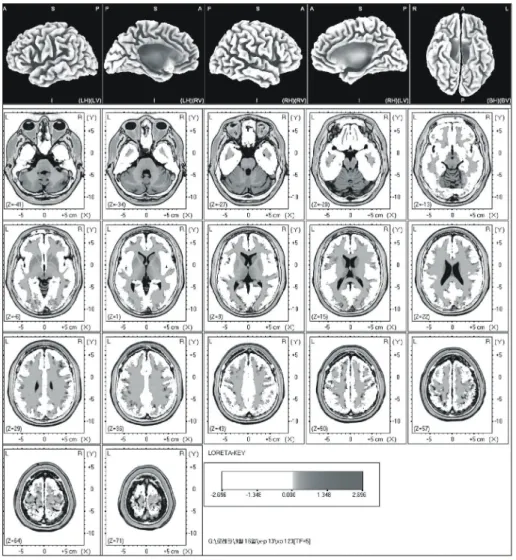
태에 비해 델타, 세타, 알파 주파수대의 통계적 비모수적 지 도 (SnPM)에서는 차이가 미약하였다. 베타 1 주파수대에서 는 좌측 변연계의 해마곁이랑 (parahippocampal gyrus of left limbic lobe)과 양쪽 관자엽의 위관자이랑 (superior temporal gyrus of both temporal lobes)에서 전류 밀도가 차이가 크게 나타났다 (Table 1) (Fig. 1). 베타 2 주파수대에서는 우측 관 자엽의 위관자이랑 (superior temporal gyrus of right temporal lobe)에서 전류 밀도가 차이가 크게 나타났다 (Table 1) (Fig.
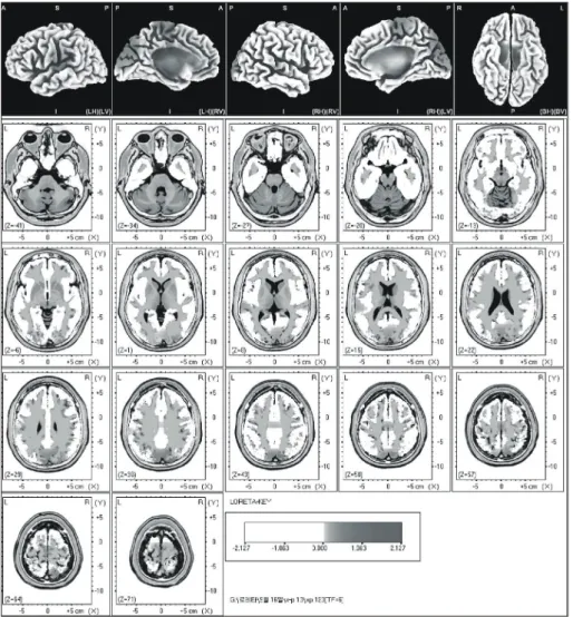
2). 베타 3 주파수대에서는 우측 마루엽의 쐐기앞소엽 (pre- cuneus of right parietal lobe), 좌측 이마엽의 중간이마이랑 (mi- ddle frontal gyrus of left frontal lobe), 우측 뒤통수엽의 중간 뒤통수고랑 (middle occipital gyrus of right occipital lobe), 양 쪽 관자엽의 위관자이랑 (superior temporal gyrus of both temporal lobe), 그리고 우측 이마옆의 위이마이랑 (superior frontal gyrus of right frontal lobe)에서 전류 밀도가 차이가 크게 나타났다 (Table 1) (Fig. 3). 위 결과들을 종합하여 보 면, 전류 밀도는 해마곁이랑 (parahippocampal gyrus), 위관자
이랑 (superior temporal gyrus), 좌측 이마엽의 중간이마이랑 (middle frontal gyrus of the left hemisphere), 위관자이랑 (su- perior temporal gyrus), 쐐기앞소엽 (precuneus), 그리고 우측 대뇌반구의 중간뒤통수고랑 (middle occipital gyrus of the right hemisphere)에서 증가하였다.
고 찰
외부 자극에 대한 뇌 반응을 보여주는 비침습적 방법들 로 인해 기능적 뇌 지도에 대한 지식을 더 많이 알 수 있다.
이전에 PET를 이용한 연구들에서 시각적 성적 자극에 의해 활성화되는 뇌영역은 앞 띠고랑 (anterior cingulate gyrus), 뒤 통수이마이랑 (orbitofrontal cortex), 꼬리핵 (caudate), 조가비 핵 (putamen) 등이라고 보고하였다.5-9 이전 연구에서 기능적 MRI를 이용하여 시각적 성적 자극 후에 국소적 뇌 혈류량
(rCBF)이 증가하는 부위는 앞, 중간 띠고랑 (anterior and mid-cingulate gyrus), 좌측 대뇌반구의 아래 관자이랑 (in-
Fig. 2. The anatomical areas of current density increment of the beta 2 (19-21 Hz) band induced by visual-sexual a- rousal. The current density is increased in the superior temporal gyrus of the right hemisphere.
ferior temporal gyrus of the left hemisphere), 아래, 중간 뒤통 수이마이랑 (inferior, medial and orbito-frontal gyrus), 아래 관 자이랑 (inferior temporal gyrus), 우측 대뇌반구의 대뇌섬 (in- sula of the right hemisphere), 꼬리핵 (caudate), 조가비핵 (pu- tamen), 양쪽 대뇌반구의 담장 (claustrum of the both hemi- sphere)등으로 보고하였다.10-20
PET 연구에서 우측 대뇌반구의 아래뒤통수이랑 (inferior occipital gyrus of the right hemisphere), 담장 (claustrum), 앞띠 이랑 (antertoior cingulate gyrus), 조가비핵 (putamen), 양쪽 대 뇌반구의 꼬리핵 (caudate nucleus of the both hemisphere)영 역을 포함하여 국소적 뇌 혈류량이 증가함에 따라 성기의 강직도가 증진되었다.5,12 기능적 MRI 연구에서, 성기의 강 직도와 연관되어 강력하게 활성화된 뇌 영역은 담장을 포 함하는 대뇌섬밑 부위 [subinsular regions (including the claus- trum)], 중간뒤통수이랑 (middle occipital gyrus), 중간관자이 랑 (middle temporal gyrus), 우측 대뇌반구의 감각, 운동, 전 운동부위 (sensory motor and pre-motor regions of the right
hemisphere), 꼬리핵 (caudate), 좌측 대뇌반구의 조가비핵 (pu- tamen of the left hemisphere), 양쪽 대뇌반구의 띠이랑 (cin- gulate gyrus of the both hemisphere)이다.13
성인 남성에서 시각적 성적자극에 의해 활성화되는 뇌 영역은 이전 PET와 fMRI를 이용한 연구에서 동일하게 앞, 중간 띠고랑 (anterior and mid-cingulate gyrus), 좌측 대뇌반 구의 아래 관자이랑 (inferior temporal gyrus of the left hemi- sphere), 아래 되통수이랑 (inferior occipital gyrus), 꼬리핵 (caudate), 좌측 대뇌반구의 조가비핵 (putamen), 아래, 중간 뒤통수이마이랑 (inferior, medial and orbito-frontal gyrus), 감 각, 운동, 전운동부위 (sensory motor and pre-motor regions), 아래, 중간 관자이랑 (inferior and middle temporal gyrus), 대 뇌섬 (insula), 대뇌섬밑 부위 (subinsular regions), 우측 대뇌 반구의 중간 뒤통수이랑 (middle occipital gyrus of the right hemisphere), 앞 띠이랑 (anterior cingulate gyrus), 띠이랑 (cin- gulate gyrus), 양쪽 대뇌반구의 담장 (claustrum of the both hemisphere)영역에서 활성화되었다.5-20
Fig. 3. The anatomical areas of current density increment of the beta 3 (22- 30Hz) band induced by visual-sexual arousal. The current density is increased in the superior temporal gyrus, the precuneus and the middle occipital gyrus of the right hemisphere, and the medial frontal gyrus of the left hemisphere.
본 연구에서는 해마곁이랑 (parahippocampal gyrus), 위관 자이랑 (superior temporal gyrus), 좌측 이마엽의 중간이마이 랑 (medial frontal gyrus of left frontal gyrus), 쐐기앞소엽 (precuneus), 그리고 우측 대뇌반구의 중간뒤통수이랑 (middle occipital gyrus of right hemisphere)부위들의 전기 밀도가 증 가하였다. Mulert 등21은 소리에 의한 대뇌 활성영역을 알아 보기 위해 LORETA와 fMRI를 사용하여 비교 분석하였다.
그 결과 일부는 동일한 대뇌 부위들이 활성화되었고 일부 는 차이가 있는 것으로 보고하였다. 이와 같이 PET와 fMRI 를 사용한 연구와 본 연구 결과를 비교하면 일부는 일치하 지만 일부는 차이가 나는 것으로 나타났다. PET와 fMRI를 사용한 연구에서는 좌측 뇌의 아래관자이랑 (inferior tem- poral gyrus)이 활성화되는 것에 비교해서 본 연구에서는 좌 측 뇌의 위관자이랑(superior temporal gyrus)이 활성화되었 다. 그리고 PET와 fMRI를 사용한 연구에서 우측 뇌의 대뇌 섬 (insular)또는 대뇌섬밑 (subinsular)부위가 활성화되는 것 에 반해서 본 연구에서는 우측 뇌의 위 관자이랑 (superior temporal gyrus)이 활성화되었다. 이는 신경 활성을 찾는 방 법의 차이로 인한 것으로 생각한다. LORETA는 대뇌의 신 경 활성을 직접적으로 측정하여 3차원으로 나타내고, fMRI 는 국부 대뇌 혈류의 양을 측정하여 간접적으로 신경활성 을 측정하므로 이러한 차이가 발생하는 것으로 생각한다.
두 가지 방법 중 어떤 방법이 신경 활성을 더 정확하게 측 정하는 지는 알려져 있지 않다. 하지만 본 저자들은 신경활 성을 직접적으로 측정하는 LORETA가 fMRI에 비해서 더 정확한 방법이라고 생각한다.
PET와 fMRI를 사용한 연구와 마찬가지로 본 연구에서도 우측 뇌의 중간 뒤통수이랑 (middle occipital gyrus)이 활성 화되었다.5-20 이는 시각적 성적자극 연관되는 occipital visual area가 활성화된 것으로 생각한다.
비디오 시청 후 성적 흥분에 대한 발기 정도 평가에서 약 간의 발기를 기록한 사람이 19명으로 나타났다. 이러한 결 과는, EEG를 측정하기 위해 시청각 성자극 시 머리에 23개 의 단자 (electrode)를 붙이고 진행되어 비디오에 대한 몰입 이 어려워서 나타난 것으로 생각한다.
본 연구는 시각적 성적자극과 뇌 영역의 활성화의 관계 를 LORETA를 이용하여 시행하였다. 시각적 자극에 의한 성적 흥분은 해마곁이랑 (parahippocampal gyrus), 위관자이 랑 (superior temporal gyrus), 우측 대뇌 반구의 중간이마이랑 (medial frontal gyrus of the left hemisphere)의 뇌 영역이 활성 화되었다. 또한 관자이랑 (temporal gyrus), 쐐기앞소엽 (pre- cuneus), 우측 대뇌 반구의 중간 뒤통수이랑 (middle occipital gyrus of the right hemisphere)도 활성화되었다. 이러한 결과 는 성인 남성에서 변연계와 좌측 뇌의 전두엽과 우측 뇌의
마루관자뒤통수부위 (parieto-temporo-occipital area)가 시각적 자극에 의한 성적 흥분에 중요한 역할을 하는 것을 시사한다.
결 론
시각 자극에 의한 성적 흥분은 대뇌 피질에서 변연계의 해마곁이랑 (parahippocampal gyrus of left limbic lobe), 양쪽 관자엽의 위관자이랑 (superior temporal gyrus of both tem- poral lobe), 우측 마루엽의 쐐기앞소엽 (precuneus of right parietal lobe), 좌측 이마엽의 중간이마이랑 (medial frontal gyrus of left frontal gyrus), 그리고 우측 뒤통수엽의 중간뒤 통수이랑 (middle occipital gyrus of right occipital lobe)이 활 성화된다. 이는 인간의 시각적 성적 자극에 의한 성적 흥분 은 이러한 부분들이 중요한 작용을 한다는 것을 시사한다.
REFERENCES
1. Pascual-Marqui RD, Michel CM, Lehmann D. Low resolution electromagnetic tomography: a new method for localizing electrical activity in the brain. Int J Psychophysiol 1994;
18:49-65
2. Kim MR, Kim KR, Ha CK, Choi SH, Lee IK. Comparative study between visual analysis and low resolution electromag- netic tomography (LORETA) method in the localizaion of epi- leptiform discharges. J Korean Neurol Assoc 2002;20:164-8 3. Talairach J, Tournoux P. Co-planar stereotaxic atlas of the
human brain. New York: Thieme; 1988;1
4. Towle VL, Bolanos J, Suarez D, Tan K, Grzeszczuk R, Levin DN, et al. The spatial location of EEG electrodes: locating the best-fitting sphere relative to cortical anatomy. Electroence- phalogr Clin Neurophysiol 1993;86:1-6
5. Stoleru S, Gregoire MC, Gerard D, Decety J, Lafarge E, Cinotti L, et al. Neuroanatomical correlates of visually evoked sexual arousal in human males. Arch Sex Behav 1999;28:1-21 6. Bocher M, Chisin R, Parag Y, Freedman N, Meir Weil Y, Lester H, et al. Cerebral activation associated with sexual arousal in response to a pornographic clip: A 15O-H2O PET study in heterosexual men. Neuroimage 2001;14:105-17 7. Redoute J, Stoleru S, Gregoire MC, Costes N, Cinotti L,
Lavenne F, et al. Brain processing of visual sexual stimuli in human males. Hum Brain Mapp 2000;11:162-77
8. Grafton ST, Arbib MA, Fadiga L, Rizzolatti G. Localization of grasp representations in humans by positron emission tomo- graphy. 2. Observation compared with imagination. Exp Brain Res 1996;112:103-11
9. Phelps ME, Mazziotta JC. Positron emission tomography:
human brain function and biochemistry. Science 1985;228:
799-809
10. Stehling MK, Turner R, Mansfield P. Echo-planar imaging:
magnetic resonance imaging in a fraction of a second. Science 1991;254:43-50
11. Longworth C, Honey G, Sharma T. Science, medicine, and the future: functional magnetic resonance imaging in neuropsy- chiatry. BMJ 1999;319:1551-4
12. Ogawa S, Lee TM, Kay AR, Tank DW. Brain magnetic reso- nance imaging with contrast dependent on blood oxygenation.
Proc Natl Acad Sci USA 1990;87:9868-72
13. Arnow BA, Desmond JE, Banner LL, Glover GH, Solomon A, Polan ML, et al. Brain activation and sexual arousal in healthy, heterosexual males. Brain 2002;125:1014-23 14. Beauregard M, Levesque J, Bourgouin P. Neural correlates of
conscious self-regulation of emotion. J Neurosci 2001;21:
RC165
15. Karama S, Lecours AR, Leroux JM, Bourgouim P, Beaudoin G, Joubert S, et al. Areas of brain activation in males and females during viewing of erotic film excerpts. Hum Brain Mapp 2005;16:1-13
16. Park K, Seo JJ, Kang HK, Ryu SB, Kim HJ, Jeong GW. A new potential of blood oxygenation level dependent (BOLD)
functional MRI for evaluating cerebral centers of penile erec- tion. Int J Impot Res 2001;13:73-81
17. Park KS, Kang HK, Seo JJ, Kim HJ, Ryu SB, Jeong GW.
Blood-oxygenation-level-dependent functional magnetic reso- nance imaging for evaluating cerebral regions of female sexual arousal response. Urology 2001;57:1189-94
18. Stehling MK, Turner R, Mansfield P. Echo-planar imaging:
magnetic resonance imaging in a fraction of a second. Science 1991;254:43-50
19. Ogawa S, Lee TM, Nayak AS, Glynn P. Oxygenation-sensitive contrast in magnetic resonance image of rodent brain at high magnetic fields. Magn Reson Med 1990;14:68-78
20. Mouras H, Stoleru S, Bittoun J, Glutron D, Pelegrini-Issac M, Paradis AL, et al. Brain processing of visual sexual stimuli in healthy men: a functional magnetic resonance imaging study.
Neuroimage 2003;20:855-69
21. Mulert C, Jager L, Schmitt R, Bussfeld P, Pogarell O, Moller HJ, et al. Integration of fMRI and simultaneous EEG : towards a comprehensive understanding of localization and time-corse of brain activity in target detection. Neuroimage 2004;22:83-94