Background and Purpose The outcome for older stroke patients who undergo endovascular revascularization remains unsatisfactory. We aimed to determine the effect of the extent of baseline ischemia on outcome according to age, testing the hypothesis that the restorative ca- pacity for recovery is only marginal in older patients.
Methods Two hundred and thirteen patients who underwent endovascular revascularization due to occlusion in the M1 segment of the middle cerebral artery (with or without internal ca- rotid artery occlusions) were selected for analysis. Patients were categorized into three age groups: group A (<66 years), group B (66–75 years), and group C (>75 years). Using pretreatment diffusion-weighted imaging (DWI), the Alberta Stroke Program Early CT Score (ASPECTS) and lesion volume were independently measured and analyzed in relation to a favorable outcome.
Results A favorable outcome was achieved in 111 of 213 patients overall: in 60 of the 94 (63.8%) patients in group A, in 36 of the 70 (51.4%) patients in group B, and in 15 of the 49 (30.6%) patients in group C (p=0.001). In older stroke patients (group C), a DWI ASPECTS
≥9 and lesion volume ≤5 mL were found to predict a favorable outcome, which was more re- strictive than the cutoffs for their younger counterparts (groups A and B; DWI ASPECTS ≥8 and lesion volume ≤20 mL).
Conclusions The age-adjusted pretreatment DWI lesion volume and ASPECTS may represent useful surrogate markers for functional outcome according to age. The use of more-restrictive inclusion criteria for older stroke patients could be warranted, although larger studies are nec- essary to confirm these findings.
Key Words stroke, reperfusion, diffusion, aging.
Impact of Baseline Ischemia on Outcome in Older Patients Undergoing Endovascular Therapy for Acute Ischemic Stroke
INTRODUCTION
The effectiveness of endovascular revascularization for stroke patients with acute large-ves- sel occlusion in the anterior circulation has been clearly established in several recent ran- domized controlled trials.1-5 However, recent increases in life expectancy mean that older stroke patients possibly constitute the population with the highest stroke incidence6 but are excluded from some of these landmark trials; this is probably due to the prognosis for this age group being expected to remain poor even after reperfusion. The rate of favorable functional outcome in older patients who undergo endovascular therapy (EVT) report- edly varies widely, from 0 to 28%, which might be at least partially due to the application of diverse patient selection criteria in previous cohort studies.7-14
Reperfusion is futile in almost 50% of older stroke patients; that is, the 3-month modi- fied Rankin Scale (mRS) score remains ≥3 despite successful reperfusion, with this per- centage being almost double that for younger patients.15 This may be partly attributed to the higher incidence of comorbidities and lower restorative ability of the brain in such patients, Yang-Ha Hwanga,b
Yong-Won Kima,b,c Dong-Hun Kanga,c,d Yong-Sun Kima,d David S. Liebeskinde
a Cerebrovascular Center, Departments of bNeurology,
c Radiology, and dNeurosurgery, Kyungpook National University School of Medicine and Hospital, Daegu, Korea
e UCLA Stroke Center,
University of California, Los Angeles, Los Angeles, CA, USA
pISSN 1738-6586 / eISSN 2005-5013 / J Clin Neurol 2017;13(2):162-169 / https://doi.org/10.3988/jcn.2017.13.2.162
Received October 27, 2016 Revised December 14, 2016 Accepted December 15, 2016 Correspondence Yang-Ha Hwang, MD, PhD Department of Neurology, Cerebrovascular Center, Kyungpook National University School of Medicine and Hospital, 130 Dongdeok-ro, Jung-gu, Daegu 41944, Korea Tel +82-53-420-5758 Fax +82-53-422-4265
E-mail [email protected]
cc This is an Open Access article distributed under the terms of the Creative Commons Attribution Non-Com- mercial License (http://creativecommons.org/licenses/by-nc/4.0) which permits unrestricted non-commercial use, distribution, and reproduction in any medium, provided the original work is properly cited.
JCN
Open Access ORIGINAL ARTICLEHwang YH et al.
JCN
which results in poor functional recovery.16 It may therefore be necessary to define stricter inclusion criteria for EVT for older stroke patients. The low restorative ability of the brain in older patients might imply that the threshold of baseline ischemia for recovery–which is one of the most-reliable sur- rogate markers of stroke outcome–17-19could be lower than that in their younger counterparts.
We hypothesized that the threshold for baseline ischemia as a predictor of good functional outcome is lower in older patients. To test this hypothesis, which was not been explored previously in the clinical literature, we examined the relation- ship between the degree of baseline ischemia and clinical outcome according to age.
METHODS
Between May 2006 and December 2014, patients were ret- rospectively selected from a prospectively maintained acute stroke registry at our institution. The criteria for inclusion in this study were as follows: 1) EVT performed due to acute anterior circulation stroke with proximal vessel occlusion [T- or L-type internal carotid artery (ICA), M1 segment of the middle cerebral artery (MCA), or ICA/M1 tandem oc- clusion], 2) arrival at our emergency center within 6 hours of symptom onset, 3) evaluable pretreatment diffusion-weight- ed imaging (DWI), and 4) prestroke mRS score of 0–2. The study protocol was approved by the local Institutional Re- view Board.
Eligible patients underwent transfemoral cerebral angi- ography with contrast agent injected into both carotid ar- teries and the dominant vertebral artery during the late ve- nous phase under local or general anesthesia, in order to define the angioarchitecture of the occluded vessel and assess the extent of collateral flow from all possible sources. If the treatable ICA or MCA occlusion persisted, EVT was initiat- ed. Treatment strategies were selected on the basis of thera- pies available at the time of angiography, and they included intra-arterial thrombolytic infusion (urokinase or recombi- nant tissue plasminogen activator), mechanical clot disrup- tion, mechanical thrombectomy with forced arterial suc- tion thrombectomy or Solitaire thrombectomy, placement of rescue intra- or extracranial stenting, or a combination of these approaches.20-22 The angiographic collateral flow grade, as observed during pretreatment angiography, was evaluated according to the American Society of Interventional and Therapeutic Neuroradiology/Society of Interventional Ra- diology Collateral Flow Grading System. This angiographic scale assigns patients to grades 0 to 4 according to the com- pleteness and rapidity of retrograde collateral filling.23 The reperfusion status was measured using the Thrombolysis in
Cerebral Infarction (TICI) scale.23
Information on demographic and clinical characteristics, medical history, admission blood pressure, and blood glucose were collected at baseline. The onset of stroke was defined as the time when the patient was last observed to be without any abnormal signs or symptoms. Before the initiation of treatment, baseline stroke severity was assessed using the National Institutes of Health Stroke Scale (NIHSS). All pa- tients underwent a CT or MRI scan at 24–48 hours after the initiation of treatment. If evidence of hemorrhage was de- tected, the subtype of the stroke was classified as hemorrhag- ic infarction, parenchymal hematoma, subarachnoid hemor- rhage, intraventricular hemorrhage, or mixed.24 Symptomatic intracranial hemorrhage was defined as any type of hemor- rhage associated with an increase in the NIHSS score of 4 or more within 24 hours of treatment.25 The DWI findings at baseline were assessed using the Alberta Stroke Program Early CT Score (ASPECTS) for DWI by two raters (Y.W.K.
and D.H.K.),26 with any disagreements resolved by consen- sus. The volume of the lesion on DWI was calculated using open-source image analysis software (OsiriX, Pixmeo, Ge- neva, Switzerland) by an experienced neurologist (Y.W.K.) who was blinded to the clinical status of the patient. Recov- ery of neurological function was assessed at 3 months using the mRS, with a favorable outcome being defined by an mRS score of 2 or less.25,27
Statistical analysis was performed using the SPSS statisti- cal package (version 20.0, SPSS Inc., Chicago, IL, USA). The statistical significance of intergroup differences was as- sessed using the chi-square test for categorical variables, one- way ANOVA for continuous variables, and the Kruskal-Wal- lis test for ordinal variables and continuous variables with skewed distributions. A receiver operator characteristic curve was plotted to calculate the sensitivity and specificity of the cutoff values for the DWI ASPECTS and lesion volume that predicted a favorable outcome. Multivariable regression anal- ysis was performed to identify independent predictors of a favorable outcome relative to the prespecified cutoff AS- PECTS and lesion volumes. The results were expressed as odds ratio (OR) estimates of the relative risk with a 95% con- fidence interval (CI). Probability values less than 0.05 were considered statistically significant.
RESULTS
Two hundred and fifty-seven patients who had proximal vessel occlusion in the anterior circulation and arrived with- in 6 hours of symptom onset were screened for inclusion in this study. Forty-four of these patients were excluded for the following reasons: nonavailability of pretreatment DWI
Baseline Ischemia on Outcome in Older Patients
JCN
(n=32), poor quality images (n=2), and prestroke mRS score of ≥3 (n=10). Thus, 213 patients were finally included in the analysis.
Clinical and imaging characteristics based on age groups
The following groups were defined based on the age at stroke onset to explore the trend between the degree of baseline ischemia and functional outcome relative to age: group A (n=94; <66 years), group B (n=70; 66–75 years), and group C (n=49; >75 years). At baseline and relative to the other two groups, group C had higher prestroke mRS and NIHSS scores, a different profile of stroke etiologies, a higher prev- alence of left hemispheric involvement, a higher prevalence of underlying hypertension, and a lower prevalence of cur- rent smoking. The details of these baseline characteristics
are provided in Table 1.
At baseline, the collateral-flow status tended to be worse in older patients [collateral flow grade 0 or 1: 26 of 49 pa- tients (53.1%) in group C vs. 66 of 164 patients (40.2%) in groups A and B; p=0.112]. The duration from arterial punc- ture to the start of EVT tended to be longer in group C (p=
0.101), which reflect the highest burden of vascular tortu- osity and atherosclerosis in older patients during the prepa- ration of EVT, but the total time spent on EVT (procedure time) was not different significantly in the groups, which may imply that the procedure itself was unrelated to the un- derlying vascular status. A favorable functional outcome was achieved in 111 of the 213 patients (52.1%) overall: in 60 of the 94 (63.8%) patients in group A, in 36 of the 70 (51.4%) patients in group B, and in 15 of the 49 (30.6%) patients in group C (p=0.001). The percentage of patients with TICI Table 1. Baseline characteristics in different age groups*
Characteristics All (n=213) Group A (n=94) Group B (n=70) Group C (n=49) p value
Age, years 67.0 (60.0–75.0) 58.5 (51.8–63.0) 70.5 (68.0–73.3) 81.0 (77.0–83.0) <0.001†
Males (%) 126 (59.2) 58 (61.7) 42 (60.0) 26 (53.1) 0.599
Prestroke mRS score (%) 0.011
0 190 (89.2) 87 (92.6) 66 (94.3) 37 (75.5)
1 20 (9.4) 6 (6.4) 4 (5.7) 10 (20.4)
2 3 (1.4) 1 (1.1) 0 (0.0) 2 (4.1)
Baseline NIHSS score 16 (12–19.5) 15 (12–19) 16 (12–20) 18 (15–20) 0.002
Baseline DWI ASPECTS 7 (6–8) 7 (6–8) 8 (5–8) 8 (6–9) 0.273†
Baseline DWI lesion volume, mL 12.50 (4.85–41.76) 14.97 (6.46–41.76) 11.83 (6.81–43.54) 7.12 (2.30–32.91) 0.179† Onset-to-door time, min 156 (59.5–237.5) 184.5 (63.5–261.8) 161 (79.3–227.3) 106 (40–195) 0.115
Left hemisphere (%) 107 (50.2) 38 (40.4) 38 (54.3) 31 (63.3) 0.025
Stroke risk factors (%)
Hypertension 131 (61.5) 46 (48.9) 49 (70.0) 36 (73.5) 0.003
Dyslipidemia 80 (37.6) 38 (40.4) 28 (40.0) 14 (28.6) 0.334
Diabetes mellitus 47 (22.1) 16 (17.0) 16 (22.9) 15 (30.6) 0.174
Atrial fibrillation 115 (54.0) 47 (50.0) 36 (51.4) 32 (65.3) 0.191
Current smoking 72 (33.8) 39 (41.5) 23 (32.9) 10 (20.4) 0.040
History of stroke/TIA 37 (17.4) 14 (14.9) 15 (21.4) 8 (16.3) 0.537
History of MI/angina pectoris 30 (14.1) 10 (10.6) 11 (15.7) 9 (18.4) 0.403
Current antiplatelet use 64 (30.0) 27 (28.7) 21 (30.0) 16 (32.7) 0.888
Current oral anticoagulation 15 (7.0) 8 (8.5) 5 (7.1) 2 (4.1) 0.617
Stroke etiologies (%) 0.025
LAA 60 (28.2) 28 (29.8) 24 (34.3) 8 (16.3)
CE 119 (55.9) 52 (55.3) 39 (55.7) 28 (57.1)
UE-NE 22 (10.3) 12 (12.8) 4 (5.7) 6 (12.2)
UE-ME, UE-IE, or OE 12 (5.6) 2 (2.1) 3 (4.3) 7 (14.3)
Use of IV tPA (%) 130 (61.0) 57 (60.6) 45 (64.3) 28 (57.1) 0.730
Data are median (interquartile range) or number (%) values.
*Age groups: group A (<66 years), group B (66–75 years), and group C (>75 years), †Kruskal-Wallis test.
ASPECTS: Alberta Stroke Program Early CT Score, CE: cardioembolism, DWI: diffusion-weighted imaging, IE: incomplete evaluation, IV tPA: intravenous tissue plasminogen activator, LAA: large artery atherosclerosis, ME: multiple etiologies, MI: myocardial infarction, mRS: modified Rankin Scale, NE:
negative evaluation, NIHSS: National Institutes of Health Stroke Scale, OE: other determined etiology, TIA: transient ischemic attack, UE: undetermined etiology.
Hwang YH et al.
JCN
grade 2 or 3 reperfusion did not differ significantly in the three groups, but the percentage of patients with futile re- perfusion was much higher in group C (55.1%, p=0.001). The rate of procedure-related complications was higher in older patients (12.2% in group C vs. 5.3 and 0% in groups A and B, respectively). The incidence rates of symptomatic intracra- nial hemorrhage and ischemic brain edema did not differ sig- nificantly in the groups. The details of angiographic and out- come characteristics are presented in Table 2.
Pretreatment DWI threshold for favorable outcome based on age groups
To determine the age-specific cutoffs for the pretreatment DWI ASPECTS and lesion volume, a receiver operating char- acteristic curve was constructed (Fig. 1). A DWI ASPECTS of 8 was found to predict a favorable outcome in groups A and B (sensitivity=51.67 and 66.67%, respectively; specifici- ty=70.59 and 64.71%, respectively), while ASPECTS=9 was identified as favorable for group C (sensitivity=60.0%, speci- Table 2. Variables regarding endovascular therapy (EVT) and clinical outcome based on age groups
Characteristics All (n=213) Group A (n=94) Group B (n=70) Group C (n=49) p value
Target arterial lesions (%) 0.109
T- or L-type ICA 61 (28.6) 19 (20.2) 25 (35.7) 17 (34.7)
M1 segment of the MCA 130 (61.0) 65 (69.1) 36 (51.4) 29 (59.2)
ICA/M1 tandem lesion 22 (10.3) 10 (10.6) 9 (12.9) 3 (6.1)
Collateral flow (%) 0.372
Poor (grade 0 or 1) 92 (43.2) 37 (39.4) 29 (41.4) 26 (53.1)
Intermediate (grade 2) 32 (15.0) 12 (12.8) 13 (18.6) 7 (14.3)
Good (grade 3 or 4) 89 (41.8) 45 (47.9) 28 (40.0) 16 (32.7)
EVT modalities (%) 0.804
Mechanical thrombectomy 161 (75.6) 68 (72.3) 55 (78.6) 38 (77.6)
Mechanical clot disruption 32 (15.0) 16 (17.0) 10 (14.3) 6 (12.2)
Thrombolytics (urokinase or tPA) 13 (6.1) 7 (7.4) 2 (2.9) 4 (8.2)
Rescue stent/angioplasty 7 (3.3) 3 (3.2) 3 (4.3) 1 (2.0)
Door-to-puncture time, min 101 (79–135) 105.5 (79–139.5) 98 (78.3–126.3) 100 (82–130) 0.815*
Puncture-to-EVT time, min 22 (17–28) 21 (15–28.3) 20.5 (18–28) 25 (19–30) 0.101*
EVT-to-reperfusion (procedure) time, min 45 (23–67) 42 (23.8–62.5) 50.5 (23.8–69.8) 37 (21–75) 0.537*
Puncture-to-reperfusion time, min 67 (46–90) 64.5 (45.8–90) 75 (50.8–90.5) 65 (48–95) 0.514*
Posttreatment reperfusion grade (%) 0.471
TICI grade 0 or 1 39 (18.3) 21 (22.3) 10 (14.3) 6 (16.3)
TICI grade 2a 55 (25.8) 20 (21.3) 19 (27.1) 16 (32.7)
TICI grade 2b or 3 119 (55.9) 53 (56.4) 41 (58.6) 25 (51.0)
Favorable outcome 111 (52.1) 60 (63.8) 36 (51.4) 15 (30.6) 0.001
Outcome based on TICI grade (%) 0.001
mRS score 0–2 with TICI grade 0 or 1 7 (3.3) 6 (6.4) 0 (0) 1 (2.0)
mRS score 0–2 with TICI grade 2 or 3 104 (48.8) 54 (57.4) 36 (51.4) 14 (28.6)
mRS score 3–6 with TICI grade 0 or 1 32 (15.0) 15 (16.0) 10 (14.3) 7 (14.3)
mRS score 3–6 with TICI grade 2 or 3 70 (32.9) 19 (20.2) 24 (34.3) 27 (55.1)
Mortality at 3 months (%) 21 (9.9) 8 (8.5) 5 (7.1) 8 (16.3) 0.214
Procedure-related complications (%) 11 (5.2) 5 (5.3) 0 (0.0) 6 (12.2) 0.012
Any ICH excluding HI (%) 0.242
PH 16 (7.5) 4 (4.3) 9 (12.9) 3 (6.1)
SAH 14 (6.6) 9 (9.6) 3 (4.3) 2 (4.1)
IVH 2 (0.9) 1 (1.1) 1 (1.4) 0 (0.0)
Mixed/other 5 (2.3) 4 (4.3) 1 (1.4) 0 (0.0)
Symptomatic ICH (%) 16 (7.5) 8 (8.5) 4 (5.7) 4 (8.2) 0.783
Ischemic brain edema (%) 35 (16.4) 16 (17.0) 14 (20.0) 5 (10.2) 0.358
Data are median (interquartile range) or number (%) values.
*Kruskal-Wallis test.
HI: hemorrhagic infarction, ICA: internal carotid artery, ICH: intracranial hemorrhage, IVH: intraventricular hemorrhage, MCA: middle cerebral artery, PH: parenchymal hematoma, SAH: subarachnoid hemorrhage, TICI: Thrombolysis in Cerebral Infarction, tPA: tissue plasminogen activator.
Baseline Ischemia on Outcome in Older Patients
JCN
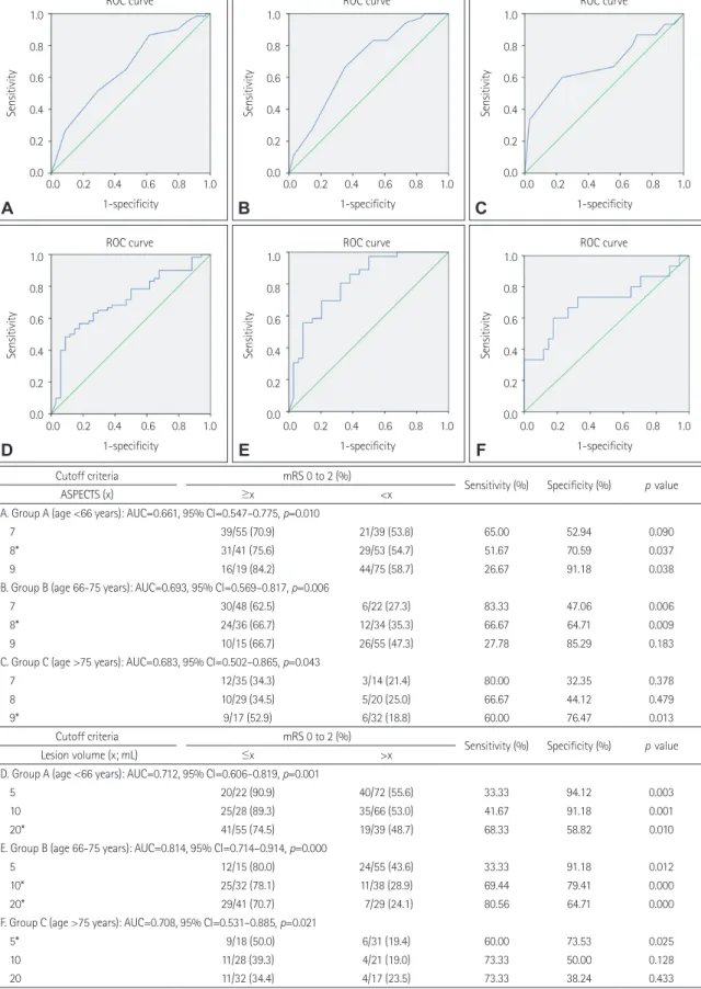
Fig. 1. Receiver operating characteristic (ROC) curves for favorable outcome and the baseline diffusion-weighted imaging (DWI) Alberta Stroke Program Early CT Score (ASPECTS) and lesion volume according to the age groups. The tables display the test characteristics for the prespecified ASPECTS and lesion volume, including optimal values (*). AUC: area under the ROC curve, CI: confidence interval, mRS: modified Rankin scale.
1.0 0.8 0.6 0.4 0.2 0.0
Sensitivity
0.0 0.2 0.4 0.6 0.8 1.0 1-specificity
ROC curve
A
1.0 0.8 0.6 0.4 0.2 0.0
Sensitivity
0.0 0.2 0.4 0.6 0.8 1.0 1-specificity
ROC curve
B
1.0 0.8 0.6 0.4 0.2 0.0
Sensitivity
0.0 0.2 0.4 0.6 0.8 1.0 1-specificity
ROC curve
C
1.0 0.8 0.6 0.4 0.2 0.0
Sensitivity
0.0 0.2 0.4 0.6 0.8 1.0 1-specificity
ROC curve
D
1.0 0.8 0.6 0.4 0.2 0.0
Sensitivity
0.0 0.2 0.4 0.6 0.8 1.0 1-specificity
ROC curve
E
1.0 0.8 0.6 0.4 0.2 0.0
Sensitivity
0.0 0.2 0.4 0.6 0.8 1.0 1-specificity
ROC curve
F
Cutoff criteria mRS 0 to 2 (%)
Sensitivity (%) Specificity (%) p value
ASPECTS (x) ≥x <x
A. Group A (age <66 years): AUC=0.661, 95% CI=0.547–0.775, p=0.010
7 39/55 (70.9) 21/39 (53.8) 65.00 52.94 0.090
8* 31/41 (75.6) 29/53 (54.7) 51.67 70.59 0.037
9 16/19 (84.2) 44/75 (58.7) 26.67 91.18 0.038
B. Group B (age 66-75 years): AUC=0.693, 95% CI=0.569–0.817, p=0.006
7 30/48 (62.5) 6/22 (27.3) 83.33 47.06 0.006
8* 24/36 (66.7) 12/34 (35.3) 66.67 64.71 0.009
9 10/15 (66.7) 26/55 (47.3) 27.78 85.29 0.183
C. Group C (age >75 years): AUC=0.683, 95% CI=0.502–0.865, p=0.043
7 12/35 (34.3) 3/14 (21.4) 80.00 32.35 0.378
8 10/29 (34.5) 5/20 (25.0) 66.67 44.12 0.479
9* 9/17 (52.9) 6/32 (18.8) 60.00 76.47 0.013
Cutoff criteria mRS 0 to 2 (%)
Sensitivity (%) Specificity (%) p value
Lesion volume (x; mL) ≤x >x
D. Group A (age <66 years): AUC=0.712, 95% CI=0.606–0.819, p=0.001
5 20/22 (90.9) 40/72 (55.6) 33.33 94.12 0.003
10 25/28 (89.3) 35/66 (53.0) 41.67 91.18 0.001
20* 41/55 (74.5) 19/39 (48.7) 68.33 58.82 0.010
E. Group B (age 66-75 years): AUC=0.814, 95% CI=0.714–0.914, p=0.000
5 12/15 (80.0) 24/55 (43.6) 33.33 91.18 0.012
10* 25/32 (78.1) 11/38 (28.9) 69.44 79.41 0.000
20* 29/41 (70.7) 7/29 (24.1) 80.56 64.71 0.000
F. Group C (age >75 years): AUC=0.708, 95% CI=0.531–0.885, p=0.021
5* 9/18 (50.0) 6/31 (19.4) 60.00 73.53 0.025
10 11/28 (39.3) 4/21 (19.0) 73.33 50.00 0.128
20 11/32 (34.4) 4/17 (23.5) 73.33 38.24 0.433
Hwang YH et al.
JCN
ficity=76.47%). A DWI lesion volume of 20 mL was selected as the criterion for predicting a favorable outcome in groups A and B (sensitivity=68.33 and 80.56%, respectively; speci- ficity=58.82 and 64.71%, respectively), while a volume of 5 mL was selected as for group C (sensitivity=60.0%, specifici- ty=73.53%). After adjusting for age, prestroke mRS score, baseline NIHSS score, final TICI grade 2b or 3 reperfusion, and procedure time, age-specific DWI ASPECTS cutoff val- ues remained statistically significant in predicting a favor- able outcome (OR=2.387, 95% CI=1.219–4.676, p=0.011).
Additionally, after adjusting for age, prestroke mRS score, baseline NIHSS score, final TICI grade 2b or 3 reperfusion, and procedure time, the age-specific DWI lesion volume cutoff values continued to remain statistically significant in predicting a favorable outcome (OR=3.595, 95% CI=1.789–
7.223, p<0.001).
DISCUSSION
The novel finding in this study was that the baseline DWI AS- PECTS and lesion volume cutoffs for a favorable outcome were much stricter in older stroke patients (≥9 and ≤5 mL, respectively) than in their younger counterparts (≥8 and ≤20 mL, respectively). Further, the rate of futile reperfusion ex- ceeded 50% in group C (age >75 years). Both of these find- ings suggest that the restorative ability of the brain is weaker in older patients (Fig. 2).
In general, the risk of stroke is closely associated with age.
A related finding is that the incidence of stroke is continuing to increase exponentially as an inevitable result of increased
life expectancy. Older stroke patients have increased mor- tality and more-severe disability compared to their younger counterparts for a similar spectrum of stroke severity and pa- thology.28 This situation can result in physicians being reluc- tant to offer EVT to older stroke patients.
In our cohort, the rate of a favorable outcome was about 31% in older patients (>75 years), and the probability of them achieving a favorable outcome was much lower com- pared to their younger counterparts despite the similarity of their baseline characteristics and reperfusion rates. Pre- vious studies have already shown that poor outcomes are more frequent in octogenarians.7-14 Poor outcomes in older patients have been attributed to higher levels of baseline disabilities, higher incidence rates of medical comorbidities and poststroke medical complications, less familial support, and withdrawal of care.29 Further, the poor restorative abili- ty of the brain in older stroke patients (>75 years) can be a contributing factor.16 Also, a tendency for the collateral flow to be worse in older patients in our cohort could be a con- tributing factor to the endovascular outcome, with it being reported that the interaction between collateral adequacy and age could contribute to an adverse outcome.30
Our hypothesis that older stroke patients have a different ischemia cutoff for a favorable outcome can be investigated using baseline brain imaging. To accurately define the cut- off value for baseline ischemia, DWI is arguably superior to CT in predicting outcome.31 Ribo et al.19 examined follow-up CT images of patients obtained after 24–36 hours to deter- mine the age-specific posttreatment infarct size predictive of a favorable outcome. They found that the target cutoff infarct volume that predicted a favorable outcome decreased with increasing age (49 mL for age <70 years, 32.5 mL for age 70–79 years, and 15.2 mL for age ≥80 years). However, that study did not examine the age-adjusted pretreatment infarct volumes. The present study examined the pretreat- ment lesion volume and ASPECTS, which were found to de- crease and increase, respectively, with increasing age.
The limitations of this study include its retrospective de- sign and the lack of control patients. Additionally, the type of acute treatment given or recommended to each individ- ual patient varied considerably since it was based on the best available treatment at that time at our institute and the will- ingness of the patient’s next of kin to proceed with the sug- gested active treatment approach. This means that the rate of TICI grade 2b or 3 reperfusion was lower than in recent endovascular trials, which may hinder the ability to define the optimal baseline ischemia threshold for clinical out- come despite TICI grades being adjusted in the statistical analyses. Another limitation is that the lower baseline in- farct volume or higher DWI ASPECTS cutoffs for a favor- Fig. 2. Schematic of the hypothetical inverse sinusoidal relationships
between advancing age and restorative brain capacity. Between points A and B, the restorative capacity decreases steeply for small age in- crease. ASPECTS: Alberta Stroke Program Early CT Score, DWI: diffu- sion-weighted imaging.
Potential brain capacity for functional recovery
DWI lesion volume: 20 mL DWI ASPECTS: 8 or more DWI lesion volume: 5 mL DWI ASPECTS: 9 or more
Advancing age
75 years
Baseline Ischemia on Outcome in Older Patients
JCN
able outcome might be related to a high prevalence of base- line intracranial atherosclerosis in our cohort. Finally, older patients were defined as those aged ≥76 years, which differs from many previous studies that analyzed patients aged
≥80 years.
In conclusion, our findings suggest that the age-adjusted pretreatment DWI lesion volume and ASPECTS are useful surrogate markers for functional outcome in older stroke pa- tients. We further suggest that these results may help to de- fine more-restrictive imaging criteria for older stroke patients in future trials. Further investigations of larger data sets from recent clinical trials would help to confirm our findings and justify their widespread clinical application.
Conflicts of Interest
The authors have no financial conflicts of interest.
Acknowledgements
This research was supported by Kyungpook National University Research Fund, 2014.
Dr. Hwang reports grants from Kyungpook National University, dur- ing the conduct of the study; Dr. Liebeskind reports grants from NIH- NINDS, other from Medtronic, other from Stryker, outside the submit- ted work.
We thank Wade Martin of Emareye for his critical English revision.
REFERENCES
1. Berkhemer OA, Fransen PS, Beumer D, van den Berg LA, Lingsma HF, Yoo AJ, et al. A randomized trial of intraarterial treatment for acute ischemic stroke. N Engl J Med 2015;372:11-20.
2. Campbell BC, Mitchell PJ, Kleinig TJ, Dewey HM, Churilov L, Yassi N, et al. Endovascular therapy for ischemic stroke with perfusion- imaging selection. N Engl J Med 2015;372:1009-1018.
3. Goyal M, Demchuk AM, Menon BK, Eesa M, Rempel JL, Thornton J, et al. Randomized assessment of rapid endovascular treatment of ischemic stroke. N Engl J Med 2015;372:1019-1030.
4. Saver JL, Goyal M, Bonafe A, Diener HC, Levy EI, Pereira VM, et al.
Stent-retriever thrombectomy after intravenous t-PA vs. t-PA alone in stroke. N Engl J Med 2015;372:2285-2295.
5. Jovin TG, Chamorro A, Cobo E, de Miquel MA, Molina CA, Rovira A, et al. Thrombectomy within 8 hours after symptom onset in isch- emic stroke. N Engl J Med 2015;372:2296-2306.
6. Hong KS, Bang OY, Kang DW, Yu KH, Bae HJ, Lee JS, et al. Stroke statistics in Korea: part I. Epidemiology and risk factors: a report from the Korean Stroke Society and Clinical Research Center for Stroke. J Stroke 2013;15:2-20.
7. Kim D, Ford GA, Kidwell CS, Starkman S, Vinuela F, Duckwiler GR, et al. Intra-arterial thrombolysis for acute stroke in patients 80 and older: a comparison of results in patients younger than 80 years. AJNR Am J Neuroradiol 2007;28:159-163.
8. Qureshi AI, Suri MF, Georgiadis AL, Vazquez G, Janjua NA. Intra- arterial recanalization techniques for patients 80 years or older with acute ischemic stroke: pooled analysis from 4 prospective studies.
AJNR Am J Neuroradiol 2009;30:1184-1189.
9. Loh Y, Kim D, Shi ZS, Tateshima S, Vespa PM, Gonzalez NR, et al.
Higher rates of mortality but not morbidity follow intracranial me- chanical thrombectomy in the elderly. AJNR Am J Neuroradiol 2010;
31:1181-1185.
10. Mazighi M, Labreuche J, Meseguer E, Serfaty JM, Laissy JP, Lavallée PC, et al. Impact of a combined intravenous/intra-arterial approach
in octogenarians. Cerebrovasc Dis 2011;31:559-565.
11. Mono ML, Romagna L, Jung S, Arnold M, Galimanis A, Fischer U, et al. Intra-arterial thrombolysis for acute ischemic stroke in octoge- narians. Cerebrovasc Dis 2012;33:116-122.
12. Chandra RV, Leslie-Mazwi TM, Oh DC, Chaudhry ZA, Mehta BP, Rost NS, et al. Elderly patients are at higher risk for poor outcomes after intra-arterial therapy. Stroke 2012;43:2356-2361.
13. Johnson JN, Haussen DC, Elhammady MS, Pao CL, Yavagal DR, Aziz-Sultan MA. Poor outcomes of elderly patients undergoing mul- timodality intra-arterial therapy for acute ischemic stroke. Clin Neu- rol Neurosurg 2014;123:136-141.
14. Parrilla G, Carreón E, Zamarro J, Espinosa de Rueda M, García-Vil- lalba B, Marín F, et al. Recanalization and mortality rates of throm- bectomy with stent-retrievers in octogenarian patients with acute ischemic stroke. Cardiovasc Intervent Radiol 2015;38:288-294.
15. Singer OC, Haring HP, Trenkler J, Nolte CH, Bohner G, Reich A, et al.
Age dependency of successful recanalization in anterior circulation stroke: the ENDOSTROKE study. Cerebrovasc Dis 2013;36:437-445.
16. Petcu EB, Sfredel V, Platt D, Herndon JG, Kessler C, Popa-Wagner A.
Cellular and molecular events underlying the dysregulated response of the aged brain to stroke: a mini-review. Gerontology 2008;54:6-17.
17. Inoue M, Olivot JM, Labreuche J, Mlynash M, Tai W, Albucher JF, et al. Impact of diffusion-weighted imaging Alberta stroke program early computed tomography score on the success of endovascular re- perfusion therapy. Stroke 2014;45:1992-1998.
18. Yoo AJ, Zaidat OO, Chaudhry ZA, Berkhemer OA, González RG, Goyal M, et al. Impact of pretreatment noncontrast CT Alberta Stroke Program Early CT Score on clinical outcome after intra-arte- rial stroke therapy. Stroke 2014;45:746-751.
19. Ribo M, Flores A, Mansilla E, Rubiera M, Tomasello A, Coscojuela P, et al. Age-adjusted infarct volume threshold for good outcome after endovascular treatment. J Neurointerv Surg 2014;6:418-422.
20. Hwang YH, Kang DH, Kim YW, Kim YS, Park SP, Suh CK. Outcome of forced-suction thrombectomy in acute intracranial internal carotid occlusion. J Neurointerv Surg 2013;5 Suppl 1:i81-i84.
21. Kang DH, Hwang YH, Kim YS, Park J, Kwon O, Jung C. Direct throm- bus retrieval using the reperfusion catheter of the penumbra system:
forced-suction thrombectomy in acute ischemic stroke. AJNR Am J Neuroradiol 2011;32:283-287.
22. Kang DH, Kim YW, Hwang YH, Park J, Hwang JH, Kim YS. Switching strategy for mechanical thrombectomy of acute large vessel occlu- sion in the anterior circulation. Stroke 2013;44:3577-3579.
23. Higashida RT, Furlan AJ, Roberts H, Tomsick T, Connors B, Barr J, et al. Trial design and reporting standards for intra-arterial cerebral thrombolysis for acute ischemic stroke. Stroke 2003;34:e109-e137.
24. Hacke W, Kaste M, Fieschi C, von Kummer R, Davalos A, Meier D, et al. Randomised double-blind placebo-controlled trial of thrombo- lytic therapy with intravenous alteplase in acute ischaemic stroke (ECASS II). Second European-Australasian Acute Stroke Study In- vestigators. Lancet 1998;352:1245-1251.
25. Saver JL, Jahan R, Levy EI, Jovin TG, Baxter B, Nogueira RG, et al.
Solitaire flow restoration device versus the Merci Retriever in pa- tients with acute ischaemic stroke (SWIFT): a randomised, parallel- group, non-inferiority trial. Lancet 2012;380:1241-1249.
26. Barber PA, Demchuk AM, Zhang J, Buchan AM. Validity and reli- ability of a quantitative computed tomography score in predicting outcome of hyperacute stroke before thrombolytic therapy. ASPECTS Study Group. Alberta Stroke Programme Early CT Score. Lancet 2000;355:1670-1674.
27. van Swieten JC, Koudstaal PJ, Visser MC, Schouten HJ, van Gijn J.
Interobserver agreement for the assessment of handicap in stroke patients. Stroke 1988;19:604-607.
28. Sharma JC, Fletcher S, Vassallo M. Strokes in the elderly-higher acute and 3-month mortality-an explanation. Cerebrovasc Dis 1999;9:2-9.
29. Chandra RV, Leslie-Mazwi TM, Mehta BP, Yoo AJ, Simonsen CZ. Clin-
Hwang YH et al.
JCN
ical outcome after intra-arterial stroke therapy in the very elderly: why is it so heterogeneous? Front Neurol 2014;5:60.
30. Arsava EM, Vural A, Akpinar E, Gocmen R, Akcalar S, Oguz KK, et al. The detrimental effect of aging on leptomeningeal collaterals in ischemic stroke. J Stroke Cerebrovasc Dis 2014;23:421-426.
31. McTaggart RA, Jovin TG, Lansberg MG, Mlynash M, Jayaraman MV, Choudhri OA, et al. Alberta stroke program early computed to- mographic scoring performance in a series of patients undergoing computed tomography and MRI: reader agreement, modality agree- ment, and outcome prediction. Stroke 2015;46:407-412.