Effects of Achyranthoside C Dimethyl Ester on Heme Oxygenase-1 Expression and NO Production
Soo Young Bang1, Ji Su Song1, Hyung-In Moon2 and YoungHee Kim1*
1Department of Molecular Biology, College of Natural Sciences, Pusan National University, Busan 609-735, Korea
2Department of Medicinal Biotechnology, College of Natural Resources and Life Science, Dong-A University, Busan 604-714, Korea Received July 6, 2015 /Revised August 8, 2015 /Accepted September 18, 2015
Achyranthoside C dimethyl ester (ACDE) is an oleanolic acid glycoside from Achyranthes japonica which has been used in traditional medicine for the treatment of edema and arthritis. In this study, we investigated the anti-inflammatory effects of ACDE in RAW264.7 macrophages. ACDE significantly induced heme oxygenase-1 (HO-1) gene expression in RAW264.7 cells, while ACDE improved LPS-in- duced toxicity of cells. And ACDE induced nuclear translocation of nuclear factor E2-related factor 2 (Nrf2), a transcription factor that regulates HO-1 expression. Further study demonstrated that ACDE-induced expression of HO-1 was inhibited by inhibitors of phosphatidylinositol 3-kinase (PI-3K) (LY294002), c-Jun kinase (JNK) (SP600125), extracellular signal regulated kinase (ERK) (PD98059) and p38 kinase (SB203580). Moreover, ACDE phosphorylated Akt, JNK, ERK, and p38 MAPK. In addition, ACDE inhibited LPS-induced NO secretion as well as inducible NO synthase (iNOS) expression in a dose-dependent manner. The inhibitory effects of ACDE on iNOS expression were abrogated by small interfering RNA (siRNA)-mediated knock-down of HO-1. Therefore, these results suggest that ACDE suppresses the production of pro-inflammatory mediator such as NO by inducing HO-1 expression via PI-3K/Akt/MAPK-Nrf2 signaling pathway. These findings could help us to understand the active principle included in the roots of A. japonica and the molecular mechanisms underlying anti-in- flammatory action of ACDE.
Key words : Achyranthoside C dimethyl ester, heme oxygenase-1, inducible nitric oxide synthase, NF-E2-related factor 2, nitric oxide
*Corresponding author
*Tel : +82-51-510-2526, Fax : +82-51-513-9258
*E-mail : [email protected]
This is an Open-Access article distributed under the terms of the Creative Commons Attribution Non-Commercial License (http://creativecommons.org/licenses/by-nc/3.0) which permits unrestricted non-commercial use, distribution, and reproduction in any medium, provided the original work is properly cited.
Journal of Life Science 2015 Vol. 25. No. 9. 976~983 DOI : http://dx.doi.org/10.5352/JLS.2015.25.9.976
서 론
Heme oxygenase-1 (HO-1)은 세포의 heme 기를 산화시켜 일산화탄소(CO), biliverdin, 철이온으로의 분해를 촉매하는 주요한 유도성 효소이다[28]. HO-1와 그 생산물들은 산화적 상처에 대응하여 몸을 보호할 수 있게 하고, 세포와 조직의 항염증성 활성에 기여한다[25]. 활성화된 대식세포에 HO-1을 발현시키거나 CO를 처리하면 tumor necrosis factor-α (TNF- α), interleukin-1β (IL-1β), IL-6, monocyte chemoattractant protein-1, macrophage inflammatory protein-1β과 같은 염증 촉진 사이토카인과 케모카인의 생산이 저해되며[25], 또한 일 산화질소 라디칼(nitric oxide, NO)과 prostaglandin E2 (PGE2)와 같은 염증촉진 매개체의 생산을 억제한다[32]. 게다 가 여러 종류의 염증치료제들이 HO-1의 발현을 유도하고, 이
를 통해서 항염증 효과를 나타낸다고 보고되고 있다. 따라서 HO-1의 발현이 유익하며 염증 치료를 위한 새로운 타겟으로 작용할 수 있을 것으로 사료된다.
HO-1는 여러 세포 유형에서 생존이나 스트레스 반응과 관 련된 신호전달 경로를 통해서 주로 전사 수준에서 조절된다.
전사인자인 NF-E2-related factor 2 (Nrf2)는 HO-1의 발현에 가장 중요한 역할을 한다[31]. 정상 상태에서 Nrf2는 세포질에 서 Kelch-like ECH-associated protein 1 (Keap1)에 결합되어 유비퀴틴 의존적인 26S 프로테아좀에 의해 분해된다[12]. 세포 가 활성화되면 Nrf2는 keap1 으로부터 방출되고 핵으로 이동 하며, Maf와 heterodimer를 이루어 HO-1과 같은 항산화 유전 자에 위치한 antioxidant response elements (AREs)에 결합한 다[12, 24].
NO는 다양한 장기에서 여러 가지 작용을 하는 자유라디칼 이다. 생물학적 매개체로 NO의 가장 눈에 띄는 생리 활동은 cGMP 의존적 혈관 확장, 신경 정보 전달, 숙주 방어, 염증 및 면역 억제, 혈액 응고이다[23]. NO는 NO synthase (NOS) 에 의해 생산되고, inducible NOS (iNOS)는 대식세포를 포함 한 다양한 세포에서 염증 사이토카인이나 박테리아의 lip- opolysaccharide (LPS)로 인해 유도된다. 특히 iNOS에 의해 합성된 NO의 과도한 양은 침입한 미생물이나 종양 세포의 성장을 저해하기 위해 염증 반응을 유도한다. 또한 외래 세포
A
B
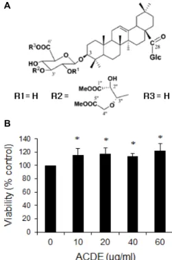
Fig. 1. Effect of ACDE on cell viability. (A) Chemical structure of ACDE isolated from the methanol-soluble fraction of the dried roots of A. japonica. (B) RAW 264.7 cells were incubated with various concentrations of ACDE in the presence of LPS for 24 hr and cell viability was measured by MTT assay. * p<0.05 vs. LPS-treated group.
에 대한 강한 염증 반응은 인접한 숙주 세포와 조직에 손상을 준다[21]. 실지로 iNOS 특이적 억제제는 여러 가지 염증 질환 을 경감시키는 효과가 있으며, 따라서 iNOS 억제제가 NO 과 잉 생산에 의해 매개되는 질환을 치료하는데 매우 유용한 약 제가 될 수 있을 것으로 추측된다[30].
우슬(Achyranthes japonica Nakai)은 여러해살이 풀이며, 한 국, 중국, 일본에 널리 분포되어 있다. 우슬의 뿌리는 그 모양 에 따라 쇠무릎이라고도 불리우며 전통적으로 부종 및 관절염 치료에 사용되었으며, 임신을 지연시키거나 피임과 낙태를 유 도하는 약으로도 사용되었다[1]. 또한 혈소판 응집을 저해하고 [34], 항균성[17] 및 항염증 활성을 갖는 것으로 보고되고 있다 [11]. 이 식물에서 Ecdysterone, inokosterone, oleanolic acid 등의 성분이 발견되었다[1, 16]. 본 저자들은 이전 연구에서 우슬 추출물이 항 염증 활성을 가지고 있음을 보고하였다[4].
그러나, 우슬의 어떤 성분이 항염증 효과를 나타내는지 밝히 기 위하여 우슬의 oleanolic acid glycoside인 Achyranthoside C dimethyl ester (ACDE)을 분리하여, ACDE의 항염증 효과 를 조사하였다.
재료 및 방법
재료
LPS (phenol extracted Salmonella enteritidis), Tween-20, 3-(4,5-dimethylthiazol-2-yl)-2,5-diphenyltetrazolium bromide (MTT)은 Sigma Chemical Co. (St. Louis, MO)에서 구매했다.
HO-1 small interfering RNA (siRNA)와 iNOS, HO-1, Nrf-2, TATA-box binding protein (TBP), α-tubulin의 antibody는 Santa Cruz Biotechnology (Santa Cruz, CA, USA)에서 구매 했다. NTERFERin siRNA Transfection Reagent Polyplus transfection (France)에서 구매했다. Dulbecco’s modified Eagle’s medium (DMEM)와 fetal bovine serum (FBS)는 Invitrogen Corporation (San Diego, CA, USA)에서 구매했다.
우슬의 methanol-soluble fraction으로부터 분리한 ACDE (Fig. 1A) [14]를 사용하였으며 사용할 때까지 빛을 차단하여 냉장 보관하였다.
세포 배양
생쥐의 대식세포인 RAW 264.7 세포를 glutamine (1 mM) 과 10% FBS가 추가된 DMEM에서 5% CO2, 37oC 환경에서 배양하였다.
Nitrite 농도 측정
Nitrite를 측정하기 위해서 100 μl를 세포배양액으로부터 분 리하고, Griess 시약 (1% sulfanilamide/0.1% N-(1-naphthyl)- ethylenediamine dihydrochloride/2.5% H3PO4)을 같은 양으 로 넣어 10분 동안 상온에서 반응했다. Nitrite 농도는 micro-
plate spectrophotometer (Bio-Rad, Hercules, USA)로 540 nm 에서 흡광도를 측정했다[6].
세포 생존 분석
Griess 반응 후 남아있는 세포를 microculture tetrazolium (MTT)-based colorimetric assay 방법으로 분석했다. MTT를 각각의 well에 첨가하고(최종농도 62.5 μg/ml) 5% CO2와 37oC에서 3시간 배양 후, 상등액을 제거하고 살아있는 세포에 서 형성된 formazan crystals을 DMSO 150 μl로 용해시켰다.
각각 well의 흡광도를 microplate reader를 사용해서 570 nm 에서 측정했다.
Western blotting 분석
세포를 phosphate buffered saline (PBS)로 3번 세척하고 lysis buffer (1% Triton X-100, 1% deoxycholate)를 넣어 lysis 했다. 단백질 농도를 Bradford reagent (Bio-Rad, Hercules, USA)를 이용하여 측정하고, 같은 양의 단백질을 10% SDS- PAGE (sodium dodecyl sulfate-polyacrylamide gel electro- phoresis)을 이용하여 전기영동하고 난 후 0.45 μm PVDF membrane에 옮겼다. Blot을 특정한 항체에 배양하고, en- hanced chemiluminescence detection system (Amersham Co.)을 이용해서 detection했다. Alpha-tubulin 또는 TATA
A B
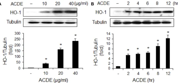
Fig. 2. Induction of HO-1 by ACDE. RAW 264.7 cells were incubated with various concentrations of ACDE for 8 hr (A), or with ACDE (40 μg/ml) for indicated times (B). Cells were harvested and equal cytosolic extracts were analyzed by Western blotting with anti-HO-1 antibody. The relative band intensity was normalized to tubulin level. * p<0.05 vs. control (untreated) group.
binding protein (TBP)은 단백질 로딩 control으로써 사용하였 다. 양적 이미지 분석은 이지미 분석 소프트웨어인 ImageJ (http://rsb.info.nih.gov/ij) 를 이용하였고, data는 control의 배수로 나타냈다.
세포핵 단백질 분리
세포핵 단백질 분리는 이전에 설명된 방법으로 하였다[3].
요약하면, 세포를 차가운 PBS에 세척하고, 1,000× g에서 5분 동안 원심분리하고, 차가운 저 삼투압 버퍼 (10 mM HEPES/
KOH, 1.5 mM MgCl2, 10 mM KCl, 0.5 mM DTT, 0.2 mM PMSF, pH 7.9) 400 μl에서 resuspended 하고, 얼음 위에서 10 min 동안 둔 후, vortex하고 15,000× g에서 30초 동안 원심 분리 했다. 침전된 핵은 차가운 버퍼 (20 mM HEPES/KOH, 1.5 mM MgCl2, 420 mM NaCl, 0.2 mM EDTA, 25% glycerol, 0.5 mM DTT, 0.2 mM PMSF, pH 7.9) 50 μl에서 resuspended 한 후, 30분 동안 얼음 위에 두면서 간간히 voltex하고, 4oC 15,000× g에서 5분 동안 원심분리 했다. 핵 단백질들이 포함된 상등액은 -70oC에서 보관했다.
siRNA를 이용한 HO-1 발현 억제
세포를 INTERFERin (Polyplus transfection, France)을 이 용하여 제조사에서 추천하는 방법에 따라 HO-1 siRNA 또는 negative control siRNA를 transfection 하였다. 그 후 세포를 단백질 발현 detection까지 48시간 동안 배양했다.
통계 분석
모든 결과는 평균 ± 표준오차로 나타내었고, 각 실험은 세 번 이상 반복하였다. 통계 처리를 위해 SPSS 소프트웨어(ver- sion 21)을 이용하여 one-way analysis of variance (ANOVA) 분석을 하였고, 세 개 또는 그 이상의 그룹을 비교하기 위해
Tukey’s post hoc test을 사용하였다. p<0.05인 경우 통계적으 로 유의하다고 표시하였다.
결과 및 고찰
세포 생존능력에 대한 ACDE의 효과
ACDE (Fig. 1A)의 세포 내 작용을 관찰하기에 앞서 ACDE 가 독성이 있는지를 MTT assay 방법으로 조사하였다. 생쥐 대식세포주인 RAW264.7 세포에 ACDE를 60 μM까지 처리하 여도 MTT 수준이 감소되지 않았으며 ACDE의 농도가 증가함 에 따라 오히려 MTT 수준이 약간 증가하는 경향이 관찰되었 다. 따라서 60 μM의 농도 범위에서는 ACDE가 세포독성을 나타내지 않으며 LPS로 인한 세포독성을 감소시킴을 알 수 있다.
ACDE의 HO-1 유도 효과
ACDE가 HO-1 발현을 유도하는지 조사하기 위하여 RAW 264.7 세포에 ACDE를 처리한 후 HO-1 발현을 Western blot- ting으로 관찰하였다. ACDE 농도가 증가함에 따라 HO-1 발 현이 증가하였고, 6시간에서 최고값을 나타내었다(Fig. 2). 이 러한 결과로 보아 ACDE는 대식세포에서 HO-1의 발현을 유 도함을 알 수 있다. HO-1은 산화 스트레스에 의해서도 유도되 는 것으로 알려져 있으므로 ACDE에 의해 세포 내 활성산소가 생성되는지를 조사하였을 때 ACDE가 활성산소를 발생시키 지 않았다(결과 미 제시). 이는 ACDE가 세포에 산화 스트레스 와 같은 세포독성을 야기해 나타나는 결과가 아니라 HO-1 특 이적으로 작용한 결과라는 것을 시사한다. 또한 ACDE가 LPS 로 인한 세포독성을 줄이는 것으로 보아, 비록 실지로 관찰하 지는 못했지만 LPS에 의해 유도되는 활성산소의 발생을 ACDE가 줄일 가능성이 있을 수 있을 것으로 추측된다.
A B
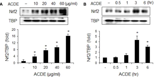
Fig. 3. Effect of ACDE on Nrf2 activation. RAW 264.7 cells were incubated with indicated concentrations of ACDE for 1 hr (A), or ACDE (40 μg/ml) for indicated times (B). Nuclear proteins were extracted and nuclear accumulation of Nrf2 was assayed by Western blotting. The relative band intensity was normalized to TBP level. * p<0.05 vs. control (untreated) group.
Nrf2 활성에 미치는 ACDE의 효과
HO-1 유전자의 프로모터 지역에 전사인자 Nrf2의 결합부 분이 포함되어있고, HO-1의 발현은 Nrf2에 의해 조절된다고 알려져 있기 때문에[31], ACDE가 Nrf2를 활성화시키는지 관 찰하였다. Nrf2의 활성화를 관찰하기 위해 ACDE를 처리하고, 핵에서 Nrf2의 축척을 Western blotting으로 관찰하였다.
ACDE를 처리하였을 때 ACDE 농도의존적으로 Nrf2가 핵에 축적되었으며, 1시간에서 최고값을 나타내었다(Fig. 3). 이 결 과는 ACDE는 Nrf2 활성을 통해서 HO-1의 발현을 유도한다 는 것을 나타낸다. 또한 Nrf2는 HO-1 뿐만 아니라 NAD(P)H quinone oxidoreductase 1 (NQO-1)과 thioredoxin-1 (Trx1) 등 의 해독성 효소와 항산화 단백질들을 조절하므로, 비록 이 연 구에서는 관찰하지 않았지만 ACDE가 HO-1 뿐만 아니라 NQO-1이나 Trx1과 같은 다른 해독성 효소를 유도할 가능성 도 있음을 추측할 수 있다.
PI3K/Akt 및 mitogen activated protein kinase (MAPK) 활성에 미치는 ACDE의 효과
포유류 세포에서 다양한 자극에 의한 HO-1 발현에 c-Jun kinase (JNK), extracellular signal regulated kinase (ERK), p38 kinase 등과 같은 MAPK와 PI3K/Akt 가 관련이 있다는 보고들이 발표되었다[18, 27]. 따라서, 이들 세포 내 신호전달 경로가 ACDE로 인해 유도되는 HO-1 발현에 관련이 있는지 알아보았다. RAW264.7 대식세포에 LY294005 (PI3K 저해제), SP600125 (JNK 저해제), PD98059 (ERK 저해제), SB203580 (p38 MAPK 저해제) 등의 단백질 인산화효소 저해제를 전처 리한 후 ACDE를 처리하고 HO-1의 발현을 관찰한 결과 이들 인산화효소 저해제에 의해 HO-1의 발현이 상당히 억제되었다 (Fig. 4A). 또한 ACDE에 의해 ERK 는 잠시 동안(20분 정도) 인산화된 반면, Akt, JNK, p38은 20분 정도에서 인산화 되어
그 상태가 60분 동안 지속되었다(Fig. 4B). 이러한 결과들은 ACDE에 의한 HO-1 발현에 PI3K/Akt와 MAPK가 주요한 신 호전달경로로 작용함을 시사한다.
PI3K/Akt와 MAPK는 다양한 자극에 의해 활성화되며 여 러 가지 세포 반응에 관여한다. 최근 프로스타글란딘, 아비산 염, 파이토케미컬(phytochemicals) 등의 여러 자극에 의해 HO-1가 발현될 때 PI3K/Akt와 MAPK가 관여한다는 보고들 이 발표되었으며[2, 10, 15, 22, 27] 이는 본 논문의 결과와 일치 한다. PI3K/Akt는 세포의 생존에 중요한 작용을 한다고 알려 져 있으므로[19] ACDE에 의해 활성화된 PI3K/Akt가 Fig. 1B 와 2에서 보듯이 HO-1의 유도를 통해 세포의 생존을 증가시킨 다고 추측할 수 있다.
대식세포에서 NO 합성과 iNOS 발현에 미치는 ACDE의 효과
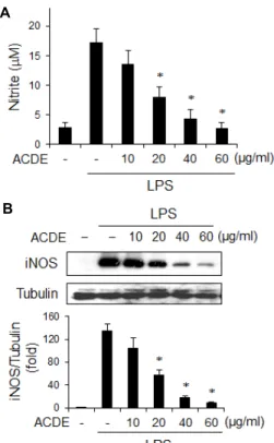
ACDE의 항염증 효과를 조사하기 위해, 대식세포에서 NO 합성에 미치는 ACDE의 효과를 조사하였다. RAW 264.7 세포 에 ACDE를 1시간 전처리한 후, LPS를 20시간 동안 처리하고 배지로 방출되는 NO의 양을 Griess 방법으로 측정하였다.
LPS를 처리한 세포들은 NO를 대량으로 생산하는 반면에, ACDE를 처리한 군은 ACDE 농도의존적으로 NO 방출을 억 제하였다(Fig. 5A). NO의 감소가 iNOS와 관련 있는지 알아보 기 위해서, iNOS의 양을 Western blotting으로 분석하였다.
ACDE의 농도가 증가함에 따라 iNOS의 발현은 현저히 감소 하였다(Fig. 5B). 이상의 결과들로 보아 ACDE는 iNOS 발현을 억제함으로써 NO 방출을 저해함을 시사한다. 대식세포에 의 한 iNOS 발현 및 NO의 다량 방출은 다양한 염증성 질환의 발병 기전에서 중요한 역할을 하는 것으로 간주된다. 대식세 포에서 iNOS 활성의 억제는 관절염과 같은 다양한 염증성 질 병을 치료하는 데 중요한 타겟이 될 수 있다. 실지로, 선택적
A
B
Fig. 4. Intracellular signaling pathway involved in HO-1 ex- pression by ACDE. (A) RAW 264.7 cells were incubated with LY294002 (20 μM), SP600125 (20 μM), PD98059 (20 μM), and SB203580 (20 μM) for 1 hr and then treated with ACDE (40 μg/ml) for 6 hr. Equal amount of cyto- solic proteins were analyzed by Western blotting with HO-1 antibody. (B) Cells were incubated with ACDE (40 μg/ml) for indicated times. Equal amount of cyto- solic proteins were analyzed by Western blotting with anti-p-Akt, -p-JNK, -p-ERK or -p-p38 antibodies, re- spectively. The relative band intensity normalized to tu- bulin (A) or unphosphorylated form (B) was indicated as fold.
A
B
Fig. 5. Effect of ACDE on the NO secretion and iNOS expression in macrophages. (A) RAW 264.7 cells were incubated with various concentrations of ACDE for 1 hr and then stimulated with LPS (0.1 μg/ml) for 20 hr. The amount of nitrite released was measured by the method of Griess.
(B) Cells were treated as mentioned above and equal cy- tosolic extracts were analyzed by Western blotting. The relative band intensity was normalized to control protein (tubulin) level. * p<0.05 vs. LPS-treated group.
iNOS 억제제를 투여하였을 때 관절염이 경감된다는 것이 보 고되었다[8, 13]. 따라서 ACDE는 대식세포에서 iNOS의 발현 수준에 영향을 미침으로써 NO 합성의 억제를 통해 관절염을 줄일 수 있을 것으로 사료된다.
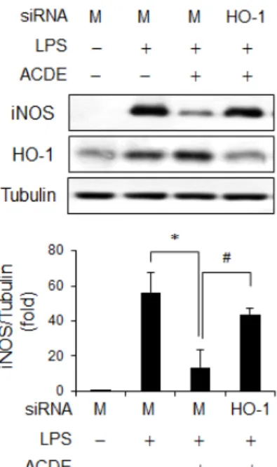
또한 ACDE가 HO-1을 유도함으로써 iNOS 발현을 억제하 는지 확인하기 위해서, HO-1발현을 억제하는 HO-1 siRNA system을 적용하였다. 세포에 HO-1 siRNA 또는 대조군 siRNA를 transfection한 후 ACDE 를 전처리하고 LPS를 처리 하여 iNOS 발현을 조사하였다. 대조군 siRNA를 처리했을 때 ACDE는 LPS로 인한 iNOS 발현을 감소시켰으나, HO-1 siRNA로 인해 HO-1 발현이 감소되었을 때는 iNOS 발현을
억제하지 못하였다(Fig. 6). 그러므로 ACDE는 HO-1 유도를 통해 iNOS 발현을 억제함을 알 수 있다. 비록 HO-1 생산물 (즉, CO, biliverdin, iron)을 본 연구에서 관찰하지 않았지만, 몇몇 연구에서 HO-1에서는 파생된 CO와 biliverdin이 염증을 억제하는 대사물질일 가능성이 있다고 지적했다[5, 9, 26, 29].
특히, 최근의 연구는 CO와 biliverdin이 생쥐에서 콜라겐이 유도한 관절염을 호전시킨다고 보고했다[5]. 게다가, HO-1은 염증을 억제할 뿐만 아니라 항산화 단백질을 유도하여 산화스 트레스로부터 세포를 보호한다고 알려져 있다[7, 20, 33]. 본 실험의 결과에서 보듯이 ACDE는 HO-1을 유도하였으며(Fig.
5), LPS로 인한 세포독성으로부터 세포를 보호하였다(Fig. 1B).
그러므로, ACDE는 LPS 뿐만 아니라 다양한 산화 스트레스들 로부터 세포를 보호하고, 항염증 효과를 나타낼 것으로 사료 된다.
요약을 하면, ACDE는 PI3K/Akt 및 MAPK와 Nrf2를 통해 HO-1 발현을 유도하며 LPS로 자극한 대식세포에서 NO 방출 과 iNOS 발현을 억제하였다. 이러한 결과들로 보아 ACDE는 염증성 질환의 조절에 중요하게 작용할 가능성이 있으며 패혈
Fig. 6. Inhibitory effect of HO-1 on iNOS expression. RAW 264.7 cells were transfected with HO-1 siRNA or control siRNA (Mock, M), after which they were pretreated with ACDE (40 μg/ml) for 1 hr and stimulated by LPS (0.1 μg/ml) for 6 hr. Protein levels of iNOS and HO-1 were analyzed by Western blotting. The relative iNOS band intensity was normalized to tubulin level. * p,#<0.05.
성 쇼크나 관절염, 염증성 장질환 같은 다양한 염증 질환을 위한 치료제로 개발될 수 있을 것으로 사료된다.
감사의 글
이 논문은 부산대학교 자유과제학술연구비(2년)에 의하여 연구되었음.
References
1. Ahn, D. K. 2003. Illustrated book of Korean medicinal herbs, pp. 308, Kyohak Publishing Co.
2. Alvarez-Maqueda, M., El Bekay, R., Alba, G., Monteseirin, J., Chacon, P., Vega, A., Martin-Nieto, J., Bedoya, F. J., Pintado, E. and Sobrino, F. 2004. 15-deoxy-delta 12,14-pros- taglandin J2 induces heme oxygenase-1 gene expression in a reactive oxygen species-dependent manner in human lymphocytes. J. Biol. Chem. 279, 21929-21937.
3. Andrews, N. C. and Faller, D. V. 1991. A rapid micro- preparation technique for extraction of DNA-binding pro- teins from limiting numbers of mammalian cells. Nucleic Acids Res. 19, 2499.
4. Bang, S. Y., Kim, J., Kim, H., Lee, Y. J., Park, S. Y., Lee, S. J. and Kim, Y. 2012. Achyranthes japonica exhibits anti-in- flammatory effect via NF-κB suppression and HO-1 in- duction in macrophages. J. Ethnopharmacol. 144, 109-117.
5. Bonelli, M., Savitskaya, A., Steiner, C. W., Rath, E., Bilban, M., Wagner, O., Bach, F. H., Smolen, J. S. and Scheinecker, C. 2012. Heme oxygenase-1 end-products carbon monoxide and biliverdin ameliorate murine collagen induced arthritis.
Clin. Exp. Rheumatol. 30, 73-78.
6. Boudard, F., Vallot, N., Cabaner, C. and Bastide, M. 1994.
Chemiluminescence and nitrite determinations by the MALU macrophage cell line. J. Immunol. Methods 174, 259- 268.
7. Castilho, A., Aveleira, C. A., Leal, E. C., Simoes, N. F., Fernandes, C. R., Meirinhos, R. I., Baptista, F. I. and Ambrosio, A. F. 2012. Heme oxygenase-1 protects retinal en- dothelial cells against high glucose- and oxidative/nitro- sative stress-induced toxicity. PLoS One. 7, e42428.
8. Cuzzocrea, S., Chatterjee, P. K., Mazzon, E., McDonald, M.
C., Dugo, L., Di Paola, R., Serraino, I., Britti, D., Caputi, A. P. and Thiemermann, C. 2002. Beneficial effects of GW274150, a novel, potent and selective inhibitor of iNOS activity, in a rodent model of collagen-induced arthritis. Eur.
J. Pharmacol. 453, 119-129.
9. Fagone, P., Mangano, K., Coco, M., Perciavalle, V., Garotta, G., Romao, C. C. and Nicoletti, F. 2012. Therapeutic poten- tial of carbon monoxide in multiple sclerosis. Clin. Exp.
Immunol. 167, 179-187.
10. Hamdulay, S. S., Wang, B., Birdsey, G. M., Ali, F., Dumont, O., Evans, P. C., Haskard, D. O., Wheeler-Jones, C. P. and Mason, J. C. 2010. Celecoxib activates PI-3K/Akt and mi- tochondrial redox signaling to enhance heme oxygen- ase-1-mediated anti-inflammatory activity in vascular endo- thelium. Free Radic. Biol. Med. 48, 1013-1023.
11. Han, B. H., Chi, H. J., Han, Y. N. and Ryu, K. S. 1972.
Screening on the anti-inflammatory activity of crude drugs.
Kor. J. Pharmacognosy 3, 205-208.
12. Itoh, K., Mimura, J. and Yamamoto, M. 2010. Discovery of the negative regulator of Nrf2, Keap1: a historical overview.
Antioxid. Redox Signal. 13, 1665-1678.
13. Jarvinen, K., Vuolteenaho, K., Nieminen, R., Moilanen, T., Knowles, R. G. and Moilanen, E. 2008. Selective iNOS in- hibitor 1400W enhances anti-catabolic IL-10 and reduces de- structive MMP-10 in OA cartilage. Survey of the effects of 1400W on inflammatory mediators produced by OA carti- lage as detected by protein antibody array. Clin. Exp.
Rheumatol. 26, 275-282.
14. Jung, S., Lee, J. H., Lee, Y. C. and Moon, H. I. 2012. Inhibitory effects of three oleanolic acid glycosides from Achyranthes japonica on the complement classical pathway. Immunophar- macol. Immunotoxicol. 34, 213-215.
15. Kietzmann, T., Samoylenko, A. and Immenschuh, S. 2003.
Transcriptional regulation of heme oxygenase-1 gene ex- pression by MAP kinases of the JNK and p38 pathways in primary cultures of rat hepatocytes. J. Biol. Chem. 278, 17927- 17936.
16. Kim, J., Kim, J. and Kang, D. 2008. A study on discriminative criteria of 6 kinds of Achyranthis Radix using HPLC/DAD:
Isolation and identification of 20-hydroxyecdysone from Achyranthes japonica NAKAI and comparison of patterns of
Achyranthis Radix from different locations by HPLC. Kor. J.
Herbol. 23, 109-116.
17. Kim, J. C., Choi, G. J., Lee, S. W., Kim, J. S., Chung, K.
Y. and Cho, K. Y. 2004. Screening extracts of Achyranthes japonica and Rumex crispus for activity against various plant pathogenic fungi and control of powdery mildew. Pest Manag. Sci. 60, 803-808.
18. Kim, K. C., Kang, K. A., Zhang, R., Piao, M. J., Kim, G.
Y., Kang, M. Y., Lee, S. J., Lee, N. H., Surh, Y. J. and Hyun, J. W. 2010. Up-regulation of Nrf2-mediated heme oxygen- ase-1 expression by eckol, a phlorotannin compound, through activation of Erk and PI3K/Akt. Int. J. Biochem. Cell Biol. 42, 297-305.
19. Koyasu, S. 2003. The role of PI3K in immune cells. Nat.
Immunol. 4, 313-319.
20. Liu, S., Hou, W., Yao, P., Li, N., Zhang, B., Hao, L., Nussler, A. K. and Liu, L. 2012. Heme oxygenase-1 mediates the pro- tective role of quercetin against ethanol-induced rat hep- atocytes oxidative damage. Toxicol. In Vitro 26, 74-80.
21. MacMicking, J., Xie, Q. W. and Nathan, C. 1997. Nitric oxide and macrophage function. Annu. Rev. Immunol. 15, 323-350.
22. Martin, D., Rojo, A. I., Salinas, M., Diaz, R., Gallardo, G., Alam, J., De Galarreta, C. M. and Cuadrado, A. 2004.
Regulation of heme oxygenase-1 expression through the phosphatidylinositol 3-kinase/Akt pathway and the Nrf2 transcription factor in response to the antioxidant phy- tochemical carnosol. J. Biol. Chem. 279, 8919-8929.
23. Moncada, S., Palmer, R. M. and Higgs, E. A. 1991. Nitric oxide: physiology, pathophysiology, and pharmacology.
Pharmacol. Rev. 43, 109-142.
24. Motohashi, H., Katsuoka, F., Engel, J. D. and Yamamoto, M. 2004. Small Maf proteins serve as transcriptional co- factors for keratinocyte differentiation in the Keap1-Nrf2 regulatory pathway. Proc. Natl. Acad. Sci. USA 101, 6379- 6384.
25. Otterbein, L. E., Bach, F. H., Alam, J., Soares, M., Tao Lu, H., Wysk, M., Davis, R. J., Flavell, R. A. and Choi, A. M.
2000. Carbon monoxide has anti-inflammatory effects in- volving the mitogen-activated protein kinase pathway. Nat.
Med. 6, 422-428.
26. Overhaus, M., Moore, B. A., Barbato, J. E., Behrendt, F. F., Doering, J. G. and Bauer, A. J. 2006. Biliverdin protects against polymicrobial sepsis by modulating inflammatory mediators. Am. J. Physiol. Gastrointest. Liver Physiol. 290, G695-703.
27. Paine, A., Eiz-Vesper, B., Blasczyk, R. and Immenschuh, S.
2010. Signaling to heme oxygenase-1 and its anti-in- flammatory therapeutic potential. Biochem. Pharmacol. 80, 1895-1903.
28. Ryter, S. W., Alam, J. and Choi, A. M. 2006. Heme oxygen- ase-1/carbon monoxide: from basic science to therapeutic applications. Physiol. Rev. 86, 583-650.
29. Sarady-Andrews, J. K., Liu, F., Gallo, D., Nakao, A., Over- haus, M., Ollinger, R., Choi, A. M. and Otterbein, L. E. 2005.
Biliverdin administration protects against endotoxin-in- duced acute lung injury in rats. Am. J. Physiol. Lung Cell.
Mol. Physiol. 289, L1131-1137.
30. Southan, G. J. and Szabo, C. 1996. Selective pharmacological inhibition of distinct nitric oxide synthase isoforms. Biochem.
Pharmacol. 51, 383-394.
31. Srisook, K., Kim, C. and Cha, Y. N. 2005. Molecular mecha- nisms involved in enhancing HO-1 expression: de-re- pression by heme and activation by Nrf2, the "one-two"
punch. Antioxid. Redox Signal. 7, 1674-1687.
32. Suh, G. Y., Jin, Y., Yi, A. K., Wang, X. M. and Choi, A.
M. 2006. CCAAT/enhancer-binding protein mediates car- bon monoxide-induced suppression of cyclooxygenase-2.
Am. J. Respir. Cell Mol. Biol. 35, 220-226.
33. Yu, J., Zhu, X., Qi, X., Che, J. and Cao, B. 2013. Paeoniflorin protects human EA.hy926 endothelial cells against gam- ma-radiation induced oxidative injury by activating the NF-E2-related factor 2/heme oxygenase-1 pathway. Toxicol.
Lett. 218, 224-234.
34. Yun-Choi, H. S., Kim, S. O., Kim, J. H., Lee, J. R. and Cho, H. I. 1985. Modified smear method for screening potential inhibitors of platelet aggregation from plant sources. J. Nat.
Prod. 48, 363-370.
초록:Heme Oxygenase-1 발현과 NO 생성에 미치는 Achyranthoside C Dimethyl Ester의 효과
방수영1․송지수1․문형인2․김영희1*
(1부산대학교 자연과학대학 분자생물학과, 2동아대학교 생명자원과학대학 의약생명공학과)
Achyranthoside C dimethyl ester (ACDE)는 우슬에서 분리한 oleanolic acid glycoside이다. 본 연구에서는 RAW264.7 대식세포에서 ACDE의 항염증 효과를 관찰하고 그 작용 기전을 연구하였다. ACDE는 세포에 독성을 유도하지 않으면서 heme oxygenase-1 (HO-1)의 발현을 유도하였다. ACDE 는 HO-1의 발현에 관여하는 전사인자 인 nuclear factor E2-related factor 2 (Nrf2)를 핵으로 이동시켰다. 또한 ACDE에 의한 HO-1의 발현은 phosphati- dylinositol 3-kinase (PI-3K) 및 mitogen activated protein kinases (MAPK) 억제제에 의해 감소되었으며, ACDE가 Akt, c-Jun kinase (JNK), extracellular signal regulated kinase (ERK), p38 kinase의 인산화를 유도하였다. 한편 ACDE는 lipopolysaccharide (LPS)로 인한 nitric oxide (NO)의 생성과 inducible NO synthase (iNOS) 발현을 억 제하였으며 HO-1 siRNA를 처리했을 때 ACDE가 iNOS의 발현을 억제하지 못하였다. 이상의 결과를 종합해보면, ACDE는 대식세포에서 PI3K/Akt 및 MAPK와 Nrf2 신호전달과정을 통해 HO-1의 발현을 유도함으로써 NO와 같은 염증매개물질의 생성을 억제한다는 것을 알 수 있다. 이러한 연구결과는 ACDE가 항염증제로 사용될 수 있 음을 시사한다.