Anti-inflammatory Effect of Scopoletin in RAW264.7 Macrophages
Su-Gyeong Lee and Moon-Moo Kim*
Department of Chemistry, Dong-Eui University, Busan 614-714, Korea
Received July 21, 2015 /Revised October 14, 2015 /Accepted October 26, 2015
Scopoletin is a component of several plant such as Erycibe obtusifolia, Aster tataricus, Foeniculum vulgare and Brunfelsia grandiflora. It was reported to have anti-angiogenesis and anti-allergy effects. In this study, the anti-inflammatory effect of scopoletin was investigated in Raw264.7 cells, mouse macro- phages. The effects of scopoletin on phagocytosis and nitric oxide (NO) production were investigated in lipopolysaccharide (LPS)-induced inflammatory responses. It was observed that scopoletin exerted inhibitory effects on both phagocytosis and NO production. In addition, scopoletin decreased the ex- pression of inducible nitric oxide synthase (iNOS) and cyclooxygenase-2 (COX-2) which were related to NO and prostaglandin E2 (PGE2) production. In particular, the expression of pro-inflammatory cy- tokines such as interleukin-1β (IL-1β), interleukin-6 (IL-6) and tumor necrosis factor-α (TNF-α). The expression levels of IL-1β, IL-6 were remarkably decreased by treatment with scopoletin. Furthermore, the content of TNFα produced by macrophage was decreased in the presence of scopoletin at 8 hr.
These results indicate that the anti-inflammatory effect of scopoletin could exert by inhibiting the ex- pression of pro-inflammatory cytokines in Raw264.7 cells stimulated with LPS. The above results sug- gest scopoletin could be a new remedial agent for anti-inflammation through inhibition of iNOS, COX-2, IL-1β, IL-6 and TNF-α expressions as well as supression of phagocytosis and NO production.
Key words :
COX-2, iNOS, phagocytosis, Raw264.7, scopoletin
*Corresponding author
*Tel : +82-51-890-1511, Fax : +82-51-890-2620
*E-mail : [email protected]
This is an Open-Access article distributed under the terms of the Creative Commons Attribution Non-Commercial License (http://creativecommons.org/licenses/by-nc/3.0) which permits unrestricted non-commercial use, distribution, and reproduction in any medium, provided the original work is properly cited.
Journal of Life Science 2015 Vol. 25. No. 12. 1377~1383 DOI : http://dx.doi.org/10.5352/JLS.2015.25.12.1377
서 론
최근 경제가 발전함에 따라 생활환경 및 식생활의 변화로 인하여 전 세계적으로 암, 당뇨병, 고혈압, 비만 및 혈관성질환 등의 생활습관 병이 차지하는 비율이 매년 증가하고 있다. 이 와 더불어, 현대 사회의 급격한 산업 발달로 인한 환경 변화 그리고 이에 따른 스트레스의 증가 등을 포함하여 다양한 요 인으로 인하여 면역조절 이상으로 유발된 염증이 지속됨으로 써 아토피, 천식 등의 만성 염증 질환이 증가하고 있다[18, 37].
염증 반응은 외부 자극에 대한 생체조직의 방어반응 하나로 서, 물리적 작용이나 유해물질, 화학적 자극, 세균감염 등에 의한 손상을 수복 재생하려는 기전이다. 염증반응은 급성염 증과 만성염증으로 나누어지는데 특히 만성염증이라고 불리 는 지속적인 염증반응은 오히려 점막손상을 촉진한다. 또한 만성염증 반응은 급성보다 오래 걸리고 지속적이며 단핵구나 대식세포, 림프구, 혈장세포 등의 침윤을 동반하는 특징이 있 으며, 조직을 파괴하고[7, 15, 40], 동맥경화, 당뇨병, 관절염 및 암과 같은 각종 만성 염증성 질환을 유발한다[22]. 염증은
염증세포인 대식세포가 외부의 자극으로 인해 활성화 되어
염증매개물질을 분비하면서 시작되는데, 염증세포는 동물 체
내 모든 조직에 분포하며 인체 내에서 선천적 면역반응을 담
당하고 인체 면역체계에서 중요한 역할을 하는 백혈구인 대식
세포(macrophage)가 주로 관여한다. 이렇게 자극된 대식세포
는 천식, 기관지염, 관절염, 다발성경화증, 동맥경화증, 뇌졸
중, 알츠하이머병이나 파킨슨병과 같은 퇴행성뇌질환 및 바이
러스 감염으로 인한 염증질환 등을 유발하고, 질환을 악화시
키게 된다[1]. 대식세포를 그람음성 균주의 세포벽 조각인 지
질다당체인 lipopolysaccharide (LPS)로 자극하게 되면 tumor
necrosis factor-α (TNF-α), interleukin-6 (IL-6), interleukin-1β
(IL-1β)와 같은 pro-inflammatory cytokine을 증가시키며, ni-
tric oxide (NO), prostaglandin E2 (PGE2) 등의 염증매개물질
을 분비한다. 또한 염증상태에서는 cyclooxygenase-2 (COX-2)
와 NO synthase (NOS)가 유도되어 과량의 PGE2, NO 등이
생성되며 이것이 질병 악화의 원인이 된다[21]. NO는 염증의
대표적인 지표물질로, 일반적인 NO는 박테리아를 죽이거나
종양을 제거하는 중요한 역할을 하지만 염증에서의 NO생성
은 염증을 유발시켜 조직의 손상, 유전자 변이 및 신경손상을
일으킨다. NO는 L-arginine 에서 NO synthase (NOS)에 의해
합성된다. NOS에는 endothelial NOS (eNOS), neuronal NOS
(nNOS), inducible NOS (iNOS)의 세가지 형태가 있으며, 이
들 중 iNOS에 의한 NO 생성이 병을 악화시키는 주요 원인이
된다[29, 36]. 염증을 매개하는 염증성 cytokine은 면역세포에
서 생성된 단백질 중재자로 외부 항원에 대한 여러 면역세포
간의 협력을 중재한다. 그래서 이들의 생성과 분비는 면역반 응조절에 있어서 매우 중요하다. 현재 12가지 이상의 cytokine 들이 규명되었고 그 중에서 IL-1, IL-6 및 TNF-α 등을 중심으로 그 기능들이 알려져 있다[26]. TNF-α는 주로 급성 염증반응에 관여하고, 면역세포를 조절하며, 인슐린 저항성을 유도하여 당대사를 저해시킨다[11]. 또한 IL-6와 같은 다른 염증성 cyto- kine을 증가시킨다[12]. 여기서 IL-6는 대식세포에서 합성되며 T 세포와 B 세포의 분화에 관여하며 IL-1과 협동적으로 작용 한다[27]. 또한 JAK-STAT (Janus kinase-signal transducer and activator of transcription) 신호전달과정을 활성화시켜 백 혈구와 후천성 면역(acquired immunity)과 관련된 림프구인 T 및 B 세포를 자극하여 급성염증을 만성염증으로 전환시킨 다[14, 39]. 이러한 매개 인자의 과잉된 생산에 의한 지속적인 염증 반응은 점막 손상을 촉진하고, 일부는 동맥경화, 당뇨병, 관절염 및 암과 같은 각종 만성 염증성 질환을 유도한다[22].
이와같이 NO 및 염증성 cytokine은 염증 반응과 밀접하게 관 련되어 있어 이들의 생성을 조절할 수 있는 물질이 염증 질환 의 예방 및 치료제로 주목 받고 있다. 특히 현재 사용되고 있는 항염증제가 위염, 신장염 및 심장 질환 같은 문제를 일으킨다 고 보고되어, 그 사용이 일부 제한되고 있다[12, 23]. 보다 안정 적인 항염증제가 필요한 실정 이므로 본 연구에서는 노화를 억제한다고 알려진 Brunfelsia grandiflora 유래의 scopoletin을 이용하여 LPS로 유도한 RAW 264.7 세포의 염증 반응을 조사 하고자 한다. Scopoletin (7- Hydroxy- 6- methoxycoumarin)은 coumarin유도체로 Erycibe obtusifolia, Aster tataricus 그리고
Foeniculum vulgare 같은 많은 약제식물에 함유되어 있다[31].또한 최근에 노화진행의 속도를 늦추는데 탁월한 효능이 보고 된 식물 중 남미 기원인 가지과의 꽃 관목에 속하는 Brunfelsia
grandiflora에도 scopoletin이 함유되어 있다고 밝혀졌다[13,33]. Scopoletin은 항염증, 항알레르기[5] 그리고 신 혈관생성 억제 등 다양한 생물학적 활성을 가지고 있다[30]. 또한 scopo- letin은 복강내 상처에서 특히 과 발현된 염증성 cytokine을 하향 조절하는 효과를 가지는 것으로 보고 되어졌다[32]. 하지 만, 이와 같은 효과의 정확한 기전은 아직 밝혀진 바가 없어 Raw264.7 세포주에서 scopoletin이 가지는 항염증 효과에 대 하여 연구하고자 하였다.
재료 및 방법
세포배양 및 재료
세포배양을 위한 Dulbecco’s Modified Eagle’s Medium (DMEM), Trypsin-EDTA, penicillin / streptomycin / am- photericin (각각 10,000 U/ml, 10,000 μg/ml 및 2,500 μg/ml), fetal bovine serum (FBS) 시약은 Gibco BRL, Paisley, Scotland 로 부터 구입하였다. Raw264.7 세포는 American Type of Culture Collection, Manassas, USA으로부터 제공받았다.
Raw264.7 세포는 5% CO
2및 37℃에서 95% 이상의 습도를 유 지한 배양기에서 10% fetal bovine serum, 2 mM glutamine and 100 μg/ml penicillin -streptomycin을 포함하는 DMEM 배지에서 배양하였다.
MTT assay
Hansen [17]의 방법에 따라 Raw264.7세포에 대한 scopo- leitn의 세포독성을 MTT (3-(4,5-dimethyl-2-yl)-2,5-diphe- nyltetrazolium bromide)를 이용하여 측정하였다.
Nitric oxide assay
Raw264.7 세포로부터 생성된 NO의 양은 Griess 시약을 이 용하여 세포 배양액 중에 존재하는 NO
2-의 형태로서 측정하였 다. Raw264.7 세포를 DMEM배지를 이용하여 1×10
5cell/well 로 조절한 후 24 well plate에 접종하고 37℃, 5% CO
2의 습윤배 양기에서 배양하였다. 세포에 scopoletin을 1,2,4,8 and 16 μM 농도를 전처리하고 1 hr 뒤 LPS를 1 μg/ml 처리한 뒤 24 hr 배양하였다. 세포배양 상등액과 동량의 Griess 시약을 혼합하 여 96 well plate 에서 10분 동안 반응시킨 후 540 nm에서 흡광도를 측정하고 그 비를 %값으로 환산 하였다.
Phagocytosis assay
Raw264.7 세포를 분리하여 DMEM배지를 이용하여 24well plate에 24 hr 배양하였다. 세포에 scopoleitn을 1, 2, 4, 8 그리 고 16 μM 농도를 전처리 하고 1 hr 후 배양액을 제거하였다.
PBS 1 g/100 ml에 녹인 뒤 30분간 가열과정을 거쳐 활성화시 킨 zymosan A 10 μl와 NBT (nitroblue tetrazolium) 20 μl 그리 고 PBS 470 μl를 처리하였다. 충분히 섞어준 뒤 620 nm에서 흡광도를 측정하고 그 비를 %값으로 환산하였다.
Western blot analysis
Raw264.7 세포에 용출 완충용액(50 mM Tris–HCl, pH
7.5, 0.4% Nonidet P-40, 120 mM NaCl, 1.5 mM MgCl
2, 2 mM
phenylmethylsulfonyl fluoride, 80 μg/ml leupeptin, 3 mM
NaF and 1 mM DTT)을 첨가하여 4℃에서 30 min 동안 처리하
였다. 10 μg의 세포용출액을 10% Tris–HCl gel에서 전기영동
후 단백질을 전기적으로 nitrocellulose membrane으로 전이
시켰다. 그 다음 10% skim milk를 nitrocellulose membrane에
전 처리하고 목적 단백질에 대한 1차 항체(anti-iNOS, anti-
COX-2, anti-IL-1β, anti-TNFα 그리고 anti β-actin)를 처리한
다음 2차 항체를 처리 후, chemiluminescent ECL kit (Amer-
sham Pharmacia Biotech Piscataway, USA)를 사용하여 목적
단백질을 검출하였다. Western blot의 band는 LAS3000
®im-
age analyzer (Fujifilm Life Science, Tokyo, Japan)를 이용하여
관찰하였다.
Fig. 1. Effect of scopoletin on viability of Raw 264.7 cells. Raw 264.7 cells were treated with scopoletin at 1, 2, 4, 8 and 16 μM and cell viability was determined using MTT as- say after 48 hr. Data are given as means of values ± S.D.
from three independent experiments. Level of significance was identified statistically using Student’s t test.
Fig. 2. Effect of of scopoletin on nitric oxide in Raw 264.7 cells.
The cells cultured in phenol red and serum-free media were pretreated with different concentrations of of sco- poletin for 1 hr and stimulated with a 1 μg/ ml of LPS for 48 hr. Conditioned medium was mixed with an equal amount of the griess reagent, and the absorbance was measured at 550 nm. The values obtained in this study were compared with value of LPS group and were calcu- lated as percentage. Data are given as means of values
± SD from three independent experiments. Level of sig- nificance was identified statistically (**, p<0.01; ***, p<0.001) using Student’s t test.
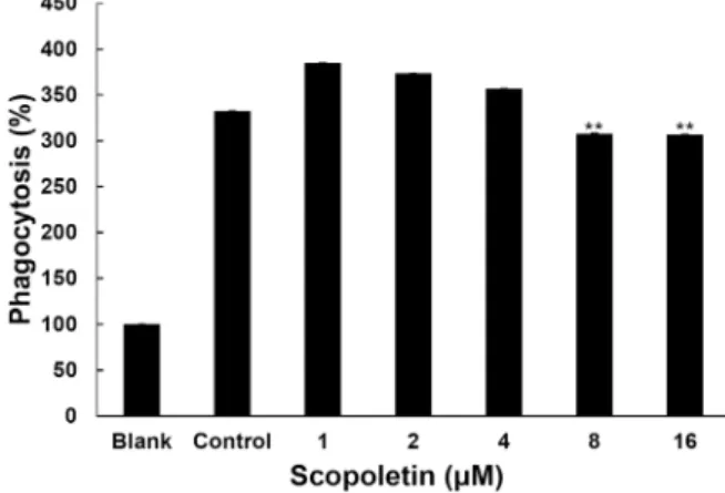
Fig. 3. Inhibitory effect of scopoletin on phagocytosis induced by zymosan A. Phagocytosis of RAW264.7 cells was evaluated by NBT. Cells cultured were pretreated with different concentrations of scopoletin for 1 hr and stimu- lated with zymosan A for 6 hr. The values obtained in this study were compared with value of zymosan group as control and were calculated as percentage. Data are given as means of values ± SD from three independent experiments. Level of significance was identified statisti- cally (**, p<0.01) using Student’s t test.
통계처리
각 실험은 3회 이상 반복실험을 통하여 그 결과를 얻어 각각 의 시료농도에 대해 평균±표준편차로 나타내었다. 각 시료농 도군에 대한 유의차 검정은 대조군과 비교하여 Student’s test 한 후 p<0.05 값을 통계적으로 유의성 있는 결과로 간주하였다.
결 과
Scopoletin의 세포 독성
scopoletin의 대식세포에 독성을 미치지 않는 농도를 결정 하기 위하여 MTT assay를 수행하였다. Fig. 1에서 보는 바와 같이 scopoletin은 농도가 증가하더라도 세포독성이 나타나지 않았다. 따라서 scopoletin은 16 μM 농도 이하의 범위에서는 세포독성이 없는 것으로 관찰되었다.
Scopoletin의 NO 생성억제 효과
LPS에 의해 Raw264.7 세포로부터 생성되는 NO에 대한 scopoletin의 억제효과를 세포배양액으로부터 Griess assay 방 법에 의해 측정하였다. Fig. 2에서 보는 바와 같이 scopoletin의 1, 2, 4, 8 그리고 16 μM 처리농도에서 NO의 생성억제 효과를 측정한 결과 8 μM 이상의 농도로 처리한 세포군에서 NO의 생성 함량은 대조군와 비교했을 때 약 20% 정도 NO의 생성이 억제되는 것으로 나타났다.
Scopoletin의 대식세포의 식균작용 억제 효과
Scopoletin의 zymosan A에 의해 활성화된 Raw264.7 세포 의 식균작용 억제능을 조사하였다. Fig. 3에서 보는 바와 같이 NBT 발색법을 이용하여 식균작용 억제능을 조사한 결과 zy- mosan A를 처리 하에서 scopoletin은 1, 2 and 4 μM 농도에서
는 식균작용을 억제시키지 못하였으나, 8 μM 이상의 농도 에
서 약 10% 정도의 식균작용 억제능을 나타내었다.
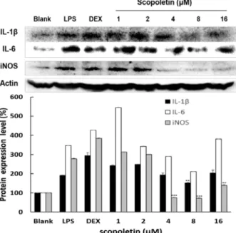
Fig. 4. Effect of scopoletin on protein expressions of iNOS, IL-6 and IL-1β in Raw264.7 cells. The cells were treated with scopoletin at 1, 2, 4, 8 and 16 μM prior to stimulation of cells with LPS at 1 μg/ml for 24 hr. Dexamethason was used as a positive control. Western blot analysis of cell lysates was performed using antibodies as indicated.
The expression level of β-actin was used as a control for normalization of target proteins. The values of scopo- letin treatment groups obtained in this study were com- pared with value of LPS group. Data are given as means of values ± SD from three independent experiments.
Level of significance was identified statistically (**, p<0.01; ***, p<0.001) using Student’s t test.
Fig. 5. Effect of scopoletin on protein expressions of TNFα and COX-2 in Raw264.7 cells. The cells were treated with sco- poletin at 1, 2, 4, 8 and 16 μM prior to stimulation of cells with LPS at 1 μg/ml for 24 hr. In case of TNFα, the cells were treated for 8 hr and 24 hr with scopoletin at 1, 2, 4, 8 and 16 μM prior to stimulation of cells with LPS at 1 μg/ ml, respectively. Aspirin was used as a positive control. Western blot analysis of cell lysates was performed using antibodies as indicated. The expression level of β-actin was used as a control for normalization of target proteins. The values of scopoletin treatment groups obtained in this study were compared with value of LPS group. Data are given as means of values ± SD from three independent experiments. Level of signifi- cance was identified statistically (**, p<0.01; ***, p<0.001) using Student’s t test.
Scopoletin의 염증관련 cytokine의 단백질 발현 조절 효과 scopoletin의 염증관련 cytokine의 단백질 수준에서의 발현 억제효과를 조사하였다. scopoletin로 전 처리된 Raw264.7 세 포에 LPS로 자극하였고, β-actin으로 단백질의 정량화를 나타 내었다. NO를 생성하는 iNOS의 발현을 조사한 결과 4 μM 이상의 농도에서 iNOS의 단백질 발현 수준을 공 시험군과 비 슷한 수준까지 억제하였다. COX-2의 단백질 수준에서의 발현 억제효과를 조사한 결과 scopoletin은 COX-2의 발현을 LPS 군보다 억제하였지만 많은 효과를 나타내지 못하였다. 또한 scopoletin은 급성염증과 만성염증을 유발한다고 알려진 IL-6 와 이와 협동적으로 작용하는 IL-1β의 단백질 발현조절 효과 를 조사한 결과, scopoletin 처리군에서 IL-6과 IL-1β 둘 다 4와 8μM 농도에서 단백질 발현 수준이 감소하였다.
Scopoletin의 시간에 따른 TNFα 단백질 발현 조절 효과 TNFα는 활성화된 macrophage에서 분비되어 염증반응을 일으키는 cytokine으로 sample 처리 후 8 hr 및 24 hr을 방치하 여 단백질 발현 조절 효과를 조사 하였다. Fig. 5에서 보는 바와
같이 scopoletin은 8 μM 농도에서 LPS 단독 처리군에 비하여 TNFα의 발현을 억제하는 것으로 나타났다.
고 찰
염증은 생체나 조직에 물리적 작용이나 화학적 물질, 세균
감염 등의 어떠한 기질적 변화를 가져오는 침습이 가해질 때
그 손상부위를 재생하려는 기전으로[3] 염증반응이 지속적으
로 또는 과도하게 일어나면 여러 염증성 질환이나 다양한 질
환의 원인이 된다[6]. 식균작용(Phagocytosis)은 선천성, 적응
성 면역반응으로 병원균 감염에 대한 첫번째 방어라인으로
작용한다[37]. 식균작용이 일어날 때 macrophage의 막이 항원
을 완전히 둘러싸서 phagosome이 되면 macrophage 안으로
들어가서 lysosome과 융합되어 phagolysosome을 형성하여
안에 있는 항원을 분해한다[2]. 또한 식균작용은 염증 매개체
의 분비와 항원의 침입으로 인해 자극된다[16]. 따라서 염증
매개체의 분비가 증가할수록 식균작용도 증가하므로 만성적 으로 지속되는 염증을 억제하기 위해서는 식균작용을 억제시 켜야 한다. 본 연구에서 scopoeltin은 LPS로 자극된 Raw264.7 세포에서 농도가 증가함에 따라 macrophage의 식균작용을 감소시키는 것으로 나타났다. 식균작용이 계속 지속된다면 염 증이 만성적으로 일어나고 있는 것 인데, scopoletin은 이러한 식균작용을 감소시켜 만성적으로 일어나는 염증을 억제 시킬 것으로 사료된다. 이전 연구에서 대식세포는 선천면역뿐만 아 니라 획득면역 등 다양한 숙주반응에 관여하여 항상성 유지에 관여하는 것으로 알려져 있으며, 염증반응 시에는 NO와 cyto- kine을 생산하여 감염초기에 생체방어에 중요한 역할을 한다 [9, 19]. 뿐만 아니라 대식세포는 cytokines을 생산하여 감염초 기에 생체방어에 중요한 역할을 한다고 보고되었다[28, 29].
일반적으로 NO의 형성은 박테리아를 죽이거나 종양을 제거 시키는 중요한 역할을 하지만, 염증 상태에서 iNOS에 의해 과잉 생산된 NO는 혈관 투과성, 부종 등의 염증 반응을 촉진 시킬 뿐만 아니라 염증 매개체의 생합성을 촉진하여 염증을 심화시키는 것으로 알려져 있다[35, 38]. PGE2는 통증, 발열에 관여하는 염증 매개체로 염증반응과 면역반응에 관여하고 신 생혈관(angiogenesis)을 촉진시켜 암 발생에 깊이 관여하고 있 는 것으로 알려져 있다[24]. Scopoletin의 NO와 PGE2 생성 억제에 대한 효과는 쥐의 귀 염증 모델에서의 연구에서 보고 된 바가 있고[10]. PGE2는 본 연구와 같은 cell인 Raw264.7에 서도 억제 효능을 나타냈다[20]. 본 연구에서 LPS로 자극된 Raw264.7 세포에서 scopoletin은 NO의 생성을 억제하는 것으 로 나타났다. 또한 NO와 PGE2는 각각 NOS와 COX-2에 의해 서 생산된다[34].
COX는 arachidonic acid를 prostaglandins로 전환하는 효 소로 COX-1과 COX-2로 나뉘어지는데, 이는 다양한 세포들에 서 각각 다른 발현 경향을 나타낸다. COX-1은 정상적인 생체 기능에 작용하는 반면, COX-2는 염증 부위에서 발현이 된다.
COX-2에 의해 생성되는 PGE2는 통증, 발열에 관여하는 염증 매개체로서 염증반응, 면역반응 등에 반응한다[24]. 또한 NO 의 합성은 3가지 NOS에 진행된다. NOS의 종류에는 neuronal NOS(nNOS/NOS I), endothelial NOS(eNOS/NOS III) 그리 고 inducible NOS(iNOS/NOSII)의 크게 세가지가 있다. iNOS 는 L-arginine을 세포 내 산화반응에 의해 N-hydroxy- L-argi- nine을 중간체로 하여 NO를 생성하는 반응을 촉매하게 된다 [28]. 본 연구에서 scopoeltin은 LPS로 자극된 Raw264.7 세포 에서 농도가 증가함에 따라 iNOS와 COX-2의 발현을 감소시 키는 것으로 나타났다. 이는 scopoletin이 NO와 PGE를 감소 시키는 것이 단백질 발현감소에서 기인한 것으로 사료된다.
염증의 초기단계에서 항원을 침입자로 인식하여 인체의 면역 세포에 신호전달을 하여 면역기능이 활성화 되도록 하거나 외부 침입자를 직접 제거하게 된다. 면역세포들은 interleukin 과 같은 염증성 cytokine을 분비하여 면역, 염증반응을 일으키
게 된다[11]. 염증성 cytokine은 면역세포에서 생성되는 단백 질 중재자로 외부 항원에 대한 여러 면역세포간의 협력을 중 재하므로 이들의 생성과 분비는 면역반응조절에 있어서 매우 중요하다. 현재 12가지 이상의 cytokine들이 규명되었으나 대 부분 최근에 밝혀진 것으로 기능이 많이 알려져 있지않으나 그 중 에서 IL-1, IL-2, IL-6 및 TNF-α 등을 중심으로 그 기능들 이 알려져 있다[26]. TNF-α는 주로 급성염증반응에 관여하고, 면역세포를 조절하며, 인슐린 저항성을 유도하여 당대사를 저 해시킨다[4]. 또한 TNF-α는 interleukins 발현을 증가시켜 염 증반응 진행에서 중요한 역할을 할 뿐 아니라 대사 증후군, 비만, 인슐린저항성의 발생에도 관여한다[41].
IL-1β는 다양한 염증성 자극 요인에 의하여 활성화된 단핵 구, 대식세포, B 세포, 수지상세포 등에서 분비되는 것으로, T 세포를 활성화 시키고 B 세포의 성숙을 돕는 역할을 하여 염증을 심화시키며, 염증반응 시 시상하부에 작용하여 발열증 상을 유도하는 cytokine이다[8, 25]. 본 연구에서 scopoeltin은 LPS로 자극된 Raw264.7 세포에서 농도가 증가함에 따라 IL-1 β, IL-6을 억제하는 것으로 나타났다. 또한 TNF-α의 발현을 8 hr 및 24 hr에서 조사한 결과 8h에서 발현이 감소하는 것으 로 나타났다. 이는 scopoletin이 염증성 cytokine인 IL-1β, IL-6, 그리고 8 hr에 발현이 일어나는 TNF-α의 발현을 감소시켜 염 증을 억제시키는 것으로 사료된다. 따라서 scopoletin이 과도 하게 일어나는 식균작용과 NO와 PGE2의 생성을 iNOS와 COX- 2 단백질 수준에서 억제하여 염증의 진행을 막고, 염증 반응을 활성화 시키는 IL-1β, TNF-α, IL-6 같은 염증성 매개인자의 발현을 억제하여 만성 염증을 억제하는 것으로 사료된다. 이 러한 연구결과를 바탕으로 scopoletin이 만성 염증을 억제시 키는 새로운 생의학 소재로 이용될 수 있으리라 기대된다.
감사의 글
이 논문은 2015년도 정부(미래창조과학부)의 재원으로 한 국연구재단의 기초연구사업 지원을 받아 수행된 것임(No.
2013R1A1A1A05005160). 동시에 이 연구는 2015년 한국여성 과학기술인지원센터의 지원을 받아 연구되었습니다.
References
1. Albina, J. and Reichner, J. 1995. Nitric oxide in inflammation and immunity. New Horiz 3, 46-64.
2. Alvarez, M. and Casadevall, A. 2006. Phagosome extrusion and host-cell survival after Cryptococcus neoformans phag- ocytosis by macrophages. Curr. Biol. 16, 2161-2165.
3. Barrett, J. T. 1983. Textbook of immunology: an introduction to immunochemistry and immunobiology: Mosby.
4. Bradley, J. 2008. TNF‐mediated inflammatory disease. J.
Pathol. 214, 149-160.
5. Cheng, A. S., Cheng, Y. H. and Chang, T. L. 2012. Scopoletin
attenuates allergy by inhibiting Th2 cytokines production in EL-4 T cells. Food Funct. 3, 886-890.
6. Cheon, M., Yoon, T., Choi, G., Kim, S., Lee, A., Moon, B., Choo, B. and Kim, H. 2009. Comparative study of extracts from rhubarb on inflammatory activity in RAW 264.7 cells.
Kor. J. Edicinal Crop Sci. 17. 109-114.
7. Cline, M. 1970. Leukocyte function in inflammation: the in- gestion, killing, and digestion of microorganisms. Ser. Hae- matol. 3, 3.
8. Delgado, A. V., McManus, A. T. and Chambers, J. P. 2003.
Production of tumor necrosis factor-alpha, interleukin 1-be- ta, interleukin 2, and interleukin 6 by rat leukocyte sub- populations after exposure to substance P. Neuropeptides 37, 355-361.
9. Ding, C., Cicuttini, F., Li, J. and Jones, G. 2009. Targeting IL-6 in the treatment of inflammatory and autoimmune diseases. Expert Opin. Investig. Drugs 18, 1457-1466.
10. Ding, Z., Dai, Y., Hao, H., Pan, R., Yao, X. and Wang, Z.
2008. Anti-inflammatory effects of scopoletin and under- lying mechanisms. Pharm. Biol. 46, 854-860.
11. Dissing Olesen, L., Ladeby, R., Nielsen, H. H., Toft Hansen, H., Dalmau, I. and Finsen, B. 2007. Axonal lesion-induced microglial proliferation and microglial cluster formation in the mouse. Neuroscience 149, 112-122.
12. Dogne, J. M., Hanson, J., Supuran, C. and Pratico, D. 2006.
Coxibs and cardiovascular side-effects: from light to shadow. Curr. Pharm. Des. 12, 971-975.
13. Filipowicz, N. and Renner, S. S. 2012. Brunfelsia (Solana- ceae): A genus evenly divided between South America and radiations on Cuba and other Antillean islands. Mol. Phyl- ogen. Evol. 64, 1-11.
14. Gabay, C. 2006. Interleukin-6 and chronic inflammation.
Arthritis Res. Ther. 8, S3.
15. Pathology of inflammatory bowel disease. Semin. Pediatr.
Surg.; 2007. Elsevier.
16. Hang, D., Choi, H. S., Kang, S. C., Kim, K. R., Sohn, E. S., Kim, M. H., Pyo, S. and Son, E. 2005. Effects of fucoidan on NO production and phagocytosis of macrophages and the proliferation of neuron cells. J. Food Sci. Nutr. 10, 344-348.
17. Hansen, M. B., Nielsen, S. E. and Berg, K. 1989. Re-examina- tion and further development of a precise and rapid dye method for measuring cell growth/cell kill. J. Immunol.
Methods 119, 203-210.
18. Heinzmann, A. and Daser, A. 2002. Mouse models for the genetic dissection of atopy. Int. Arch. Allergy Immunol. 127, 170-180.
19. Jeong, J. B., Hong, S. C., Jeong, H. J. and Koo, J. S. 2012.
Anti-Inflammatory effects of ethyl acetate fraction from cni- dium officinale makino on LPS-stimulated RAW 264.7 and THP-1 cells. Hangug Jaweon Sigmul Haghoeji 25, 299-307.
20. Kim, H. J., Jang, S. I., Kim, Y. J., Chung, H. T., Yun, Y. G., Kang, T. H., Jeong, O. S. and Kim, Y. C. 2004. Scopoletin suppresses pro-inflammatory cytokines and PGE 2 from LPS-stimulated cell line, RAW 264.7 cells. Fitoterapia 75, 261-266.
21. Lee, S. J. and Lim, K. T. 2008. Phytoglycoprotein inhibits interleukin-1β and interleukin-6 via p38 mitogen-activated protein kinase in lipopolysaccharide-stimulated RAW 264.7 cells. Naunyn-Schmiedeberg's Arch. Pharmacol. 377, 45-54.
22. Ljung, T., Lundberg, S., Varsanyi, M., Johansson, C., Schmidt, P. T., Herulf, M., Lundberg, J. O. and Hellstrom, P. M. 2006.
Rectal nitric oxide as biomarker in the treatment of in- flammatory bowel disease: responders versus nonresponders.
World J. Gastroenterol. 12, 3386.
23. Makins, R. and Ballinger, A. 2003. Gastrointestinal side ef- fects of drugs. Expert Opin. Drug Saf. 2, 421-429.
24. Masferrer, J. L., Zweifel, B. S., Manning, P. T., Hauser, S.
D., Leahy, K. M., Smith, W. G., Isakson, P. C. and Seibert, K. 1994. Selective inhibition of inducible cyclooxygenase 2 in vivo is antiinflammatory and nonulcerogenic. Proc. Natl.
Acad. Sci. USA 91, 3228-3232.
25. Masters, S. L., Simon, A., Aksentijevich, I. and Kastner, D.
L. 2009. Horror autoinflammaticus: the molecular patho- physiology of autoinflammatory disease. Annu. Rev. Immu- nol. 27, 621.
26. Meydani, S. N. 1990. Dietary modulation of cytokine pro- duction and biologic functions. Nutr. Rev. 48, 361-369.
27. Mizel, S. B. 1989. The interleukins. FASEB J. 3, 2379-2388.
28. Murphy, M. P. 1999. Nitric oxide and cell death. Biochimica et Biophysica Acta (BBA)-Bioenergetics 1411, 401-414.
29. Nathan, C. 1992. Nitric oxide as a secretory product of mam- malian cells. FASEB J. 6, 3051-3064.
30. Pan, R., Dai, Y., Gao, X. H., Lu, D. and Xia, Y. F. 2011.
Inhibition of vascular endothelial growth factor-induced an- giogenesis by scopoletin through interrupting the autophos- phorylation of VEGF receptor 2 and its downstream signal- ing pathways. Vascul. Pharmacol. 54, 18-28.
31. Pan, R., Gao, X., Lu, D., Xu, X., Xia, Y. and Dai, Y. 2011.
Prevention of FGF-2-induced angiogenesis by scopoletin, a coumarin compound isolated from Erycibe obtusifolia Benth, and its mechanism of action. Int. Immunopharmacol.
11, 2007-2016.
32. Pan, R., Gao, X. H., Li, Y., Xia, Y. F. and Dai, Y. 2010. Anti‐
arthritic effect of scopoletin, a coumarin compound occur- ring in Erycibe obtusifolia Benth stems, is associated with decreased angiogenesis in synovium. Fundam. Clin. Pharmacol.
24, 477-490.
33. Plowman, T. 1977. Brunfelsia in ethnomedicine. Bot Mus Lealf Harv Univ. 289-320.
34. Posadas, I., Terencio, M. C., Guillén, I., Ferrándiz, M. L., Coloma, J., Payá, M. and Alcaraz, M. J. 2000. Co-regulation between cyclo-oxygenase-2 and inducible nitric oxide syn- thase expression in the time-course of murine inflammation.
Naunyn-Schmiedeberg's Arch. Pharmacol. 361, 98-106.
35. Shih, M. F., Cheng, Y. D., Shen, C. R. and Cherng, J. Y.
2010. A molecular pharmacology study into the anti-in- flammatory actions of Euphorbia hirta L. on the LPS-in- duced RAW 264.7 cells through selective iNOS protein inhibition. J. Nat. Med. 64, 330-335.
36. Stuehr, D. J., Cho, H. J., Kwon, N. S., Weise, M. F. and Nathan, C. F. 1991. Purification and characterization of the
초록:대식 세포인 Raw264.7 cell에서 scopoletin의 항염증 효과
이수경․김문무*
(동의대학교 화학과)
Scopoletin은 Erycibe obtusifolia, Aster tataricus, Foeniculum vulgare 그리고 Brunfelsia grandiflora라는 식물들에서 발견된 성분이다. 이것은 신 혈관형성 억제와 항알러지 효과를 가지고 있다고 알려져 있다. 본 연구에서는 대식세 포인 Raw264.7 cells 에서 scopoletin의 항염증 효과에 대하여 조사되었다. 먼저 Scopoletin이 LPS로 염증이 유도 된 Raw264.7 cells에서 nitric oxide (NO) 생성과 식균작용(phagocytosis)에 대하여 어떠한 효과가 있는지 평가하 였다. Scopoletin은 NO생성과 식균작용을 감소시키는 것으로 나타났다. 뿐만 아니라 NO와 prostaglandin E2 (PGE2)의 생성과 관련된 inducible nitric oxide synthase (iNOS)와 COX-2의 단백질 발현을 감소시키는 것으로 나타났다. 특히 염증성 cytokine인 interleukin-1β (IL-1β), interleukin-6 (IL-6) 그리고 TNF-α의 발현을 조사 하였 는데, IL-1β and IL-6의 발현은 scopoletin의 처리에 의해 농도 의존적으로 감소하였다. tumor necrosis factor-α (TNF-α)는 scopoletin을 8 hr 처리하였을 때 감소하는 것으로 나타났다. 이러한 결과는 scopoletin이 iNOS, COX-2, IL-1β, IL-6, TNF-α 또한 phagocytosis 그리고 NO생성을 감소시켜 항염증 효능을 나타내어, 향후에 항염 증제로서 활용의 잠재성이 있다고 판단된다.
cytokine-induced macrophage nitric oxide synthase: an FAD-and FMN-containing flavoprotein. Proc. Natl. Acad. Sci.
USA 88, 7773-7777.
37. Sung, Y. Y., Kim, D. S., Yang, W. K., Nho, K. J., Seo, H.
S., Kim, Y. S. and Kim, H. K. 2012. Inhibitory effects of Drynaria fortunei extract on house dust mite antigen-in- duced atopic dermatitis in NC/Nga mice. J. Ethnopharmacol.
144, 94-100.
38. Tsatsanis, C., Androulidaki, A., Venihaki, M. and Margioris, A. N. 2006. Signalling networks regulating cyclooxygenase-
2. Int. J. Biochem. Cell Biol. 38, 1654-1661.
39. Wang, X., Luo, Y., Liao, W. B., Zhang, J. and Chen, T. M.
2013. Effect of osteoprotegerin in combination with inter- leukin-6 on inhibition of osteoclast differentiation. Chin. J.
Traumatol. 16, 277-280.
40. Willoughby, D. 1975. Heberden Oration, 1974. Human ar- thritis applied to animal models. Towards a better therapy.
Ann. Rheum. Dis. 34, 471.
41. Ye, J. 2008. Regulation of PPARγ function by TNF-α.
Biochem. Biophys. Res. Commun. 374, 405-408.