http://crossmark.crossref.org/dialog/?doi=10.14474/ptrs.2017.6.4.208&domain=pdf&date_stamp=2017-12-25
Received: 26 September, 2017 Revised: 19 November, 2017 Accepted: 26 November, 2017 Corresponding author: Gyu Chang Lee
Department of Physical Therapy, Kyungnam University, 7 Gyeongnamdaehak-ro, Masanhappo-gu, Changwon 51767, Republic of Korea Tel: 82-55-249-2739 Fax: 82-55-999-2173 E-mail: [email protected]
This is an Open-Access article distributed under the terms of the Creative Commons Attribution Non-Commercial License (http://creativecommons.org/licenses/
by-nc/4.0) which permits unrestricted non-commercial use, distribution, and reproduction in any medium, provided the original work is properly cited.
Copyright © 2017 Korean Academy of Physical Therapy Rehabilitation Science
https://doi.org/10.14474/ptrs.2017.6.4.208 pISSN 2287-7576
eISSN 2287-7584
Phys Ther Rehabil Sci 2017, 6 (4), 208-213 www.jptrs.org
Osteokinematic analysis during shoulder abduction using the C-arm
Seung Hoo Lee
a, Younghoon Kim
b, Dong Geon Lee
a, Kyeong-Bong Lee
c, Gyu Chang Lee
daDepartment of Physical Therapy, Graduate School of Kyungnam University, Changwon, Republic of Korea
bKim Institute, Lancaster, CA, USA
cPhysical Therapy Part, Department of Physical and Rehabilitation Medicine, Samsung Medical Center, Seoul, Republic of Korea
dDepartment of Physical Therapy, Kyungnam University, Changwon, Republic of Korea
Objective: Despite reliable evidence of abnormal scapular motions increases, there is not yet sufficient evidence of abnormal humeral translations. This study aims to analyze the motion of the humeral head toward the scapula when the shoulder is actively abducted using the C-arm.
Design: A case report.
Methods: The participant was a healthy man without any limitation and pain during shoulder movement. The participant’s shoulder was abducted; this movement in the frontal plane was measured using a C-arm (anterior-posterior view) and was ana- lyzed with computer-aided design. The starting posture was 15°, and as the participant abducted his shoulder measurements were taken and analyzed at 30°, 60°, 90°, 120°, 150°, and ending at 165°. A line was drawn perpendicularly to the line connecting the humeral head axis to the glenoid, and another line was drawn perpendiculary to the line connecting the scapular axis to the glenoid.
The distance between the two lines measured is defined as the e value.
Results: At the starting posture (15°), the central axis of the humeral head was located 1.92 mm inferior to the central axis of the scapula. The humeral head was superiorly translated from the starting posture to 120°, and then, showed an inferior translation to the ending posture (165°).
Conclusions: The results of this study showed that the humeral head moved upward from the starting posture (15°) up to 120° in- dicating, superior translation, and it moved downward when the posture was past 120°, indicating inferior translation.
Key Words: C-arm, Kinematic analysis, Shoulder abduction
Introduction
Generally, 34% of the population complains of shoulder pain more than once in a lifetime [1], and its prevalence is 20.9%, placing it as the second common impairment in the musculoskeletal system [2]. In addition, shoulder dysfunc- tion is continuous and repetitive [3]. An important key to shoulder function is the rotator cuff [4]. In addition, for the stabilization of the humeral head toward the glenoid during shoulder abduction, normal function of the rotator cuff is necessary [5,6]. Rotator cuff injury affects 30% to 40% of
the population over the age of 60 years and has a significant impact on medical expenses [7]. Repairs for rotator cuff in- jury continue to increase, and physical therapy for this is im- portant [8,9]. In addition, assessment of pain after the repair using a pain scale, such as the visual analog scale and range of motion (ROM) is important [10]. Self-exercise, joint mo- bilization and continuous passive movement are often used to enhance ROM [11].
For joint mobilization, physical therapists often use the concepts of Kaltenborn, Maitland, and Mulligan [12].
Kaltenborn and Evjenth [13] proposed joint mobilization ac-
cording to the convex-concave rule on glide. The con- vex-concave rule is considered a critical theory in physical therapists’ treatment decision-making [14]. The humeral head (convex) glides in the opposite direction when it moves to the glenoid (concave). This refers to the inferior glide of the humeral head in shoulder abduction [15]. Joint mobi- lization is an approach that makes a motion in the reduced di- rection to restore the normal movement, and the direction of the glide is determined considering the shape of the joint sur- face [15]. However, a recent study raises a question about this principle [16]. A previous study of a cadaver noted that 40% of the fibular joint surface of tibiofibular joints was plane; 57% convex and concave; and 3% convex [17]. This study shows the difficulty in the application of the con- vex-concave rule according to the joint surface [16]. In addi- tion, a study of the joint translation noted that superior trans- lation of the humeral head occurred (average 2 mm) in shoulder abduction [18]. Nishinaka et al. [19] reported that in shoulder abduction from 0° to 80°, the humeral head moved upward of approximately 1.7 mm and that from 80°
to 150°, the humeral head moved upward of less than 1 mm.
In addition, Graichen et al. [20] reported that in shoulder ab- duction from 30° to 120°, there was a superior translation of the humeral head. In shoulder abduction from 120° to 150°, there was an inferior translation of the humeral head.
In addition, a dispute over joint mobilization based on the controversial convex-concave rule is also in progress [16].
Kaltenborn’s convex-concave rule is a familiar concept in joint treatment techniques and arthrokinematics. However, the weighting of the evidence according to the Elwood [21]
classification and the agency for health care policy and re- search [22] classification guidelines indicated that the evi- dence was weak and limited. The indirect method, using Kaltenborn’s convex-concave rule as applied to the gleno- humeral joint, may need to be reconsidered [15].
Despite reliable evidence of abnormal scapular motions increases, there is not yet sufficient evidence of abnormal humeral translations [23-25]. Shoulder kinematics was ana- lyzed using standard radiographs [4,5,26-28], magnetic res- onance imaging (MRI) [29-32], ultrasound [33] and com- puted tomography (CT) [34]. Of the researchers, Poppen and Walker [5] defined the axis of the humerus and scapula using standard radiographs and analyzed the motion, accordingly. In most previous studies, images were shot in static positions, and dynamic glenohumeral arthrokine- matics was not described [35]. In addition, because the two-dimensional (2D) radiographic approach was used in
both of these studies, descriptions of humeral head trans- lations were limited to static measures in the superior-in- ferior direction and were subject to possible projection er- rors [36]. It is difficult to measure dynamic humeral head translation, but the results of a 3D analysis during the clin- ical shoulder motion are beginning to be reported [19,37,38].
It seems that treatment and rehabilitation of shoulder in- juries will be enhanced with better knowledge of gleno- humeral mechanics during dynamic activities in healthy and pathologic joints [19]. The number of studies projecting the body’s motion and analyzing joint motion using the C-arm (GE OEC 9800 plus; GE OEC Medical Systems Inc., Salt Lake City, UT, USA, 2012) that can move and shoot X-ray images continue to increase [39].
Thus, through this study, we visually recorded the move- ment of the humerus toward the scapula when the shoulder is abducted dynamically and consecutively. This study aims to analyze the motion of the humeral head toward the scap- ula when the shoulder is abducted actively using the C-arm.
Methods Subjects
This study recruited one healthy man through the bulletin board in Kyungnam University. The participant’s character- istics were as follows: age, 28 years; height, 183 cm; and weight, 85 kg. The participant who did not have any pain in the shoulder joint, did not have a history of any injury on the muscles around the joint and the skeleton, and could perform shoulder abduction within normal ROM was selected.
Persons who had congenital disorders in the shoulder joint or surrounding muscles and skeleton, those who had side ef- fects (vomiting, diarrhea or dizziness) in an X-ray exam, those who had adhesive capsulitis, degenerative arthritis and active inflammatory disease were excluded. The participant joined voluntarily and signed an agreement to participate.
This study was conducted after the approval of Kyungnam University Ethics Commission (IRB No. 1040460-A-2016- 011).
Measure
The C-arm used in this study was 1,024×1,024 pixels. The
image processing speed was 16-bit, which can enlarge im-
ages from 9 to 12 inches. The measurement was taken in the
anterior-posterior direction. The participant was asked to sit
on a chair in an anatomical position, rotating 30° from the
X-ray beam (30° from the frontal plane). The 30° rotation
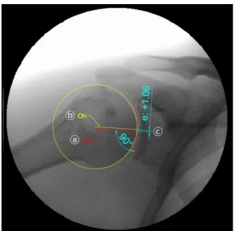
Figure 1. Radiographic analysis. a: axis of the scapula, b: axis of the humeral head (Oh), c: the e value.
was to make the scapular plane and X-ray beam on a straight line. The shoulder was located at least within 2 cm from an image intensifier to minimize distortion. In order to mini- mize exposure to the X-ray, the participant was asked to wear a lead dress on the parts other than those used for the experiment. The height of the C-arm was adjusted so that the entire ROM of the proximal axis of the shoulder joint and humerus was displayed. Then, the participant raised the arm sideways. A radiologist with five years of clinical experi- ence had made these measurements. Beginning from the starting posture (15°), measurements were taken at intervals of 30°, 60°, 90°, 120°, 150°, and the ending posture (165°) as the participant abducted the shoulder. A line was drawn per- pendicularly to the line connecting the humeral head axis to the glenoid, and another line was drawn perpendicularly to the line connecting the scapular axis to the glenoid. The dis- tance measured between the two lines was defined as the e value. The measurements were taken dynamically and con- secutively, and the results were saved in video and picture files, which could be observed later. The picture files were captured (135°), and a professional designer took these and conducted computer-aided design (CAD) work.
The standard definitions of the humeral translation and the angle of the humerus were based on Poppen and Walker [5] (Figure 1). In order to show anatomical characteristics, AutoCAD software (Autodesk Inc., San Rafael, CA, USA, 2014) was used, and an image-transforming operation was conducted by a designer with an 8-year experience in CAD.
The designer was not aware of the contents of the study. The glenoid was described by drawing lines on the top and bot- tom edges of the glenoid, and the center was defined as the axis of the scapula (Os) (Figure 1; ⓐ). A circle was drawn around the humeral head, and the center was defined as the axis of the humeral head (Oh) (Figure 1; ⓑ) [5]. Lines were drawn, respectively, perpendicular to the lines from the Os and Oh to the glenoid. The distance measured between the two lines was defined as the e value (Figure 1; ⓒ) [5].
Variable e can be expressed by the glide of the humeral head toward the glenoid or the distance of the translations. At the center of the scapula, the center of the humeral head can move upward or downward [4]. Using this figure, the trans- lations of the humeral head toward the glenoid were ana- lyzed by the angle.
Results
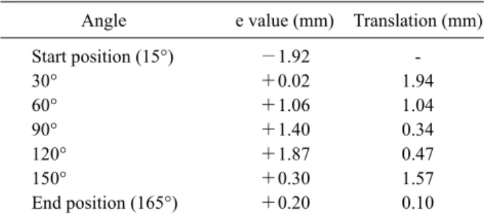
The starting posture (15°) entailed having the participant
sit comfortably in a chair in an anatomical position. At this angle, the central axis of the humeral head was located to be 1.92 mm lower than the central axis of the scapula (inferior).
When the shoulder was abducted to 30°, the e value was +0.02, and the humeral head was translated superiorly of 1.94 mm from the starting posture. When the shoulder was abducted from 30° to 60°, the e value was +1.06, and it the humeral head was translated superiorly of 1.04 mm. When the shoulder was abducted from 60° to 90°, the e value was +1.40, and the humeral head was translated superiorly of 0.34 mm.
When the shoulder was abducted from 90° to 120°, the e val- ue was +1.87, and it there was a superior translation of 0.47 mm of the humeral head. When the shoulder was abducted from 120° to 150°, the e value was +0.30, and the humeral head was inferiorly translated of 1.57 mm. When the should- er was abducted from 150° to 165°, the e value was +0.20, and the humeral head was inferiorly translated of 0.10 mm.
From the starting angle to 120°, the humeral head was supe- riorly translated relative to the scapula and then was trans- lated inferiorly (Table 1).
Discussion
The motion of the humeral head at the shoulder joint sur- face was studied for the first time by Poppen and Walker [5].
This study defined the axis of the humeral head and the axis
of the scapula, and it conducted static experimental research
Table 1. Change in e value by angle
Angle e value (mm) Translation (mm)
Start position (15°) −1.92 -
30° +0.02 1.94
60° +1.06 1.04
90° +1.40 0.34
120° +1.87 0.47
150° +0.30 1.57
End position (165°) +0.20 0.10
using X-ray [5]. Later studies, too, have depended on cadav- eric simulations [18], 2D imaging [5], static 3D imaging [40-42] and conventional motion measurement systems [36]. Cadaveric experiments can provide accurate measures of joint position or motion, but are unable to duplicate the complex motions or forces associated with in vivo con- ditions [43]. The shoulder joint was experimented in 3D us- ing MRI [41], CT [40], and biplane radiography [42], but currently, these technologies are limited to static analyses.
This study conducted serial radiography in shoulder ab- duction using the C-arm that can conduct dynamic experi- ments, and followed the definitions of the axis of the hum- eral head and the axis of the scapula by Poppen and Walker [5]. In addition, the digital image was analyzed using CAD.
Bey et al. [43] found that when the shoulder was ab- ducted, the humeral head was translated superiorly then in- feriorly in both the repaired rotator cuff and the contralateral shoulder. The differences in the previously mentioned study is that it was conducted with a patient after supraspinatus surgery and static 3D imaging and biplane radiography were used; the patient was also holding a weight as the experiment was conducted. These findings were the same as the result of Graichen et al. [41] which was the inferior translation of the humeral head from 30° to 150°. However, Graichen et al.
[41] measured passive ROM, which is different from the measurement of active ROM in this study. Boyer et al. [37]
reported on the superior translation of the humeral head from 0° to 45° and its inferior translation from 45° to 90° in a 3D evaluation of the humeral head motion.
According to Poppen and Walker [5] the humeral head was superiorly translated as the shoulder was abducted in both groups with and without a problem in the shoulder. A later study by Deutsch et al. [4] revealed significant findings of the superior translation of the humeral head in impinge- ment syndrome.
In this study, the healthy participant actively abducted the shoulder, which was measured at every 30°. It was found
that the humeral head moved upward from the starting pos- ture (15°) up to 120°, indicating superior translation, and it moved downward when the posture was past 120°, indicat- ing inferior translation. A previous study showed the same result of the humeral head moving upward from 30° to 120°
and moving downward from 120° to 150° during shoulder abduction [20].
A study found that during active shoulder abduction, the superior translation of the humeral head from the inferior to the center of the glenoid is an average of 1.7 mm [19].
Another study found that the superior translation of the hum- eral head increases regardless of the angle [35]. Both studies investigated healthy participants without any shoulder disease. Chen et al. [26] also, investigated healthy partici- pants. In the group before exercise, the superior translation of the humeral head moved in shoulder abduction from 0° to 135°, but it was not significant. In the group after the ex- ercise, the superior translation of the humeral head was dis- covered (2.8 mm) in shoulder abduction from 0° to 90°, and inferior translation was discovered (0.3 mm) from 90° to 135° [26]. A later study also found that the superior trans- lation of the humeral head was discovered (average, 0.79 mm) in shoulder abduction after the fatigue of the supra- spinatus, infraspinatus, and teres minor muscles [35].
According to Schomacher [16] the reasoning model of the convex-concave rule is that the axis of motion should be considered stationary for simplification; however, it was re- ported that the rolling component in human joints shifts the axis. This report refutes the problem of the first approach to the convex-concave rule, as well as the problem in the set- ting of the direction of the joint motion. In addition, he ar- gued that it is important to find the actual motion limitation and execute the joint mobilization rather than deciding the joint mobilization only from a pathological perspective [16].
In other words, if there is a limitation in shoulder abduction, it is important to find and execute the actual limitation direc- tion of the glide, rather than executing the inferior glide of the humeral head without a test. Lazennec et al. [17] found that 55% of the tibial joint surface was plane; 40%, convex;
and 5%, concave. This is an example that demonstrates how difficult it is to apply the convex-concave rule according to the surface of the joint [16]. Studies supporting this chal- lenge have also concluded that superior translation of the humeral head occurs in shoulder abduction.
A limitation of this study is that it is a pilot study in which
only one young, healthy man participated; therefore, it is dif-
ficult to generalize the results to other age groups, women,
or people with shoulder dysfunction. Another limitation is that it did not measure direct humeral head translation within the joint, so it is difficult to understand the exact data values.
It would be necessary to further conduct the study with more participants and investigate the actual motion by shooting with the scale, which is the basis in shooting with a C-arm in the future. We believe that for shoulder kinematics, dynamic C-arm measurement and analysis can provide important in- formation to increase the understanding of shoulder func- tion. In addition, it is judged that this information will lead to a better strategy for rehabilitation.
Conflict of Interest
The authors declared no potential conflicts of interest with respect to the authorship and/or publication of this article.
References
1. Green S, Buchbinder R, Glazier R, Forbes A. Interventions for shoulder pain. Cochrane Database Syst Rev 2000;2:CD001156.
2. Picavet HS, Schouten JS. Musculoskeletal pain in the Nether- lands: prevalences, consequences and risk groups, the DMC(3)- study. Pain 2003;102:167-78.
3. Winters JC, Jorritsma W, Groenier KH, Sobel JS, Meyboom-de Jong B, Arendzen HJ. Treatment of shoulder complaints in gen- eral practice: long term results of a randomised, single blind study comparing physiotherapy, manipulation, and cortico- steroid injection. BMJ 1999;318:1395-6.
4. Deutsch A, Altchek DW, Schwartz E, Otis JC, Warren RF.
Radiologic measurement of superior displacement of the hum- eral head in the impingement syndrome. J Shoulder Elbow Surg 1996;5:186-93.
5. Poppen NK, Walker PS. Normal and abnormal motion of the shoulder. J Bone Joint Surg Am 1976;58:195-201.
6. Poppen NK, Walker PS. Forces at the glenohumeral joint in abduction. Clin Orthop Relat Res 1978;(135):165-70.
7. Milgrom C, Schaffler M, Gilbert S, van Holsbeeck M. Rotator- cuff changes in asymptomatic adults. The effect of age, hand dominance and gender. J Bone Joint Surg Br 1995;77:296-8.
8. Osborne JD, Gowda AL, Wiater B, Wiater JM. Rotator cuff re- habilitation: current theories and practice. Phys Sportsmed 2016;44:85-92.
9. Sambandam SN, Khanna V, Gul A, Mounasamy V. Rotator cuff tears: an evidence based approach. World J Orthop 2015;6:902- 18.
10. Yi A, Villacis D, Yalamanchili R, Hatch GF 3rd. A comparison of rehabilitation methods after arthroscopic rotator cuff repair: a systematic review. Sports Health 2015;7:326-34.
11. Thomson S, Jukes C, Lewis J. Rehabilitation following surgical repair of the rotator cuff: a systematic review. Physiotherapy 2016;102:20-8.
12. Noten S, Meeus M, Stassijns G, Van Glabbeek F, Verborgt O, Struyf F. Efficacy of different types of mobilization techniques in patients with primary adhesive capsulitis of the shoulder: a sys- tematic review. Arch Phys Med Rehabil 2016;97:815-25.
13. Kaltenborn FM, Evjenth O. Manual mobilization of the ex- tremity joints: basic examination and treatment techniques.
Oslo: O.N. Bokenhandel; 1989.
14. Kirby K, Showalter C, Cook C. Assessment of the importance of glenohumeral peripheral mechanics by practicing physiotherapists.
Physiother Res Int 2007;12:136-46.
15. Brandt C, Sole G, Krause MW, Nel M. An evidence-based re- view on the validity of the Kaltenborn rule as applied to the gle- nohumeral joint. Man Ther 2007;12:3-11.
16. Schomacher J. The convex-concave rule and the lever law. Man Ther 2009;14:579-82.
17. Lazennec JY, Besneard J, Cabanal J. L'articulation péronéo-tibiale supérieure une anatomie et une physiologie mal connues: quel- ques réflexions physiologiques et thérapeutiques. Annales de ki- nésithérapie 1994;21:1-5.
18. Harryman DT 2nd, Sidles JA, Clark JM, McQuade KJ, Gibb TD, Matsen FA 3rd. Translation of the humeral head on the glenoid with passive glenohumeral motion. J Bone Joint Surg Am 1990;72:1334-43.
19. Nishinaka N, Tsutsui H, Mihara K, Suzuki K, Makiuchi D, Kon Y, et al. Determination of in vivo glenohumeral translation using fluoroscopy and shape-matching techniques. J Shoulder Elbow Surg 2008;17:319-22.
20. Graichen H, Hinterwimmer S, von Eisenhart-Rothe R, Vogl T, Englmeier KH, Eckstein F. Effect of abducting and adducting muscle activity on glenohumeral translation, scapular kine- matics and subacromial space width in vivo. J Biomech 2005;38:
755-60.
21. Elwood JM. Critical appraisal of epidemiological studies and clinical trials. Oxford: Oxford University Press; 2007.
22. Ejnisman B, Andreoli CV, Soares BG, Fallopa F, Peccin MS, Abdalla RJ, et al. Interventions for tears of the rotator cuff in adults. Cochrane Database Syst Rev 2004;1:CD002758.
23. Ludewig PM, Cook TM. Alterations in shoulder kinematics and associated muscle activity in people with symptoms of shoulder impingement. Phys Ther 2000;80:276-91.
24. Lukasiewicz AC, McClure P, Michener L, Pratt N, Sennett B.
Comparison of 3-dimensional scapular position and orientation between subjects with and without shoulder impingement. J Orthop Sports Phys Ther 1999;29:574-83; discussion 584-6.
25. Warner JJ, Micheli LJ, Arslanian LE, Kennedy J, Kennedy R.
Scapulothoracic motion in normal shoulders and shoulders with glenohumeral instability and impingement syndrome. A study using Moiré topographic analysis. Clin Orthop Relat Res 1992;
(285):191-9.
26. Chen SK, Simonian PT, Wickiewicz TL, Otis JC, Warren RF.
Radiographic evaluation of glenohumeral kinematics: a muscle fatigue model. J Shoulder Elbow Surg 1999;8:49-52.
27. Rossi F, Ternamian PJ, Cerciello G, Walch G. Posterosuperior glenoid rim impingement in athletes: the diagnostic value of tra- ditional radiology and magnetic resonance. Radiol Med 1994;
87:22-7.
28. Yamaguchi K, Sher JS, Andersen WK, Garretson R, Uribe JW, Hechtman K, et al. Glenohumeral motion in patients with rotator
cuff tears: a comparison of asymptomatic and symptomatic shoulders. J Shoulder Elbow Surg 2000;9:6-11.
29. Bonutti PM, Norfray JF, Friedman RJ, Genez BM. Kinematic MRI of the shoulder. J Comput Assist Tomogr 1993;17:666-9.
30. Graichen H, Stammberger T, Bonel H, Haubner M, Englmeier KH, Reiser M, et al. Magnetic resonance-based motion analysis of the shoulder during elevation. Clin Orthop Relat Res 2000;
(370):154-63.
31. Rhoad RC, Klimkiewicz JJ, Williams GR, Kesmodel SB, Udupa JK, Kneeland JB, et al. A new in vivo technique for three-dimen- sional shoulder kinematics analysis. Skeletal Radiol 1998;27:92-7.
32. Shibuta H, Tamai K, Tabuchi K. Magnetic resonance imaging of the shoulder in abduction. Clin Orthop Relat Res 1998;(348):
107-13.
33. Collins RA, Gristina AG, Carter RE, Webb LX, Voytek A.
Ultrasonography of the shoulder. Static and dynamic imaging.
Orthop Clin North Am 1987;18:351-60.
34. Baratelli M, Cabitza P, Parrini L. Role of computerized tomog- raphy in the evaluation of the stability of the glenohumeral articulation. Radiol Med 1984;70:714-7.
35. Teyhen DS, Miller JM, Middag TR, Kane EJ. Rotator cuff fa- tigue and glenohumeral kinematics in participants without shoulder dysfunction. J Athl Train 2008;43:352-8.
36. Ludewig PM, Cook TM. Translations of the humerus in persons with shoulder impingement symptoms. J Orthop Sports Phys Ther 2002;32:248-59.
37. Boyer PJ, Massimini DF, Gill TJ, Papannagari R, Stewart SL, Warner JP, et al. In vivo articular cartilage contact at the gleno- humeral joint: preliminary report. J Orthop Sci 2008;13:359-65.
38. Mahfouz M, Nicholson G, Komistek R, Hovis D, Kubo M. In vivo determination of the dynamics of normal, rotator cuff-defi- cient, total, and reverse replacement shoulders. J Bone Joint Surg Am 2005;87 Suppl 2:107-13.
39. Suero EM, Hawi N, Citak M, Decker S, Brandes J, Meller R, et al. Intraoperative imaging of the shoulder: a comparison of two- and three-dimensional imaging techniques. Technol Health Care 2015;23:171-7.
40. Baeyens JP, Van Roy P, De Schepper A, Declercq G, Clarijs JP.
Glenohumeral joint kinematics related to minor anterior in- stability of the shoulder at the end of the late preparatory phase of throwing. Clin Biomech (Bristol, Avon) 2001;16:752-7.
41. Graichen H, Stammberger T, Bonel H, Englmeier KH, Reiser M, Eckstein F. Glenohumeral translation during active and passive elevation of the shoulder - a 3D open-MRI study. J Biomech 2000;33:609-13.
42. Paletta GA Jr, Warner JJ, Warren RF, Deutsch A, Altchek DW.
Shoulder kinematics with two-plane x-ray evaluation in patients with anterior instability or rotator cuff tearing. J Shoulder Elbow Surg 1997;6:516-27.
43. Bey MJ, Kline SK, Zauel R, Lock TR, Kolowich PA. Measuring dynamic in-vivo glenohumeral joint kinematics: technique and preliminary results. J Biomech 2008;41:711-4.