http://dx.doi.org/10.13065/jksdh.2015.15.03.523 ISSN 2288-2294(Online) J
사람구강편평상피암세포에서 유지놀에 의한 세포자멸사 유도 효과
김용호⋅박봉수
부산대학교 치의학전문대학원 구강해부학교실
The effect of eugenol on the induction of apoptosis in HSC-2 human oral squamous cell carcinoma
Yong-Ho Kim⋅Bong-Soo Park
Department of Oral Anatomy, School of Dentistry, Pusan National University
*Corresponding Author: Bong-Soo Park, Department of Oral Anatomy, School of Dentistry, Pusan University, Beomeo-ri, Mulgeum-eup, Yangsan-si, Gyeongsangnam-do, 626-810, Korea, Tel : +82-51-510-8242, Fax : +82-51-510-8241, E-mail : [email protected] Received: 2 March 2015; Revised: 4 May 2015; Accepted: 26 May 2015
한국치위생학회
ABSTRACT
Objectives: Oropharynx tumors(oral cancer), are caused by tobacco, alcohol consumption, and high-risk human papillomavirus(HPV) infection. Oral squamous cell carcinoma(OSCC) is the most common type of oral cancer and frequently arises from the mucosa of the oropharynx and oral cavity. Despite advances in the diagnosis and treatment(chemotherapy, radiotherapy, and surgery) of oral cancer, over the past two decades, the overall survival rates remains at about 60%.
Methods: We pretreated HSC-2 cells with various doses of exposed the cells to eugenol and then we measured cell viability by MTT assay.
Results: Cell proliferation was markedly inhibited after eugenol treatment compared to the control. The majority of HSC-2 cells in the control groups showed normal morphology with round regular nuclei. In contrast, apoptotic bodies were seen in the 0.5 mM, 1 mM, 2 mM group. However, the pretreatment with eugenol increased HSC-2 cells apoptosis according to dose-dependency. PI staining quantitatively confirmed the anti-apoptotic effects of propofol. The expression levels of cleaved caspase 3, and Bak significantly increased in HSC-2 cells.
Conclusions: These findings indicate that eugenol could be a potential anti-cancer agent for human OSCC and provide valuable data for the development of a novel anticancer strategy.
Key Words: apoptosis, cleaved caspase 3, eugenol, oral cancer, OSCC 색인: 구강암, 구강편평상피세포암, 세포자멸사, 유지놀, 캐스파제 3
서 론19)
구강편평상피세포암(Oral Squamous Cell Carcinoma, OSCC)은 전 세계적으로 6번째로 호발하는 암으로 머리와 목 부위에 발생하는 인체에 치명적인 악성종양 중의 하나이다1).
Copyrightⓒ2015 by Journal of Korean Society of Dental Hygiene
This is an open-access article distributed under the terms of the Creative Commons Attribution Non-Commercial License (http://creativecommons.org/
licenses/by-nc/3.0/), which permits unrestricted non-commercial use, distribution, and reproduction in medium, provided the original work is properly cited.
구강암은 노령층에서 호발하였으나 최근 들어 스트레스, 흡 연, 음주의 증가로 인해 구강암 환자의 연령이 낮아지고 있는 추세이다. OSCC는 조기 발견율 이 낮아 생존율은 대게 50%
이하로 알려져 있으며 발생률과 사망률이 세계적으로 증가하 여 중요한 문제가 되고 있다2). OSCC의 주요 발병 원인으로 는 흡연, 음주, 사람 유두종 바이러스 감염 등이 있으며 OSCC는 발병 시 치료가 매우 어려운 암 중의 하나로 잘 알려 져 있다3,4). 다양한 임상적인 치료방법은 주로 외과적인 수술 과 항암화학요법, 면역치료, 내시경 치료, 방사선을 사용한 치 료를 하며 성공적으로 치료를 마친 이후에도 환자는 저작, 연
하, 발음 등의 기능적 장애와 안면이 변형되는 등의 다양한 부작용을 초래하여 삶의 질을 크게 저하시킨다. 따라서 최근 의 연구는 암세포만 선택적으로 사멸(cell death) 시키기 위한 연구가 많이 이루어지고 있으며 특히 다양한 생리활성물질을 정제하여 이들의 항암효능을 평가하는 연구가 주로 이루어지 고 있다. 이전 연구에서 천연물질 또는 합성화합물질을 사용 하여 악성종양의 세포사멸을 규명한 기존의 연구는 다수 있 지만 OSCC와 관련해서는 많은 연구가 이루어지지 않았다5). Eugenol (4-allyl(−2-mthoxyphenol))은 주로 인도네시아 열 대지역에 서식하는 정향나무의 꽃봉오리인 정향(clove)에서 정향 정유(Syzgiumaromaticum) 형태로 추출되는 천연물질 로 주로 치과 의료 분야에서 다양한 방법의 치료제로 사용되 고 있으며 일부 마취 효과를 가지는 물질로 알려져 있다. 또한 eugenol은 진통 작용 및 항균작용이 있는 것으로 알려져 있으 며 항바이러스, 항염증 기능을 하는 것으로도 보고된 바 있다
6-10). 뿐만 아니라 여러 악성종양에서 항암작용을 하는 것으로
도 알려져 있다. 유지놀의 항암효과는 B16 흑색종 이종이식 모델에서 암세포증식을 억제하며 흑색종세포, 백혈병세포, 간 암세포를 비롯한 다양한 암세포에서 세포사멸을 유도하였다 고 보고되었다11-16). 하지만 유지놀에 의한 구강암 예방과 치 료 효과에 대한 연구는 명확하게 밝혀져 있지 않았다. 이에 본 연구는 기존의 항암 연구자들에 의해 보고된 많은 실험 결과를 바탕으로 eugenol을 이용하여 사람 구강암 세포주인 HSC-2세포에서 세포자멸사 표지 인자 Bcl-xl, Cleaved caspase 3, Bak의 기능을 조사, 평가하고 새로운 구강암 치 료제 개발을 위한 기초연구 자료를 제공할 목적으로 수행되 었다.
연구방법
1. 실험재료
본 실험에 사용된 HSC-2 세포 주는 전북대학교 구강병리학 교실 조성대 교수에 의해 제공받았다. HSC-2 배지는 Mininum Essential Medium/Eagle’s Balanced Salt Solution (MEM/
EBSS) 배지에 4 mM L-글루타민, 1.5 g/l 탄산수소나트륨, 4.5 g/l 글루코스 and 1.0 mM 피루빈산나트륨과 함께 10% 비활성 소태아혈청(FBS)와 1% 페니실린(GIBCO-BRL, Rockville, MD, USA)를 첨가하였으며 5% 가습된 37°C로 유지된 인큐베 이터에서 배양하였다.
2. 세포 생존율(cell viability) 측정
HSC-2세포의 세포생존율은 MTT분석을 사용하여 측정 하였다. HSC-2세포를 96-well plate에 각각 4x10⁴/well 으 로 배양하였다. eugenol을 처리한 후 배지를 제거하고 MTT 100 μl(500 mg/ml)를 각 웰에 첨가하여 세포를 37°C에서
4시간 배양하였다. 보라색 포르마잔 결정이 형성되고 DMSO(Duchefa, Haarlem, NED)로 용해시켜 피펫으로 96-well로 옮겼다. 흡광도 측정은 ELISA reader(Tecan, Männedorf, Switzerland)를 사용하였다.
3. DAPI 염색
Eugenol을 처리한 세포를 4% formaldehyde 용액에 고 정시킨 후 PBS로 세척하고 1 μg/ml Hoechst 33342로 37°C에서 10분간 염색 후 다시 PBS로 세척하여 형광현미 경(Carl Zeiss, Goettingen, Germany)로 핵의 형태변화를 관찰하였다.
4. 유세포 계측(Flow cytometric analysis)
HSC-2세포에 eugenol을 24시간 처리한 후에 포집하여 1% 소혈청알부민(BSA)-PBS로 2회 세척하고 30분간 4°C에 서 배양하였다. DNA hypoploidy를 정량하고 propidium iodide(Sigma-Aldrich, USA)로 염색하여 형광을 측정하였다.
세포주기 및 세포자멸사 분석은 CYTOMICS FC500 flow cytometer(Beckman Coulter, CA, USA)를 사용하였다.
5. 단백질 전기영동분석(Western blot analysis) HSC-2세포에 차가운 PBS에 2회 세척 후 포집한 세포 를 solubilizing buffer[50mM Tris-Cl, Cl (pH 7.6), 0.5%
Triton X-100, 2 mM PMSF, 2 μl/ml aprotinin and 2 μ l/ml leupeptin]에 4°C에서 1시간 반응하였다. 4°C 14,000 RPM으로 15분간 원심분리 후 상층액을 옮겼다. Bradford protein assay(Bio-Rad, Richmond, CA, USA)로 단백질 정 량 후 12.5% SDS-polyacrylamide del electrophoresis (SDS-PAGE)를 시행하였다. 전기영동이 끝난 GEL의 단백 질은 electrotransfer system을 이용하여 membrane(Filter Type:PVDF)으로 이동시키고 blocking buffer(5% skim milk)와 상온에서 2시간 반응하였다. Cleaved caspase 3, Bak, Bcl-xl및 GAPDH에 대한 항체는 1:1,000으로 희석하 여 nitrocellulose membrane과 4°C에서 6시간 반응하였으 며, 각 항체에 대한 이차항체 anti-rabbit IgG와 anti-mouse IgG는 1:3,000으로 상온에서 2시간 반응한 후 enhanced chemilluminescence(ECL) kit(Pierce, Rockford, IL, USA) 를 사용하여 현상하고 Alpha Imager HP(Alpha Innotech, Santa Clara, USA)를 사용하여 관찰하였다.
6. 통계 분석
결과분석을 위한 통계처리는 SPSS(Statistical Package for the Social Sciencesversion 17.0, Chicago, IL, USA) program 을 이용하여 분산분석(ANOVA)하였다. Tukey' posthoc correction 으로 각 조건들 간의 다중범위 검증을 하였고, 유의성 검증은
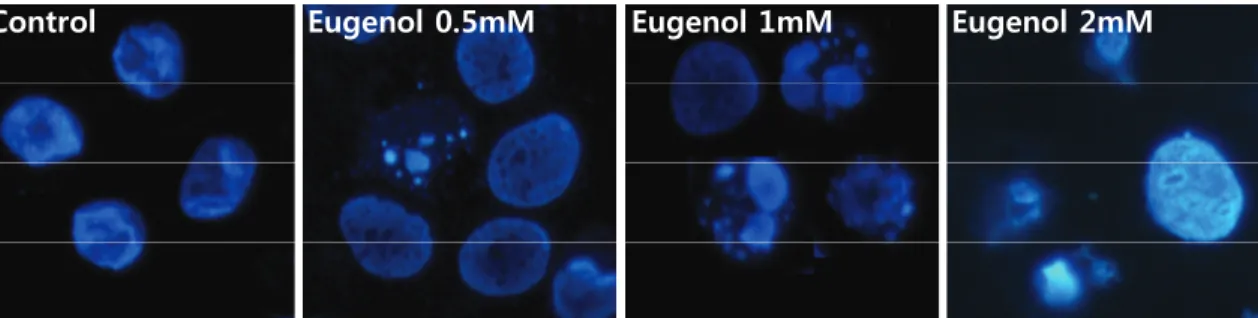
Control Eugenol 0.5mM Eugenol 1mM Eugenol 2mM
Fig. 3. Nucleus condensation signally was observed on HSC-2 cells stimulated with eugenol. Eugenol induced apoptotic HSC-2 cells were increased by eugenol concentration.
Probability(p<0.05)에서 유의한 것으로 판정하였다.
연구 결과
1. Eugenol의 구강암세포 사멸효과
Eugenol에 의한 세포 증식 억제 능력을 평가하기 위하 여 HSC-2세포에 농도별로(0, 0.5 mM, 1 mM, 2 mM) 24 시간 동안 처리 한 후 MTT assay를 시행하였다17,18). 사 용된 유지놀의 화학적 구조는<Fig. 1>에 제시되어 있다.
Fig. 1. Chemical structure of eugenol
0 20 40 60 80 100
Control 0.5 mM 1 mM 2 mM
% of cell viability
*
*
*
*p<0.05 versus untreated samples.
Fig. 2. Eugenol induced the cell cytotoxicity on HSC-2 cell specific as a dose-dependent manner. Cells were stimulated with eugenol at different concentrations for 24h and % viable cells were calculated using MTT assay. The values are denoted as mean SE of three independent experiments (n=3).
HSC-2세포에 eugenol을 농도별로(0, 0.5 mM, 1 mM, 2 mM) 처리하였을 때 대조군과 0.5 mM 농도에서는 구강암세포 사멸효과가 거의 없었으나 1 mM과 2 mM 에서는 eugenol 농도가 증가함에 따라 구강암세포의 생존율이 농도의존적으로 저하되어 69.8%와 51.3%로 각각 나타났다<Fig. 2>.
2. Eugenol 처리에 따른 세포자멸사 유도효과
Eugenol을 처리한 HSC-2세포에 apoptosis가 유도되는 지 확인하기 위하여 DAPI 염색법을 이용하여 관찰하였다.
eugenol을 농도별로(0, 0.5 mM, 1 mM, 2 mM) HSC-2세 포에 처리하여 24시간 동안 배양한 후 핵의 형태학적 변화 를 조사하였다. 그 결과, 각 농도별 apoptotic body가 관찰 되는 비율은 대조군에 비해 농도의존적으로 비율이 증가하 였다<Fig. 3>. 그리고 eugenol 처리된 HSC-2세포는 대조 군에서는 관찰되지 않은 apoptosis의 특징 중 하나인 DNA 단편화에 의한 세포핵이 붕괴되는 것이 관찰되었다.
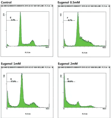
3. 구강암세포의 세포주기 분석
Eugenol에 유도된 apoptosis를 정량적으로 비교하기 위 하여 DNA flow cytometry 분석을 하여 세포주기 중 apoptosis가 일어난 세포그룹에 해당하는 sub-G1기의 비율 을 조사하였다. eugenol 처리 농도의 증가에 따라 각각의 세포주기 해당되는 비율의 차이는 큰 변화가 없었지만 apoptosis가 발현된 것으로 평가 할 수 있는 sub-G1기에 해 당하는 세포의 비율은 매우 증가함을 알 수 있었다. 특히 eugenol을 처리하지 않았을 경우에 sub-G1기의 세포의 빈 도는 4.3%였지만 eugenol처리 농도가 증가할수록 sub-G1 기에 해당하는 세포 비율이 증가하여 0.5 mM의 농도에서 는 8.7%가 관찰되었고 1 mM 농도에서는 17.6%, 2 mM 농도에서는 58.6%에 해당하는 세포가 sub-G1기로 관찰되 었다<Fig. 4>.
0 100 200 300 400
Control 0.5 mM 1 mM 2 mM
Cleaved caspase 3 protein expression
Cleaved caspase 3
0 100 200 300
Control 0.5 mM 1 mM 2 mM
Bak protein expression
Bak Cleaved caspase 3
Bak Bcl-xl
GAPDH
17 kDa 20 kDa 30 kDa 37 kDa Control 0.5 mM 1 mM 2 mM
* *
*
* *
*
* *
0 30 60 90 120
Control 0.5 mM 1 mM 2 mM
Bcl-xl protein expression
Bcl-xl
*p<0.05 versus untreated samples.
Fig. 5. Apoptosis is induced after eugenol in HSC-2 cells. Western blot analysis of the expression levels of apoptosis protein, Cleaved caspase 3, Bcl-xl, Bak in HSC-2 cells treated with indicated dose of eugenol 24 h. The values are denoted as mean SE of three independent experiments(n=3).
Control Eugenol 0.5mM
Eugenol 1mM Eugenol 2mM
Fig. 4. The ratio of apoptotic cells was determined by flow cytometry analysis. To indentify the eugenol-induced HSC-2 cells apoptosis, FACS analysis were performed by PI staining.
4. 세포자멸사 유도 단백질 분석
Eugenol에 의한 HSC-2세포의 증식 억제 및 형태변화는 apoptosis유도와 연관이 있음을 알 수 있었다. eugenol에 의한 유도된 HSC-2세포의 apoptosis의 기전과 신호전달 경로를 규명 하기 위해 내인성, 외인성 세포사멸 경로 하위단계에 있으며 신호 전달에 필수적인 역할을 하는 Cleaved caspase 3의 변화를 확인하였다. Cleaved caspase 3는 세포자멸사 유도에 핵심적인 역할을 하는 인자로서 세포내의 핵과 mitochondria의 바깥막에 불활성 상태로 존재하다가 apoptosis를 유도하는 자극에 의해 활성화된다고 알려져 있다. Cleaved caspase 3의 western blot분 석에서 eugenol의 처리에 따른 Cleaved caspase 3의 증가가 관찰되었으며 Cleaved caspase 3의 발현 증가는 upstream으로 알려진 p53과 연관성이 있을 것으로 생각된다. eugenol에 의해 유도된 apoptosis의 기전 해석을 위하여 apoptosis 조절유전자 로 알려진 pro-apoptotic 인자인 Bak의 발현양은 농도의존적으 로 단백질의 발현이 현저하게 증가하는 것으로 나타났다 (p<0.05). 그러나 anti-apoptotic인자인 Bcl-xl의 경우 단백질 수준에서는 큰 변화가 관찰되지 않았지만 다소 감소한 것으로 나타났다<Fig. 5>.
총괄 및 고안
OSCC는 구강에 발생하는 대표적인 악성종양으로 전 세계에 서 매년 600,000명 이상이 발생하며 대략 35,000명이 사망한다 고 보고되고 있다19-21). Eugenol(4-allyl-2-methoxyphenol)은 Syzigium aromaticum(정향) 페놀성분의 생리활성물질의 하나 로 항산화, 항염증, 항암작용에 효과가 있다고 알려져 있다.
그러므로 구강암 예방과 치료에 다양한 식물들에서 추출된 천연 물질들을 사용하여 암의 예방과 치료를 하고자 노력하고 있으며 또한 기존의 항암제와 함께 천연물질 추출물을 사용할 경우 항암기능을 활성화 시키는 기전을 알아내기 위한 연구가 많이 진행되고 있다22,23). Eugenol은 각종 암에 대해 항암활성을 가지 고 있는 것으로 보고되고 있다. eugenol은 정상세포에는 손상을 주지 않는 천연물질로 알려져 있으나 암조직인 흑색종과 골육종 등 에서는 eugenol을 처리할 경우 농도의존적, 시간의존적으로 암세포의 증식을 저하시켰으며 Cleaved caspase 3와 PARP가 증가하는 것을 보여주었다15,22). 백혈병세포(HL-60) 에서 밝혀 진 eugenol의 항암기전 또한 ROS의 증가와 더불어 미토콘드리 아 막전위와 Cleaved caspase 3 활성화에 의한 세포사멸이 관찰되었다24,25). 위암, 피부종양 등에서도 eugenol에 의한 세포 사멸 기전은 많이 연구되었고 이 또한 eugenol이 p53 단백질에 핵심적인 역할을 하며 Cleaved caspase 3의 활성화를 유도하여 세포사멸을 일으킨다고 설명하고 있다12,26). 이전의 연구결과에 도 알 수 있듯이 eugenol은 여러 악성종양에 효과가 있음을 제시해주고 있으며 본 연구결과에서도 마찬가지로 구강조직에
발생하는 구강암세포에서도 eugenol처리에 따른 세포사멸 유 도를 확인함으로써 구강암에도 적용 가능한 치료물질로서 가능 성을 제시하였다고 할 수 있다. DAPI 염색에 의한 핵의 붕괴는 일반적인 세포자멸사 과정 중에 관찰되며 이는 eugenol이 세포 자멸사를 유도한다는 것을 형태적으로 관찰 할 수 있다. FACS 실험결과 또한 농도의존적으로 세포사멸이 증가하였다는 것을 뒷받침 해주고 있다. 본 연구는 구강암을 치료하는 치료제 후보 군으로서 eugenol 효과와 기전을 평가하기 위해 이루어졌다.
Eugenol은 구강암세포의 사멸에는 유의성 있는 결과를 얻었지 만 다양한 신호경로 기전을 확인 하지는 못하였다. 추가적인 실험과 향후 동물실험까지 진행하여 구강암세포 사멸 효과에 대한 명확한 기전을 알 수 있다면 eugenol을 다양한 암조직에 적용 할 수 있을 것이라 생각된다. 위의 결과로 평가해볼 때 Eugenol은 구강암에 치료에 적용 가능한 효과적인 항암물질 후보군으로 생각된다.
결 론
본 연구는 eugenol을 이용하여 사람 구강암세포주인 HSC-2 세포에서 eugenol에 의한 세포자멸사를 확인하고 세포자멸사 표지단백질인 Bcl-xl, Cleaved caspase 3, Bak의 기능을 조사 하기 위하여 실험하였다.
1. 구강편평상피세포암 HSC-2세포에 eugenol을 처리하 였을 때 구강암세포의 생존율이 농도의존적으로 감소 하였다(p<0.05).
2. Eugenol에 의해 유도된 구강암세포의 죽음은 세포핵 응축으로 대표되는 세포자멸사에 의한 것으로 생각된다. 3. Eugenol은 내인적, 외인적 두 종류의 세포자멸사 과정
중 bak에서 caspase 3 경로를 거치는 내인적 세포자멸사 과정으로 생각된다.
구강암은 발생 부위가 구강악안면에 국한되어 발생하며 안모의 변형을 초래하여 한 인간의 삶에 큰 상처를 남긴다. 그러므로 스트레스를 줄이고 구강위생상태를 청결히 유지하 는 것이 구강암 예방에 매우 중요하다고 할 수 있다. 본 연 구 결과 eugenol은 구강암 치료에 적용 가능한 효과적인 천 연물질로 사료된다.
References
1. Chen SF, Nien S, Wu CH, Liu CL, Chang YC, Lin YS.
Reappraisal of the anticancer efficacy of quercetin in oral cancer cells. J Chin Med Assoc 2013; 76(3): 146-52.
2. Zhang X, Liu Y, Gilcrease MZ, Yuan XH, Clayman GL,
Adler-Storthz K, et al. A lymph node metastatic mouse model reveals alterations of metastasis-related gene expression in metastatic human oral carcinoma sublines selected from a poorly metastatic parental cell line. Cancer 2002; 95(8):
1663-72. http://dx.doi.org/10.1002/cncr.10837.
3. Chen YJ, Chang JT, Liao CT, Wang HM, Yen TC, Chiu CC, et al. Head and neck cancer in the betel quid chewing area: recent advances in molecular carcinogenesis. Cancer Sci 2008; 99(8): 1507-14. http://dx.doi.org/10.1111/ j.1349- 7006.2008.00863.x.
4. Adelstein DJ, Li Y, Adams GL, Wagner H, Jr, Kish JA, Ensley JF. An intergroup phase III comparison of standard radiation therapy and two schedules of concurrent chemoradiotherapy in patients with unresectable squamous cell head and neck cancer. J Clin Oncol 2003; 21(1): 92-8.
http://dx.doi.org/10.1200/jco.2003.01.008.
5. Boulikas T, Vougiouka M. Recent clinical trials using cisplatin, carboplatin and their combination chemotherapy drugs (review). Oncol Rep 2004; 11(3): 559-95.
http://dx.doi.org/10.3892/or.11.3.559.
6. Butler MS. The role of natural product chemistry in drug discovery. J Nat Prod 2004; 67(12): 2141-53. http://dx.
doi.org/10.1021/np040106y.
7. Sondak VK, Sabel MS, Mule JJ. Allogeneic and autologous melanoma vaccines: where have we been and where are we going?. Clin Cancer Res 2006; 12(7 Pt 2): 2337s-41s.
http://dx.doi.org/10.1158/1078-0432.ccr-05-2555.
8. Pramod K, Ansari SH, Ali J. Eugenol: a natural compound with versatile pharmacological actions. Nat Prod Commun 2010; 5(12): 1999-2006.
9. Stich HF, Stich W, Lam PP. Potentiation of genotoxicity by concurrent application of compounds found in betel quid:
arecoline, eugenol, quercetin, chlorogenic acid and Mn2+.
Mutat Res 1981; 90(4): 355-63. http://dx.doi.org/10.1016/
0165-1218(81)90058-6.
10. Benencia F, Courreges MC. In vitro and in vivo activity of eugenol on human herpesvirus. Phytother Res 2000; 14(7):
495-500. http://dx.doi.org/10.1002/1099-1573(200011) 14:
7<495::aid-ptr650>3.0.co;2-8.
11. Al-Sharif I, Remmal A, Aboussekhra A. Eugenol triggers apoptosis in breast cancer cells through E2F1/survivin down-regulation. BMC Cancer 2013; (1)3: 600. http://dx.doi.
org/10.1186/1471-2407-13-600.
12. Park BS, Song YS, Yee SB, Lee BG, Seo SY, Park YC, et al. Phospho-ser 15-p53 translocates into mitochondria and interacts with Bcl-2 and Bcl-xL in eugenol-induced apoptosis. Apoptosis 2005; 10(1): 193-200. http://dx.doi.
org/10.1007/s10495-005-6074-7.
13. Slamenova D, Horvathova E, Wsolova L, Sramkova M, Navarova J. Investigation of anti-oxidative, cytotoxic, DNA-damaging and DNA-protective effects of plant volatiles eugenol and borneol in human-derived HepG2, Caco-2 and VH10 cell lines. Mutat Res 2009; 677(1-2):
46-52. http://dx.doi.org/10.1016/j.mrgentox.2009.05.016.
14. Okada N, Hirata A, Murakami Y, Shoji M, Sakagami H, Fujisawa S. Induction of cytotoxicity and apoptosis and inhibition of cyclooxygenase-2 gene expression by eugenol -related compounds. Anticancer Res 2005; 25(5): 3263-9.
15. Ghosh R, Nadiminty N, Fitzpatrick JE, Alworth WL, Slaga TJ, Kumar AP. Eugenol causes melanoma growth suppression through inhibition of E2F1 transcriptional activity. The Journal of biological chemistry 2005; 280(7):
5812-9. http://dx.doi.org/10.1074/jbc.m411429200.
16. Pisano M, Pagnan G, Loi M, Mura ME, Tilocca MG, Palmieri G, et al. Antiproliferative and pro-apoptotic activity of eugenol-related biphenyls on malignant melanoma cells.
Mol Cancer 2007; (6): 8.
17. Lee AR, Park, SY, Lee, KH. The protective effect of coenzyme Q10 on cytotoxicity of regin monomer of odontoblast caused by TEGDMA. J Korean Soc Dent Hyg 2014; 14(5): 775-81. http://dx.doi.org/10.13065/jksdh.2014.
14.05.775.
18. Huh MK, Kim HJ. Antibacterial effect on leaf-extract from Nelumbo nucifera against oral microorganism. J Korean Soc Dent Hyg 2014; 14(1): 117-22.
19. Chen PN, Hsieh YS, Chiang CL, Chiou HL, Yang SF, Chu SC. Silibinin inhibits invasion of oral cancer cells by suppressing the MAPK pathway. J Dent Res 2006; 85(3):
220-5. http://dx.doi.org/10.1177/154405910608500303.
20. Siegel R, Naishadham D, Jemal A. Cancer statistics, 2012.
CA Cancer J Clin 2012; 62(1): 10-29. http://dx.doi.org/
10.3322/caac.20138.
21. Parkin DM, Bray F, Ferlay J, Pisani P. Global cancer statistics, 2002. CA Cancer J Clin 2005; 55(2): 74-108. http://dx.
doi.org/10.3322/canjclin.52.1.23.
22. Schneider-Stock R, Boltze C, Lasota J, Miettinen M, et al. High prognostic value of p16INK4 alterations in gastrointestinal stromal tumors. J Clin Oncol 2003; 21(9):
1688-97. http://dx.doi.org/10.1200/jco.2003.08.101.
23. Adams JM, Cory S. Bcl-2-regulated apoptosis: mechanism and therapeutic potential. Curr Opin Immunol 2007; 19(5):
488-96. http://dx.doi.org/10.1016/j.coi.2007.05.004.
24. Jaganathan SK, Supriyanto E. Antiproliferative and molecular mechanism of eugenol-induced apoptosis in
cancer cells. Molecules 2012; 17(6): 6290-304. http://dx.doi.
org/10.3390/molecules17066290.
25. Yoo CB, Han KT, Cho KS, et al. Eugenol isolated from the essential oil of Eugenia caryophyllata induces a reactive oxygen species-mediated apoptosis in HL-60 human promyelocytic leukemia cells. Cancer Lett 2005; 225(1):
41-52. http://dx.doi.org/10.1016/j.canlet.2004.11.018.
26. Kaur G, Athar M, Alam MS. Eugenol precludes cutaneous chemical carcinogenesis in mouse by preventing oxidative stress and inflammation and by inducing apoptosis. Mol Carcinog 2010; 49(3): 290-301. http://dx.doi.org/10. 1002/
mc.20601.