226
F OREST S OCIETY
Effects of Pruning Season on Compartmentalization of Pruning Wounds in Acer palmatum and Pinus strobus
Kyu Hwa Lee* and Kyung Joon Lee
Department of Forest Sciences, Seoul National University, Seoul 151-921 Korea
Abstract : This study was conducted to examine the effects of pruning season on the compartmentalization of pruning wounds in Acer palmatum and Pinus strobus . A total of eighty five field-grown trees for each species were allocated to five different seasons, early- and late-winter, mid-spring, mid- and late-summer, for pruning treatments. Wound closure rate (WCR) of the two species for one year after treatment, area of discolored stem tissue on the medial longitudinal surface and cambial dieback length under the pruning wound of A. palmatum were measured. Changes of total phenols and variations of extractives, holocellulose and lignin at the treated branch unions were examined. In WCR of A. palmatum , late-winter (March, 39.8%) and mid-spring (May, 39.7%) were higher than any other seasons, while early-winter (November, 28.4%) was significantly lower than late-winter and mid-spring. P. strobus showed similar results with A.
palmatum . The WCR of early-winter (57.2%) was the lowest significantly among the five seasons, and mid-spring (73.5%) and late-winter (71.4%) showed higher a WCR than other seasons. In the discolored/
wound area ratio of A. palmatum , early-winter (73.2%) was the highest by far, and mid- (July) and late- summer (September, 36.7%, respectively) were the lowest among the five seasons. In the length of cambial dieback, two dormant seasons, early- and late-winter were longer than any other seasons. Phenol contents at the treated branch union were changed in line with the seasonal fluctuation of the tree. Total phenols in the below core of the treated union were higher than those of the branch union with living branch, while little differences were seen in the above core. At the branch core of the treated union, phenols of A . palmatum decreased one month after the treatments, but P . strobus maintained similar to or a little higher than those at the controls. The major changes in chemical composition at pruning wounds were extractives and lignin increased by less than 20% in A . palmatum , while extractives in P . strobus remarkably increased by 70%.
Key words : wound closure, total phenols, holocellulose, lignin, discolored area, cambial dieback
Introduction
Tree pruning is performed for various purposes. In horticulture, the major objectives of pruning are to influ- ence the productivity of fruit trees and appearances of ornamental trees, while the aim in silviculture is to pro- duce knot-free lumber. Pruning in arboriculture is an integral part of the tree maintenance of any landscape site. Correct pruning helps maintain vigorous plants and can aid the supply of additional energy for development of flowers, fruit and limbs. But it is inevitable for trees to get injuries by the pruning practice, and those pruning wounds are to be compartmentalized by the trees.
According to the CODIT model (Shigo and Marx, 1977), lesions in functional sapwood are bounded by
four walls laid down in the wood, envisaged as essen- tially static barriers preventing the spread of infection.
Walls 1 to 3, formed in wood present at the time of wounding, are equivalent to reaction zones rich in phe- nolic compounds, but wall 4 is distinct, comprising a tis- sue laid down de novo by the cambium in the vicinity of wounds, and is the most durable of the four kinds of compartmentalization walls.
The phloem as well as the sapwood of trees has an effective defense system to secure their vital function of transport and storage, to restrict the embolism of air and, in later stages, also the spread of pathogens. The response capacity is primarily influenced by the physi- ological state, e.g. the prevailing season (Dujesiefken et al ., 2005; Kozlowski and Pallardy, 1997; Leben, 1985;
Neely, 1970; Shortle, 1979). So it is very important to ascertain the most effective time for the pruning of indi- vidual species, considering its growing environments.
*Corresponding author
E-mail: [email protected]
Meanwhile, recommendable times for pruning are dif- ferent from person to person. Winter is recommended as the best season, while some people prohibit pruning dur- ing the winter period, especially in early winter. Pruning in late winter or early spring is generally recommended, but spring pruning after budbreak is undesirable. This is because the bark is at its most tender moment and dam- age to the bark is most likely. Mid-summer is an accept- able pruning time for some trees known as “bleeders”, such as Ulmus sp., Betula sp. and Acer sp., but root growth is slowed temporarily when live branches are removed in the summer (Gilman, 2002; Harris et al ., 2004; Hartman et al ., 2000).
Pruning in the late summer is considered undesirable because it removes foliar carbohydrates that would be sent to the tree before the leaves fall, and new growth which will not be sufficiently hardened to prevent winter damage may be encouraged. In the fall, a number of important decay fungi have been observed to produce their largest numbers of spores. Evergreens are consid- ered to be pruned any time the wood is not frozen (Gil- man, 2002; Harris et al ., 2004; Hartman et al ., 2000;
Hensley, 1979; Lee and Lee, 2001; Shigo, 1989).
Among the several considerations in decision of prun- ing season, enclosing speed of pruning wounds is the most important because the decay process continues as long as the wound remains open (Shigo and Larson, 1969). But most researches on wound closure were per- formed with the artificial wounds made on the stems for silvicultural purposes. The objective of this study was to examine the effects of pruning season on the compart- mentalization of pruning wounds, and the trees selected for this experiment were Acer palmatum , a deciduous hardwood, and Pinus strobus , a evergreen conifer, both of which are planted broadly in the urban forests of Korea.
Materials and Methods
1. Seasonal onsets and timing of pruning treatments Choi et al. (2006) reported the long-term spatial pat- terns of seasonal onsets defined by daily temperatures in South Korea for the period of 1973-2004. According to the study, two experimental sites, Yeoju, Gyeonggi, and Jincheon, Chungbuk, had similar spatial patterns of sea- sonal onsets with each other, and their seasonal onsets were March 21 for spring, May 30 for summer, September 17 for fall, and November 16 for winter (Table 1).
Pruning treatments were made during five different seasons. Early- and late-winter and mid-summer were recommendable seasons for pruning, and mid-spring and late-summer were the undesirable seasons. Pruning treat- ments for each season were made on March 1, 2008 for late-winter, May 16, 2008 for mid-spring, July 18, 2008
for mid-summer, September 8, 2007 for late-summer, and November 24, 2007 for early-winter (Table 2).
2. Tree species for the study
Eighty five field-grown A. palmatum (Japanese maple) and the same number of P. strobus (eastern white pine) were used for this study. Eighty five trees of each spe- cies were allocated seventeen trees to every five pruning seasons. Two trees out of the seventeen were used to monitor the changes of phenolic compounds at the prun- ing wounds. Of the two, one tree was harvested one month after pruning treatment and the other one was harvested six months after the treatment.
A. palmatum growing in Yeoju, Gyeonggi, Korea (37.10.841 N, 127.38.900 E) were thirteen years old, 10.9 cm thick in average diameter at 30 cm above the ground and 360 cm tall in average height. P. strobus growing in Jincheon, Chungbuk, Korea (36.49.543 N, 127.27.020 E) were eleven years old, 11.9 cm thick in average diameter at 30 cm above the ground and 460 cm tall in average height. During the experimental period, the annual average temperature of both sites weas around 12
oC and those annual precipitations were between 796 and 1,764 mm (Korea Meteorological Administration, 2009).
3. Experimental design
Fifteen trees of each species were allocated to treat- ment season according to the randomized block design.
The stands of each species were divided into three blocks, which were border, intermediate between border and interior, and interior of the stand. Each block for the season consisted of five trees.
4. Pruning cuts
Pruning cuts were conformable with an American National Table 1. Seasonal onsets at the two experimental sites, Yeoju, Gyeonggi and Jincheon, Chungbuk, defined by daily temperatures for the period of 1973-2004 in South Korea (Choi et al. , 2006).
Seasons Spring Summer Fall Winter Onset dates March 21 May 30 September 17November 16 Table 2. Dates of the pruning treatment by season based on seasonal onsets of the experimental sites.
Pruning
seasons Dates of Pruning
treatment Remarks
Late-winter March 1, 2008 Recommendable season
Mid-spring May 16, 2008 Undesirable season
Mid-summer July 18, 2008 Recommendable season
Late-summer September 8, 2007 Undesirable season
Early-winter November 24, 2007 Recommendable season
Standard (American National Standard Institute, 2001).
A pruning cut was made close to the trunk or parent limb, without cutting into the branch bark ridge (BBR) or collar, or leaving a stub (Lee and Lee, 2009). In case where the branch collar was not readily apparent, the cut bisected the angle between its BBR and an imaginary line perpendicular to the branch. A branch that was too large to support with one hand was precut to avoid split- ting of the wood or tearing of the bark. The proper angle of cut depends on the branch collar. Conifers have flat branch collars and a straight cut close to the collar was made for P. strobus (Shigo, 1989).
The size of the branch for pruning cut was between 1 cm and 4 cm in diameter at 1.5 cm above the BBR. Four to twenty branches per tree were removed depending on the branch density of the tree.
5. Measurements
1) Branch and stem diameter, area of pruning wound and length of cambial dieback
Prior to pruning, diameters of branches and stems at 1.5 cm above the BBR were gauged with a cal- iper. Immediately after each cut, horizontal width and vertical length of the pruning wound crossing the center of branch were measured to calculate the wound area. The vertical length of cambial dieback under the pruning wound was also measured from the sample of A. palmatum to examine the variation of cambial dieback under the wound by pruning sea- son (Figure 1).
2) Discolored area
The samples from the harvested A. palmatum were dissected with a cleaver along the medial longitudinal plane and shaved with a sharpened knife to measure dis- colored areas formed after branch removal. The area of discolored stem tissue on the medial longitudinal surface was estimated by the length of pruning wound and the deepest penetration of discolored wood into the trunk (Lee et al ., 2009). In case of P. strobus , which differed from A. palmatum , the whole area of the branch core after the pruning cut was darkened with increased resin concentration, and measuring discolored areas formed after branch removal was meaningless.
6. Determination of total phenols
Phenolic compounds are concentrated at the reaction zone to resist the spread of organisms into the trunk.
They were determined three times, one month, six months and twelve months after treatments. Eight branch union samples were obtained from one tree in each treat- ment at each point of time after treatments. The eight samples consisted of four with pruning wounds and the other four with living branches. The 0.06 g Fresh Weight (FW) of wood tissues was collected with a chisel from three separated parts of each sample, which were core of the branch, above core and below core (Lee et al ., 2009).
A total of 0.24 g of FW shavings for each part col- lected from four samples were extracted in 4 mL of 100% methanol for 24 hours at 4
oC. After the extraction, the wood tissues were dried at 105±1
oC and then weighed. The methanol solution was filtered with 0.50
µ m of pore hydrophobic syringe filter. Total phenols were estimated by the Folin-Ciocalteu method as described by Bonello and Pearce (1993). For each sample, 15 µ L of methanol extract was diluted to 1500 µ L of water. To this solution, 750 µ L of Folin and Ciocalteu’s reagent (Sigma Chemical Co.), diluted two-fold with water, was added and left for 3 minutes, followed by the addition of 750 µ L of 1 M aqueous Na
2CO
3. The solution was shaken and left for 1 hour before the absorbance was measured at 725 nm with a spectrophotometer.
Concentrations of total phenols were calculated with reference to a gallic acid standard curve (10-200 µ g/mL) by applying the regression analysis and results were expressed as gallic acid equivalent (mg) per g Dry Weight (DW).
7. Determinations of extractives, holocellulose and lignin Chemical analyses for the tissues of the branch core, and the above and below core were performed to exam- ine the changes of their chemical properties one year after treatments. Two trees from each treatment were Figure 1. A diagram of the sample of branch union with
pruning wound one year after pruning. The branch bark
ridge (BBR) was protected when the cut was made. A
ring of woundwood is closing the wound, but cambial
dieback (D-D’) under the pruning wound is still open.
selected for the analysis. A total of sixteen branch union samples, eight from each tree, were collected from each treatment, and eight samples of each tree consisted of four with pruning wounds and the other four with living branches.
The samples were dissected with a cleaver along the medial longitudinal plane, and the parts beyond an imag- inary line perpendicular to the stem pith at 1.5 cm above the BBR and an imaginary line perpendicular to the stem pith at 1.5 cm below the end of branch pith were trimmed off (Lee et al ., 2009). The trimmed samples were air- dried, split into three parts (core of branch, above and below core) and then ground through a 40 mesh screen.
T he content of the extractives was determined by the soxhlet method (Technical Association of Pulp and Paper Industry, 1994). Two grams of sample in a thimble filter was extracted by 150 mL of ethanol/benzene mix- ture (1:2 v/v) using a soxhlet extractor with reflux con- denser at 80
oC for 6 hours. The extracted solution was evaporated under reduced pressure. The extractive was dried at 105±1
oC and then weighed. The extractive-free sample left in the thimble filter was used for analyses of holocellulose and Klason lignin.
The content of holocellulose was determined as the delignified residue by NaClO
2(Wise et al ., 1946). The 1.25 g of extractive-free sample in 250 mL flask was treated with 75 mL of distilled water, 0.5 g of NaClO
2and 0.1 mL of CH
3COOH at 80
oC for 1 hr. This pro- cedure was repeated 2 more times. The solution was fil- tered by using a glass filter (1G3, Iwaki, Japan), and then washed with 250 mL of cold distilled water and 25 mL of acetone, successively. The filtrated residue was dried at 105±1
oC and then weighed.
Klason lignin was analyzed according to the standard NREL procedures (National Renewable Energy Labora- tory, 2005). The 0.3 g of extractive-free sample in 50 mL flasks was hydrolyzed with 3 mL of 72% H
2SO
4at 30
oC for 1 hr. The hydrolysate was then transferred to 100 mL flask and diluted to 4% H
2SO
4by adding 84 mL of distilled water. The flasks were sealed and autoclaved
for 1 hr at 121
oC. The solution was then filtered by using a glass filter (1G4, Iwaki, Japan). The filtrated res- idue was dried at 105±1
oC and then weighed.
8. Data analysis
Some indicators were introduced for this study (Table 3). The wound closure rate (WCR) and the discolored/
wound area ratio (D/W ratio) were introduced to exam- ine the variations of ability to compartmentalize the pruning wounds after the pruning treatments. The WCR represented the enclosing extent of the pruning wound during the year after treatments, and the D/W ratio was computed by comparing the area of discolored stem tis- sue on the medial longitudinal surface one year after treatments with the area of pruning wound at the time of pruning. In the CODIT model, the WCR indicated vital- ity of the cambium around the wound which formed wall 4 after the tree was wounded, while the D/W ratio expressed the ability of a tree to limit the spread of wood discolorations and decays with wall 1, 2 and 3 which were already present in the wood at the time of the pruning.
The two-way ANOVA for the block design was used to calculate the ratio of the variance among the means to the variance within the samples. Duncan’s multiple com- parison procedure was adopted to test the differences among the treatments. SAS 9.1 (SAS Institute Inc., USA) was used for the analysis, and the significance level, α -value, was 0.05 unless otherwise specified.
Results
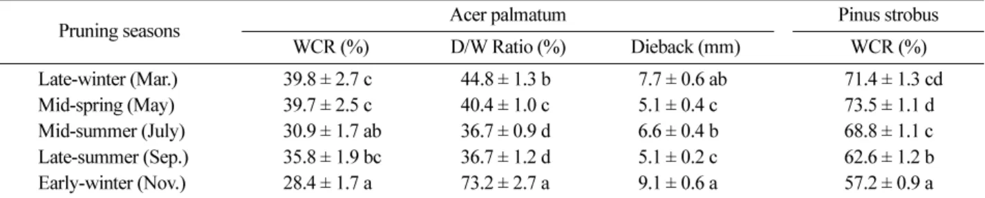
1. Wound closure and discolored area
Table 4 shows means and statistical differences in the WCR of both species, and the D/W Ratio and length of cambial dieback by pruning season one year after prun- ing treatments for A. palmatum . In the WCR of A. pal- matum , late-winter (March, 39.8%) and mid-spring (May, 39.7%) were higher than any other seasons, while early- winter (November, 28.4%) was the lowest and signifi- Table 3. Indicators to analyze the changes in the area of pruning wound, and development of discolored wood on the medial longitudinal surface after treatments.
Indicators Formulae
Wound Closure Rate (WCR, %)
PWA
y0: area of pruning wound when pruning : area of pruning wound one year after pruning Discolored/Wound Area Ratio
(D/W ratio, %)
DA
y+1: area of discolored stem tissue on the medial longitudinal surface one year after pruning PWA
y0: area of pruning wound when pruning
W D
---ratio DA
y 1+PWA
y0---
=
cantly different with late-winter and mid-spring ( P <0.01).
Late-summer (September, 35.8%) was higher than mid- summer (July, 30.9%), but the difference was not sig- nificant.
P. strobus showed similar results with A. palmatum . The WCR of early-winter (57.2%) was the lowest sig- nificantly among the five seasons ( P <0.01), and mid- spring and later-winter showed a higher WCR than other seasons. But the mid-summer (68.8%) was higher than late-summer (62.6%), which differed from A. palmatum . Comparing the WCR of the two species, P. strobus was much higher than A. palmatum in general.
In the D/W ratio of A. palmatum , early-winter (73.2%) was the highest by far, and mid- and late-summer were the lowest among the five seasons. Late-winter (44.8%) was also significantly higher than mid-/late-summer ( P <
0.05). In the length of the cambial dieback, two dormant seasons, early- and late-winter (9.1 mm and 7.7 mm, respectively), were longer than any other seasons. Mid- summer (6.6 mm) was the longest among the growing
seasons, but the differences were not significant.
2. Wood chemical properties
Table 5 shows the changes of total phenols at the three parts (above core, branch core, and below core) between the treated branch union with pruning wound and the control one with living branch by pruning season in both A . palmatum and P . strobus . On the whole, contents of total phenols showed seasonal fluctuation, and phenol contents at the treated branch union were changed in line with the seasonal fluctuation of the tree. There were lit- tle differences in phenol contents at the above cores between treated and control branch unions, but the con- tents at the below cores were higher at the treated branch union, especially in P . strobus . At the branch core, there was a minor difference between the two species. In case of A . palmatum , phenol contents of the treated union increased one month after the treatments, and then decreased below the levels of the controls. But in P . stro- bus , the phenol contents at the treated union were similar Table 4. Variations of wound closure rate (WCR) of Acer palmatum and Pinus strobus , and discolored/wound area ratio (D/W ratio) and length of cambial dieback (Dieback) of A . palmatum by pruning season one year after pruning treatments. mean ± standard error. Means with the same letter are not significantly different at P < 0.05.
Pruning seasons Acer palmatum Pinus strobus
WCR (%) D/W Ratio (%) Dieback (mm) WCR (%)
Late-winter (Mar.) 39.8 ± 2.7 c 44.8 ± 1.3 b 7.7 ± 0.6 ab 71.4 ± 1.3 cd Mid-spring (May) 39.7 ± 2.5 c 40.4 ± 1.0 c 5.1 ± 0.4 c 73.5 ± 1.1 d Mid-summer (July) 30.9 ± 1.7 ab 36.7 ± 0.9 d 6.6 ± 0.4 b 68.8 ± 1.1 c Late-summer (Sep.) 35.8 ± 1.9 bc 36.7 ± 1.2 d 5.1 ± 0.2 c 62.6 ± 1.2 b Early-winter (Nov.) 28.4 ± 1.7 a 73.2 ± 2.7 a 9.1 ± 0.6 a 57.2 ± 0.9 a Table 5. Changes of total phenols at the three parts (above core, branch core, and below core) between treated branch union with pruning wound (Tr.) and control one with living branch (Co.) by pruning season in Acer palmatum and Pinus strobus . (unit: gallic acid equivalent (mg) per g DW).
Pruning seasons Late-winter (Mar.) Mid-spring (May) Mid-summer (July) Late-summer (Sep.) Early-winter (Nov.) Month(s) after pruning 1M 6Ms 12Ms 1M 6Ms 12Ms 1M 6Ms 12Ms 1M 6Ms 12Ms 1M 6Ms 12Ms
Acer palmatum Above
core Tr. 65 57 59 85 75 49 77 80 75 51 76 56 149 102 27
Co. 67 53 53 75 66 57 70 85 80 52 76 52 158 95 28
Branch
core Tr. 72 33 58 86 21 29 86 79 57 79 80 44 150 72 141
Co. 68 50 57 74 63 57 63 65 81 41 76 58 170 90 162
Below
core Tr. 72 76 67 102 99 60 84 100 88 56 88 82 152 119 66
Co. 71 54 65 76 69 56 72 88 72 49 85 56 155 99 34
Pinus strobus Above
core Tr. 22 25 45 45 25 39 26 63 27 16 66 27 46 47 60
Co. 22 21 39 40 27 35 27 54 23 20 72 29 40 44 34
Branch
core Tr. 930 946 1,127 5,430 1,424 4,190 940 1,235 973 762 1,027 1,014 1,249 4,402 2,123 Co. 989 913 1,067 5,250 1,167 4,523 918 1,143 881 788 1,009 939 1,238 3,917 1,477 Below
core Tr. 27 33 89 67 27 139 28 108 90 33 52 45 40 68 148
Co. 22 23 54 45 25 92 25 55 24 21 36 25 39 42 37
Figure 2. Variations of extractives, holocellulose and lignin in branch union at the three parts (ac: above core, c: branch core, bc: below core) between branch union with pruning wound (Treated) and control one with living branch (Control) by pruning season in Acer palmatum . LW: Late-winter (March), MSp: Mid-spring (May), MSu: Mid-summer (July), LSu: Late-summer (September), EW: Early winter (November).
Figure 3. Variations of extractives, holocellulose and lignin at the three parts (ac: above core, c: branch core, bc: below
core) between branch union with pruning wound (Treated) and control one with living branch (Control) by pruning
season in Pinus strobus . LW: Late-winter (March), MSp: Mid-spring (May), MSu: Mid-summer (July), LSu: Late-
summer (September), EW: Early winter (November).
to or a little higher than those at the controls.
Figure 2 and 3 show variations of extractives, holo- cellulose and lignin at the three parts (above core, branch core and below core) between the treated branch union with pruning wound and the control one with liv- ing branch by pruning season in A . palmatum and P . strobus , respectively. In A . palmatum , extractives and lignin at the branch core of the treated branch union (2.6% and 25.9% in average, respectively) were higher than those at the same part of the control one (2.2% and 22.6% in average, respectively), but holocellulose con- tents at the treated union (70.5% in average) were lower than those at the control one (72.1% in average). In P . strobus , differences in holocellulose and lignin between the treated and control branch union (65.6% vs. 66.4%, and 26.5% vs. 26.7% in average, respectively) were not significant. But contents of extractives at the branch core increased sharply after pruning treatments (from 16.8%
at the control to 30.0% at treated in average).
Discussion
No other research on the effects of pruning season has been done for A . palmatum and P . strobus . The results of the present study, detailing wounds in late-winter and early growing season would be more effectively com- partmentalized than those in the autumn and early-win- ter, are generally consistent with the findings for other tree species (Dujesiefken et al ., 2005). Neely (1970) reported that wounds made in late winter (March) and in May healed much more than wounds made in fall (Octo- ber) and in July, respectively, in the experiment on white ashes, honey locusts, and pin oaks. In Eucalyptus cam- aldulensis , wounds made in spring closed most rapidly, and the slowest closure was recorded for wounds made during late fall and early winter. Wounds made in late winter and summer closed at similar rates as the results of P . strobus in the present study (Perry and Hickman, 1987). In red maple ( Acer rubrum L.), wounds made in the spring, March to May, also caused greater wound occlusion than wounds made in the autumn, mid Sep- tember to early November (Leben, 1985). Debell et al . (2006), however, found that pruning just prior to the beginning of the growing season (March 1) resulted in longer time for the formation of clear wood than pruning during the growing season (May 1 and June 15) for red alder. The time of year during which the cambium is active varies with climate, species, etc., which might cause such differences among experiments (Winget and Kozlowski, 1965).
Researches on the speed of wound closure by tree spe- cies were limited. Neely (1970; 1973; 1983) reported the differences among species in the rate of wound closure.
According to the experiments, tulip trees and pin oaks were the fastest in wound closure, while American elm and soft maple trees were faster than white ashes and honey locusts. In the present study, P. strobus was faster in wound closure than A. palmatum , which showed a variation in healing ability in previous studies.
A certain correlation was found between the D/W ratio and the length of cambial dieback, except in mid- summer pruning. Greater phloem dieback increased the wound size, and the larger area of exposed xylem apparently resulted in larger volumes of discolored wood (Blanchette, 1992). The cambial growth was very responsive to envi- ronmental stresses (Kozlowski and Pallardy, 1997), and wounds made in October or early winter suffered from considerable tissue dieback beyond the original wound margins (Neely, 1970; Perry and Hickman, 1987). Longer cambial dieback in wounds made in early-winter also was related to the lower WCR of the treatment, as shown by the results of the present study (Leben, 1985).
Another important consideration that directly influences bark dieback was moisture loss from the tree (Blanchette, 1992). Loss of moisture and replacement with air in xylem could affect discoloration processes and subse- quent invasion by fungi (Boddy and Rayner, 1983;
Rayner, 1986). The amount to which air would enter the xylem could vary at different times of the year depend- ing upon whether water columns were under positive or negative hydrostatic pressure when the xylem was wounded (Rayner, 1986). In the present study, a longer cambial dieback length and a higher D/W ratio in early- and late-winter treatments might be caused by a longer winter and less precipitation than an average year after treatments in November 2007 and March 2008, respec- tively.
Mid-summer pruning (July 2008) in the present study showed a relatively longer cambial dieback, still shorter than winter treatments, and a lower or equal D/W ratio when comparing with other treatments in a growing sea- son. Wounds made in the summer (June) did not suffer from extensive dieback like wounds made in early-win- ter (Perry and Hickman, 1987), but the correlations between the length of cambial dieback and discolored wood column were not significant (Leben, 1985). Fur- ther study is necessary to examine the reasons, but changes of cambial electrical resistance (CER) would be helpful to understand the result (Lee et al ., 2009). The CER one month after treatment (10.8 k Ω on August 16) was higher compared to one month before treatment (9.5 k Ω on June 16), and a less vigorous cambial zone might affect the cambial dieback after treatment.
A tree begins to form a protective chemical shield, a reac-
tion zone with mostly phenolic compounds in angiosperms
and resins in conifers, around and immediately behind
the wound, and the barrier could eventually be eroded and retreated by fungal activities (Prior, 1976; Shigo, 1979). Levels of the phenolic contents at the treated union changed in accordance with seasonal variation in the production of phenolic compounds (Barry et al ., 2000; Jalal et al. , 1982; Mireku and Wilkes, 1989) as the case of the above core, while more discussion is neces- sary for the changes of the contents at the core and the below core of the both treatments.
The vascular cambium and the growth rings it pro- duces are continuous from trunk to branch, but cells formed by the cambium in the upper junction of branch and trunk are oriented in a right angle to the normal ori- entation in the trunk and branch, while the branch xylem is oriented downward at the branch base and encircles it to form a collar which meets on the trunk below the branch. Thus conduction into and out of the branch fol- lowed the pathway of the branch collar and there was no local direct conduction between the trunk xylem above a branch and within a branch (Shigo, 1985). Consequently, the phenol contents at the above core were not affected by the treatments during a year in the present study, and the contents at the branch core and the below core changed in accordance with the extension of discolored sapwood, which proceeded further in the axial rather than radial or tangential wood alignments (Deflorio et al. , 2007).
In Eucalyptus nitens , total phenols at the reaction zone after wounding increased slowly, reaching a maximum after 21 days, and then would decrease with the exten- sion of the discolored area and the retreat of reaction zones rich in phenols as time went by (Barry et al. , 2001). But in Scots pine, there was no significant dif- ference between healthy roots and roots infected with Cylindrocarpon destructans in total phenols (Bonello and Pearce, 1993). Total phenols at the branch core of A . palmatum and P . strobus of the present study changed consistently with the previous studies on hardwoods and conifers. Higher total phenols at the below core in the present study would be caused by a downward extension of discolored area along the conduction pathway.
In Scots pine, there was little difference in lignin between healthy roots and roots infected with Cylindrocarpon destructans (Bonello and Pearce, 1993). But resins con- tribute to defense in conifers by forming mechanical bar- riers in resin-soaked tissues (Pearce, 1987; Prior, 1976;
Shain, 1979), and oleoresin was accumulated in the reac- tion zone and infected sapwood to the extent of about 30% of the moisture-free weight of the tissue (Shain, 1967), as shown by the results of the present study.
Formation of lignin is one of the defense mechanisms in the wood tissues to protect trees from wounding and microbial invasion (Yamada, 2001). Geiger et al. (1986)
reported an increase of 25-30% in lignin-like material in the wood of Hevea brasiliensis taproots close to the infection front of Rigidoporus lignosus . Increases of lig- nin contents at the core of the treated union by about 15%, when comparing with the core of the control in the present study, would result from lignification of exposed xylem tissues as a defense mechanism of the trees. The content of holocellulose is a relative proportion per unit weight, and the higher lignin content is accompanied by the lower relative content of holocellulose.
This study showed a little different response in com- partmentalizing the pruning wounds between A . palma- tum and P . strobus . This suggests each tree species has its own pattern to compartmentalize the wounds accord- ing to its phenological cycle. Therefore, more pruning researches on major tree species in urban forest are required to keep the forest healthy. One useful informa- tion from this study is that the area with a cold winter should avoid an early-winter pruning.
Literature Cited
1. American National Standard Institute. 2001. American National Standard for Tree Care Operations-Tree, Shrub, and Other Woody Plant Maintenance-Standard Prac- tices (Pruning). ANSI A300 (Part 1)-2001 Pruning.
American National Standard Institute, Washington, DC.
2. Barry, K.M., Pearce, R.B., Evans, S.D., Hall, L.D. and Mohammed, C.M. 2001. Initial defence responses in sapwood of Eucalyptus nitens (Maiden) following wound- ing and fungal inoculation. Physiological and Molecu- lar Plant Pathology 58: 63-72.
3. Barry, K.M., Pearce, R.B. and Mohammed, C.M. 2000.
Properties of reaction zones associated with decay from pruning wounds in plantation-grown Eucalyptus nitens . Forest Pathology 30: 233-245.
4. Blanchette, R.A. 1992. Anatomical responses of xylem to injury and invasion by fungi. pp. 76-95. In: Blanchette, R.A. and Biggs, A.R. (ed.). Defense Mechanisms of Woody Plants against Fungi. Springer-Verlag, Berlin.
5. Boddy, L. and Rayner, A.D.M. 1983. Origins of decay in living deciduous trees: the role of moisture content and a re-appraisal of the expanded concept of tree decay. New Phytologist 94: 623-641.
6. Bonello, P. and Pearce, A.D.M. 1993. Biochemical defence responses in primary roots of Scots pine challenged in vitro with Cylindrocarpon destructans . Plant Pathology 42: 203-211.
7. Choi, G.Y., Kwon, W.T. and Robinson, D.A. 2006. Sea- sonal onset and duration in South Korea. Journal of the Korean Geographical Society 41: 435-456.
8. DeBell, D.S., Harrington, C.A., Gartner, B.L. and Sin-
gleton, R. 2006. Time and distance to clear wood in
pruned red alder saplings. In: Deal, R.L. and Har-
rington, C.A. (ed.). pp. 103-113. Red Alder - a state of knowledge. General Technical Report PNW-GTR-669.
Pacific Northwest Research Station, USDA, Portland, OR.
9. Deflorio, G., Barry, K.M., Johnson, C. and Mohammed, C.L. 2007. The influence of wound location on decay extent in plantation-grown Eucalyptus globulus and Eucalyptus nitens . Forest Ecology and Management 242: 353-362.
10. Dujesiefken, D., Liese, W. and Shortle, W. 2005. Response of beech and oaks to wounds made at different times of the year. European Journal of Forest Research 124:
113-117.
11. Geiger, J.P., Rio, B., Nicole, M. and Nandris, D. 1986.
Biodegradation of Hevea brasiliensis wood by Rigi- doporus lignosus and Phellimus noxius . European Jour- nal of Forest Pathology 16: 147-159.
12. Gilman, E.F. 2002. An Illustrated Guide to Pruning (second edition). Delmar, Albany, NY.
13. Harris, R.W., Clark, J.R. and Matheny, N.P. 2004. Arbo- riculture (4th ed.). Prentice Hall, Upper Saddle River, NJ.
14. Hartman, J.R., Pirone, T.P. and Sall, M.A. 2000. Pirone's Tree Maintenance (7th ed.). Oxford University Press, Inc., New York.
15. Hensley, D.L. 1979. Pruning - why, when and how. Journal of Arboriculture 5: 239-240.
16. Jalal, M.A.F., Read, D.J. and Haslam, E. 1982. Phenolic composition and its seasonal variation in Calluna vul- garis . Phytochemistry 21: 1397-1401.
17. Korea Meteorological Administration. 2009. Weather information. Available from http://www.kma.go.kr/sfc/
sfc_03_01.jsp (cited 24 September 2009).
18. Kozlowski, T.T. and Pallardy, S.G. 1997. Physiology of Woody Plants. Academic Press, San Diego, CA.
19. Leben, C. 1985. Wound occlusion and discoloration col- umns in red maple. New Phytologist 99: 485-490.
20. Lee, K.H. and Lee, K.J. 2009. Impact of pruning inten- sity on tree growth and closure of pruning wounds of Pinus strobus L. and Acer palmatum Thunb. Journal of Korean Forest Society 98: 584-592.
21. Lee, K.H. Lee, K.J., Gwak, K.S. and Choi, I.G. 2009.
Impact of transplanting on tree growth and compart- mentalization of pruning wounds in Acer palmatum Thunb. Journal of Korean Forest Society 98: 618-629.
22. Lee, K.J. and Lee, S.J. 2001. Arboriculture (1st ed.).
Seoul National University Press, Seoul.
23. Mireku, E. and Wilkes, J. 1989. Seasonal variation in the ability of the sapwood of Eucalyptus maculata to compartmentalize discolouration and decay. Forest Ecology and Management 28: 131-140.
24. National Renewable Energy Laboratory. 2005. Biomass Analysis Technology Team Laboratory Analytical Pro- cedure. National Renewable Energy Laboratory, US Department of Energy.
25. Neely, D. 1970. Healing of wounds on trees. Journal of
the American Society for Horticultural Science 95:
536-540.
26. Neely, D. 1973. Tree wound healing and radial growth correlations. HortScience 8: 384-385.
27. Neely, D. 1983. Tree trunk growth and wound closure.
HortScience 18: 99-100.
28. Pearce, R.B. 1987. Antimicrobial defences in secondary tissues of woody plants. pp. 219-238. In: Pegg, G.F.
and Ayres, P.G. (ed.). Fungal Infection of Plants. Cam- bridge University Press, Cambridge, UK.
29. Perry, E. and Hickman, G. 1987. Wound closure in eucalyptus. Journal of Arboriculture 13: 148-152.
30. Prior, C. 1976. Resistance by Corsican pine to attack by Heterobasidion annosum . Annals of Botany 40: 261-279.
31. Rayner, A.D.M. 1986. Water and the origins of decay in trees. pp. 321-341 In: Ayres, P.G. and Boddy, L. (ed.).
Water, Fungi and Plants. Cambridge University Press, Cambridge, UK.
32. Shain, L. 1967. Resistance of sapwood in stems of loblolly pine to infection by Fomes annosus . Phytopathology 57: 1034-1045.
33. Shain, L. 1979. Dynamic responses of differentiated sap- wood to injury and infection. Phytopathology 69: 1143- 1147.
34. Shigo, A.L. 1979. Tree Decay - an Expanded Concept.
USDA Forest Service, Durham, NH.
35. Shigo, A.L. 1985. How tree branches are attached to trunks. Canadian Journal of Botany 63: 1391-1401.
36. Shigo, A.L. 1989. Tree Pruning: a Worldwide Photo Guide for the Proper Pruning of Trees. Shigo and Trees, Associates, Durham, NH.
37. Shigo, A.L. and Larson, E.H. 1969. A Photo Guide to the Patterns of Discoloration and Decay in Living Northern Hardwood Trees. USDA Forest Service, Upper Darby, PA.
38. Shigo, A.L. and Marx, H.G. 1977. Compartmentaliza- tion of Decay In Trees. USDA Forest Service.
39. Shortle, W.C. 1979. Compartmentalization of decay in red maple and hybrid poplar trees. Phytopathology 69:
410-413.
40. Technical Association of Pulp and Paper Industry. 1994.
Solvent extractives of wood and pulp; S.L. (T-204 OM-88).
41. Winget, C.H. and Kozlowski, T.T. 1965. Seasonal basal area growth as an expression of competition in north- ern hardwoods. Ecology 46: 786-793.
42. Wise, L.E., Murphy, M., and D'Addieco, A.A. 1946.
Chlorite holocellulose, its fractionation and bearing on summative wood analysis and on studies on the hemi- celluloses. Paper Trade Journal 122: 35-43.
43. Yamada, T. 2001. Defense mechanisms in the sapwood of living trees against microbial infection. Journal of Forest Research 6: 127-137.
(Received January 25, 2010; Accepted February 26, 2010)