26
Copyrights © 2014 The Korean Society of RadiologyINTRODUCTION
The cytomegalovirus (CMV) is a DNA virus and a member of the herpes virus group. Symptomatic CMV infections usually occur in patients with severe immunodeficiency such as with acquired immunodeficiency syndrome, organ transplantation, hematologic malignancy, chemotherapy for malignancy, or ste- roid therapy (1). A CMV enterocolitis is the most common gas- trointestinal manifestation in an immunocompromised patient.
In immunocompetent hosts, symptomatic CMV enterocolitis is rare but may present with gastrointestinal bleeding, perforation or peritonitis. Radiologic findings of CMV enterocolitis are characterized by mural thickening of the small bowel and colon and frequently by segmental involvement and single halo en- hancement pattern on CT (2). Recently, we treated a patient with
CMV enteritis presenting with small bowel obstruction. The post contrast CT scan showed a focal narrowing at the jejunal loop and a thrombosis of the superior mesenteric artery (SMA). Herein, we describe a rare case of CMV manifestation with enterocolitis and the correlated imaging findings with pathologic findings.
CASE REPORT
A 51-year-old man with a 3-week-history of periumbilical pain visited our hospital. He lost 22 pounds body weight in one month. On physical examination, the patient appeared to be acutely ill with a blood pressure of 135/80 mm Hg, heart rate of 75 beats/min and a body temperature of 37°C. There was no spe- cific previous medical history. The abdomen was distended with direct tenderness in the periumbilical area. The white blood cell
Case Report
pISSN 1738-2637 / eISSN 2288-2928 J Korean Soc Radiol 2014;71(1):26-29 http://dx.doi.org/10.3348/jksr.2014.71.1.26
Received March 20, 2014; Accepted May 8, 2014 Corresponding author: Kyoung Ah Kim, MD Department of Diagnostic Radiology, CHA Bundang Medical Center, CHA University, 59 Yatap-ro, Bundang-gu, Seongnam 463-712, Korea.
Tel. 82-31-780-5422 Fax. 82-31-780-5381 E-mail: [email protected]
This is an Open Access article distributed under the terms of the Creative Commons Attribution Non-Commercial License (http://creativecommons.org/licenses/by-nc/3.0) which permits unrestricted non-commercial use, distri- bution, and reproduction in any medium, provided the original work is properly cited.
Cytomegalovirus (CMV) infection is usually associated with immunocompromised patients. The gastrointestinal (GI) tract may be affected by the CMV anywhere from the esophagus to the colon and cause GI bleeding, peritonitis or perforation. We encountered an immunocompetent patient with CMV enteritis presenting with small bowel obstruction and showing focal concentric small bowel narrowing and superior mesenteric artery (SMA) thrombosis on a post contrast enhanced CT scan.
We surgically proved focal CMV enteritis. To the best of our knowledge, this is the first report on a case of CMV enteritis presenting with small bowel obstruction and SMA thrombosis complication in an immunocompetent host.
Index terms Cytomegalovirus Small Bowel Obstruction Small Bowel Follow-Through Computed Tomography
Cytomegalovirus Enteritis in an Immunocompetent Patient Causing Small Bowel Obstruction and Superior Mesenteric Artery
Thrombosis: A Case Report
비면역저하 환자에서 소장폐색으로 발현된 거대세포 위장관염과 상장간막 동맥혈전증의 병발: 증례 보고
Jong-Won Park, MD, Kyoung Ah Kim, MD, Sang-Wook Yoon, MD, Dae Jung Kim, MD
Department of Diagnostic Radiology, CHA Bundang Medical Center, CHA University, Seongnam, Korea
Jong-Won Park, et al
27
jksronline.org J Korean Soc Radiol 2014;71(1):26-29
exclude the possibility of small bowel malignancy such as ade- nocarcinoma because of the patient’s age and significant weight loss. Also we could not recommend a capsule endoscopy due to the persistent symptom of bowel obstruction. A segmental resec- tion of the small bowel was done and a segmental stricture was pathologically confirmed (Fig. 1E). The immunohistochemical stain was consistent with a CMV infection (Fig. 1F).
DISCUSSION
CMV is a herpes virus that usually does not cause symptoms in an immunocompetent host. However, a CMV infection can pres- ent as asymptomatic viremia or CMV syndrome with viremia and symptoms including fever and malaise, or even as tissue-in- vasive disease, such as colitis, hepatitis, pneumonitis, myocarditis, meningoencephalitis and rarely retinitis in immunocompromised count was 12.01 × 10³/μL and the C-reactive protein was 4.54
mg/dL, showing an inflammatory condition.
A plain radiograph of the abdomen showed dilated small bowel loops (Fig. 1A). A double contrast small bowel follow- through showed a beaklike narrowing with passage disturbance of contrast material without mucosal irregularity at the distal je- junum (Fig. 1B). Also the proximal portion of jejunal loops was dilated.
The contrast-enhanced computed tomography (CT) scans showed a mild focal luminal narrowing without mass at the dis- tal jejunum and a relatively long segmental partial thrombosis in SMA (Fig. 1C). Due to relatively long segmental SMA throm- bosis (Fig. 1D), we diagnosed a benign stricture associated with infection such as enteritis or focal ischemic change as there was no visible mass, mural thickening, edema or perienteric infiltra- tion and a mucosal enhancement was unclear. But we could not
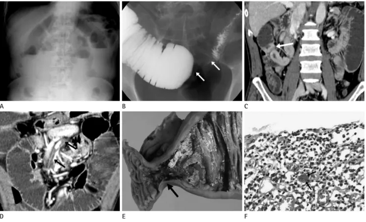
Fig. 1. Partial small bowel obstruction in a 51-year-old man.
A. A plain abdominal radiograph shows dilatated small bowel loop with air-fluid level suggesting partial mechanical ileus.
B. Small bowel follow-through shows focal luminal narrowing (white arrows) at jejunal loop and proximal bowel loop dilatation.
C. Coronal reformatted post contrast enhanced CT scan shows focal concentric luminal narrowing with somewhat shouldering at small bowel (white arrow).
D. Post contrast enhanced coronal reformatted CT image shows partial superior mesenteric artery thrombosis (black arrows).
E. Gross specimen shows short segmental stricture at small bowel loop without ischemic change (black arrow).
F. Histologic specimen shows cytomegalic cells with intranuclear inclusion (black arrows), confirmed cytomegalovirus enteritis (H&E stain, × 400).
E B
D A
F C
CMV Enteritis in Immunocompetent Patient Causing SBO and SMA Thrombosis
28
J Korean Soc Radiol 2014;71(1):26-29 jksronline.orginduced vasculopathy and thrombosis are rare conditions. The few published reports on these conditions focus either on immu- nocompromised transplant recipients who are receiving high- dose immunosuppressive agents or on HIV-infected patients (9).
There are few reports involving immunocompetent adults in whom no hemostatic abnormalities can be found similar to our patient (10). Our case showed a CMV enteritis, presenting as a small bowel obstruction accompanied by SMA thrombosis in an immunocompetent individual with no underlying disease.
Regarding the superior mesenteric artery thrombosis, recent studies showed that the cytomegalovirus can accelerate a vascular disease by inflammation and smooth muscle cell migration from the vessel media to the intima and by proliferation that culmi- nates in vessel narrowing. In our case, distal SMA different from proximal SMA thrombosis was opacified on the contrast en- hanced CT and we hypothesized the SMA thrombosis occurred at a significant time point before so that peripheral branches could be retrogradely perfused by collaterals. Although our treatment options could include antibiotics or heparinization, we performed a segmental resection of the small bowel to rule out the possibility of malignancy. The histology confirmed the CMV infection. The significance of SMA thrombosis was not fully evaluated in this study, but we supposed the SMA vascu- lopathy as a result of the CMV infection.
In conclusion, we experienced an extremely rare case with CMV small bowel involvement and SMA at the same time. It was successfully treated with surgical intervention and compli- mentary antiviral medication.
REFERENCES
1. Sakamoto I, Shirai T, Kamide T, Igarashi M, Koike J, Ito A, et al. Cytomegalovirus enterocolitis in an immunocompe- tent individual. J Clin Gastroenterol 2002;34:243-246 2. Grundy JE. Virologic and pathogenetic aspects of cytomeg-
alovirus infection. Rev Infect Dis 1990;12 Suppl 7:S711- S719
3. Keates J, Lagahee S, Crilley P, Haber M, Kowalski T. CMV enteritis causing segmental ischemia and massive intesti- nal hemorrhage. Gastrointest Endosc 2001;53:355-359 4. Chae EY, Lee SS, Chung JW, Kim HJ, Park SH, Kim AY, et al.
Cytomegalovirus enterocolitis in apparently immunocom- subjects. The involvement of the gastrointestinal tract is a relative-
ly common manifestation of a tissue-invasive CMV disease (1).
CMV can cause lesions throughout the gastrointestinal tract from the mouth to the anus. The lesions are various, but ulcer- ative lesions causing mainly colitis are most common. Other pathological lesions caused by CMV infection of the gastroin- testinal tract could be perforations, hemorrhagic proctocolitis, inflammatory pseudotumor, appendicitis, toxic megacolon or pneumatosis intestinalis. Less than 10% of the CMV gastroen- teritis cases show small bowel gastroenteritis (2, 3). In an immu- nocompetent patient, CMV enteritis can cause symptoms like in an immunocompromised host. A focal small bowel stenosis like in the presented case is a rare finding of CMV enteritis.
In one study were the CT findings of CMV enterocolitis in immunocompetent hosts similar to those reported in patients with acquired immunodeficiency syndrome (4). The most com- mon CT finding of CMV enterocolitis was segmental and con- centric mural thickening involving the colon was present in most patients and the small bowel was involved in approximately 30%. Isolated enteritis was noted in one patient of 12 patients and a focal involvement of rectum was seen in one case also. A stenosing and ulcerative CMV infection of the colon resembled a neoplasm clinically, macroscopically and radiologically. A case of gastric antral obstruction due to mass has also been described caused by CMV infection (5, 6).
We considered a malignant neoplasm as one of the possible differential diagnosis because of the focal involvement of the small bowel. However, there was no definite radiologic evidence of neoplasm such as mass-like lesion, enlargement of mesenteric lymph node or desmoplastic reaction. Adhesion could be a pos- sible diagnosis based on the fact that the lesion showed a dra- matic luminal narrowing with no visible cause. So, we put more weight on small bowel obstruction caused by inflammatory condition or post ischemic change.
Although the mechanism remains unclear, a causative associ- ation between CMV infection and thromboembolic disease has been proposed, most frequently described in the context of im- munosuppression after organ transplantation (7). The proposed mechanism in most instances is that of a thrombotic microangi- opathy. It is suggested that viruses are able to alter the phenotype of endothelium from anticoagulant to procoagulant, thus pro- moting the adhesion of neutrophils and platelets (8). But CMV-
Jong-Won Park, et al
29
jksronline.org J Korean Soc Radiol 2014;71(1):26-29
tation 1998;66:294-297
8. Vercellotti GM. Effects of viral activation of the vessel wall on inflammation and thrombosis. Blood Coagul Fibrinoly- sis 1998;9 Suppl 2:S3-S6
9. Koskinen PK, Nieminen MS, Krogerus LA, Lemström KB, Mattila SP, Häyry PJ, et al. Cytomegalovirus infection and accelerated cardiac allograft vasculopathy in human car- diac allografts. J Heart Lung Transplant 1993;12:724-729 10. Abgueguen P, Delbos V, Chennebault JM, Payan C, Pichard
E. Vascular thrombosis and acute cytomegalovirus infec- tion in immunocompetent patients: report of 2 cases and literature review. Clin Infect Dis 2003;36:E134-E139 petent hosts: evaluation of the radiologic findings and clin-
ical features. J Comput Assist Tomogr 2010;34:892-898 5. Lewis-Jones HG, Ward RG, Garvey C. Cytomegalovirus in-
fection masquerading as colonic neoplasia. Br J Radiol 1990;63:573-574
6. Elta G, Turnage R, Eckhauser FE, Agha F, Ross S. A submu- cosal antral mass caused by cytomegalovirus infection in a patient with acquired immunodeficiency syndrome. Am J Gastroenterol 1986;81:714-717
7. Madalosso C, de Souza NF Jr, Ilstrup DM, Wiesner RH, Krom RA. Cytomegalovirus and its association with hepat- ic artery thrombosis after liver transplantation. Transplan-
비면역저하 환자에서 소장폐색으로 발현된 거대세포 위장관염과 상장간막 동맥혈전증의 병발: 증례 보고
박종원 · 김경아 · 윤상욱 · 김대중
거대세포 바이러스(cytomegalovirus) 감염은 주로 면역저하 환자에서 일어나며 대부분의 장기에 이환 가능하나 식도부터 직장에 이르기까지 위장관계에 이환되어 위장관 출혈, 복막염, 혹은 심한 경우 위장관 천공까지 일으키는 것으로 알려져 있다. 최근 우리는 알려진 면역저하 원인이 없는 건강한 환자가 소장의 폐색과 상장간막 동맥의 혈전으로 내원하여 수술 적으로 치료한 결과 거대세포 위장관염으로 확진하였다. 면역저하가 없는 환자에서 소장의 거대세포 장염과 동맥혈전이 병발되는 것은 매우 드문 증례로 저자들이 아는 한 처음으로 보고하고자 한다.
CHA의과학대학교 분당차병원 영상의학과