submit.radiology.or.kr
대한영상의학회지 2012;67(1):13-1613 서론
Carotidynia는 편측의 경동맥 분기부 주변에 특발성의 다양 한 동통과 압통을 주소로 하는 증후군으로 1927년 Fay(1)에 의해 처음 기술되었다. 그러나 carotidynia를 어떻게 특징 지우 고 분류할 것인가에 대해서는 이견이 존재하지만, 최근에는 경 동맥 주변에 구조적 이상들이 보고되면서 독립된 질환으로 보 는 견해들이 많다. Carotidynia는 임상양상의 정확한 이해를 바 탕으로 대부분 문진과 이학적 검사 등을 통해 진단하지만, 경 부통을 증상으로 하는 다양한 질환들과의 감별이 어려운 경우 들이 있다. 경부통은 비특이적인 증상으로, 이를 호소하는 환자 에서 먼저 원인이 될 수 있는 좀더 흔한 질환들을 진단에서 제 외한 후 carotidynia를 진단할 수 있기 때문에, 반드시 영상검사 가 필요한 경우가 있다. 또한 경부통은 임상에서 자주 접하는 증상으로 영상검사 없이 간단한 검사와 증상에 대한 보존적 치 료만을 시행하기 때문에 영상검사를 통하여 carotidynia를 경험 할 수 있는 기회는 드물다. 저자들은 임상소견과 더불어 다양한 영상검사들을 통해 진단된 carotidynia의 1예를 보고한다.
증례 보고
49세 남자 환자로 3일 전부터 시작된 인후통, 좌측 경부의 찌 릿한 통증 및 충만감, 두통 등을 주소로 내원하였다. 환자는 고 혈압과 뇌 경색의 과거력을 갖고 있는 것 외에 다른 특이 사항 은 없었다. 환자는 좌측 경동맥 분기부 주변에 심한 박동성 감 각과 통증을 호소하였고, 오한감이나 발열 등의 전신증상은 없 었다. 통증은 좌측 경동맥 분기부 주변을 누를 때 더욱 심화되 었고, 좌측 귀 부위까지 방사되는 양상을 보였다. 좌측 경부에 촉지되는 종물은 없었다. 후두경 소견을 포함한 이비인후과적 이상 소견은 보이지 않았다. 내원 당시의 백혈구 수치는 정상이 었지만, erythrocyte sedimentation rate (ESR) 32 mm/hr(정상 0~9 mm/hr), C-reactive protein (CRP) 6.46 mg/L(정상 0~3 mg/L)로 증가되어 있었다.
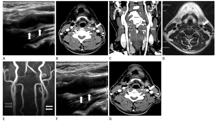
입원 당시 시행한 초음파 검사에서 좌측 총경동맥 말단 부위 와 경동맥 분기부, 내경동맥 근위부에 동맥벽의 바깥쪽으로 돌출하는 비후된 저에코음영이 보였고, 경동맥 내경은 약간 좁 아져 있었다(Fig. 1A). 같은 날 시행한 횡단면 CT 검사 및 3D
Case Report
pISSN 1738-2637
J Korean Soc Radiol 2012;67(1):13-16
Received March 27, 2012; Accepted May 15, 2012 Corresponding author: Heung Cheol Kim, MD Department of Radiology, Hallym University College of Medicine, Chuncheon Sacred Heart Hospital, 77 Sakju-ro, Chuncheon 200-704, Korea.
Tel. 82-33-240-5158 Fax. 82-33-242-7085 E-mail: [email protected]
Copyrights © 2012 The Korean Society of Radiology
Carotidynia is a rare disease representing various aspects of pain around the carotid artery bifurcation. The pain in the carotid bifurcation area is a nonspecific symptom, and various diseases causing similar symptoms in the corresponding area should be distinguished. Mostly, carotidynia is diagnosed by history taking and physical exam- ination, but it is not easy to distinguish carotidynia from other diseases without im- aging study. Therefore, there are quite a number of inadequate treatments due to a considerable number of misdiagnosis, as infectious diseases. In US, CT scans, and MRI examinations, the authors experienced a patient who showed the outer thick- ening wall of blood vessels around the carotid artery, near the carotid artery bifur- cation, and was diagnosed as carotidynia with clinical findings. In the follow-up that was carried out two weeks later, the thickness of the lesions was significantly decreased. Imaging studies are helpful in the differential diagnosis because caroti- dynia shows relatively characteristic imaging findings.
Index terms
Carotidynia Neck Pain Headache Carotid Artery Steroid TherapyThe Findings of Ultrasonography, CT and Magnetic Resonance Imaging of Carotidynia: A Case Report
Carotidynia의 초음파, CT와 자기공명영상 소견: 1예 보고
Jung Min Kim, MD, Heung Cheol Kim, MD
Department of Radiology, Hallym University College of Medicine, Chuncheon Sacred Heart Hospital, Chuncheon, Korea
Carotidynia의 초음파, CT와 자기공명영상 소견
submit.radiology.or.kr
대한영상의학회지 2012;67(1):13-16
14
고찰
Carotidynia는 Fay에 의해 경동맥 분기부 주변에 통증을 의 미하는 용어로 처음 사용되었고, 점차 독립된 질환으로 간주되 면서 1988년에 International Headache Society (이하 IHS)는 carotidynia에 대한 4가지의 진단기준을 마련하였었다(Table 1)(2). 그러나 Biousse와 Bousser (3)은 이러한 진단기준들에 대해 이견을 제시하면서 carotidynia에 대한 진단기준들과 일 치하지 않았던 다양한 임상증상들을 보고하였다. 이러한 견해 로 인해 2004년 IHS는 두통분류에서 carotidynia를 제외시켰 지만 저자들에 따라 carotidynia를 여러 원인에 의한 단순 경부 통으로 간주하는 경우와 특정 질환의 일종으로 보는 경우 등 논란은 지속되고 있다. 그러나 최근 여러 문헌들을 통해 carot- 재구성 조영증강 관상면 CT 영상에서 병변은 초음파와 동일한
범위를 침범하고 있었고, 경동맥 주변에 국한하는 저음영 병변 및 같은 부위에 경미한 조영증강 소견을 보였다. 병변은 혈관 외벽의 전내측이 다른 부위에 비해 좀더 두꺼워져 있었다(Fig.
1B, C). Magnetic resonance angiography (이하 MRA) 검사 에서는 CT에서 보이는 병변과 동일한 부위에 T2 강조영상에 서 경계가 잘 그려지는 고신호 강도를 보였다(Fig. 1D). MRA 에서는 병변부위의 총경동맥 및 내경동맥의 내경은 반대측에 비해 감소되어 보였다(Fig. 1E).
환자는 비스테로이드성 항염증약물로 보존적 치료를 시행하 였다. 입원 14일 후 시행한 CT와 초음파 검사에서 각각 경동맥 분기부 주변 병변의 두께는 현저히 감소하였고, 같은 시기에 환자의 임상증상도 함께 호전되었다(Fig. 1F, G).
E A
F B
G
C D
Fig. 1. A 49-year-old man with left neck pain with tenderness.
A. Initial ultrasonography show hypo-echoic thickening of outer wall surrounding the left distal common carotid artery and more prominent thickening in medial perivascular area (arrows). Because ultrasonographic scanning is conducted from lateral to medial direction of neck, the ar- rows indicating area correspond to medial perivascular region of distal common carotid artery.
B. Initial contrast-enhanced axial CT scan shows perivascular thickening with mild enhancement surrounding the left distal common carotid ar- tery (arrow).
C. Three-dimensional-reformated coronal image of initial contrast-enhanced CT shows mild luminal narrowing and perivascular thickening with mild enhancement surrounding the left distal common carotid artery and proximal internal carotid artery (arrows), compared to the opposite side (hollow arrows).
D. Initial axial T2-weighted image shows well marginated perivascular hight signal intensity surrounding the left distal common carotid artery (arrow).
E. On MRA image, there is mild luminal narrowing in affected artery (arrows), compared to the opposite side (hollow arrows). However, there is not evident as the CT findings.
F, G. Follow-up ultrasonography (F) and CT (G) after 2 weeks show marked resolution of abnormal perivascular thickening (arrows).
김정민 외
submit.radiology.or.kr
대한영상의학회지 2012;67(1):13-1615
경동맥 분기부 주변에 통증을 증상으로 하는 carotidynia는 드문 질환으로, 대부분 염증성 질환으로 오인하여 항생제 투여 등의 불필요한 치료를 하는 경우가 많다. 비슷한 증상을 유발 할 수 있는 다양한 질환들과 감별하기 위해서는 carotidynia의 임상적 양상에 대한 충분한 이해가 필요하며, 더불어 특징적인 영상소견이 진단에 도움이 될 것으로 보인다.
참고문헌
1. Fay T. Atypical neuralgia. Arch Neurol Psychiatry 1927;18:
309-315
2. Classification and diagnostic criteria for headache disor- ders, cranial neuralgias and facial pain. Headache Classifi- cation Committee of the International Headache Society.
Cephalalgia 1988;8 Suppl 7:1-96
3. Biousse V, Bousser MG. The myth of carotidynia. Neurolo- gy 1994;44:993-995
4. Upton PD, Smith JG, Charnock DR. Histologic confirmation of carotidynia. Otolaryngol Head Neck Surg 2003;129:443- 444
5. Burton BS, Syms MJ, Petermann GW, Burgess LP. MR im- aging of patients with carotidynia. AJNR Am J Neuroradiol 2000;21:766-769
6. Kuhn J, Harzheim A, Horz R, Bewermeyer H. MRI and ultra- sonographic imaging of a patient with carotidynia. Cepha- lalgia 2006;26:483-485
7. Kosaka N, Sagoh T, Uematsu H, Kimura H, Miyayama S, Noguchi M, et al. Imaging by multiple modalities of pa- tients with a carotidynia syndrome. Eur Radiol 2007;17:
2430-2433
8. Pipitone N, Versari A, Salvarani C. Role of imaging studies in the diagnosis and follow-up of large-vessel vasculitis:
an update. Rheumatology (Oxford) 2008;47:403-408 idynia의 특징적인 영상소견들과 일부 병리소견이 보고되면서,
경동맥 부위에 구조적 이상소견을 동반하는 특정질환으로 보 는 견해들이 늘고 있다(4-8). Upton 등(4)은 carotidynia로 진 단받은 환자의 조직검사에서 일반적인 혈관염과는 달리 경동 맥의 외막(adventitia)에 염증을 나타내는 특이소견을 보고함으 로써 위의 주장을 더욱 뒷받침하였다.
Carotidynia의 증상은 편측 경부에 경동맥 분기부를 중심으 로 인접한 총경동맥이나 내경동맥을 따라 간헐적 혹은 지속적 통증이 있고, 통증 부위에 압통, 팽대, 박동증가 등을 동반한다 (9). 환부에 압박으로 통증이 재현될 수 있고, 경동맥 주변 동 측 구조물 즉 귀, 인후 및 머리까지 방사통이 유발될 수 있다 (10). 인후통이 가장 흔한 증상이며 이통과 합쳐 전체 증상의 약 87%를 차지한다(10). 이러한 증상들은 비스테로이드성 항 염증약물이나 스테로이드에 의해 잘 반응하며, 대부분 2주 이 내에 소실되지만, 경우에 따라 몇 주에서 몇 달까지 지속될 수 있다(10). 진단은 대부분 병력청취와 이학적 검사만으로도 가 능할 수 있지만, 임상양상을 정확히 인식하고 있지 않으면 대부 분 오진하기 쉬우며, 결과적으로 부적절한 치료를 하게 될 가 능성이 높다. Emmanuelli 등(10)에 의하면 carotidynia로 진단 된 환자들에서 많은 수가(43.8%) 감염성 질환으로 오진하여 항생제 치료를 받았던 것으로 보고하였다. 환자의 대부분은 합 병증 없이 증상이 소실되지만, 일부에서는 반복적으로 수년간 지속될 수 있다(3, 10).
영상진단 방법으로는 이중초음파, 조영증강 CT 및 MRI 등 이 있다. Carotidynia의 이중초음파 소견은 경동맥 주위로 돌출 하는 양상의 저에코음영이 보이며, 경동맥의 내강은 변화가 없 거나 약간 감소하며 정상 도플러 파형을 보인다. CT와 MRI에 서 경동맥 주변에 비후가 관찰되는데, 이 부위에 조영 전 CT 검사에서는 저음영으로 나타나고, MRI 검사에서는 T2 강조영 상에서 고신호 강도를 보이며, 두 검사방법에서 모두 같은 부 위에 조영증강을 보인다. 추적 관찰에서 이러한 영상소견들은 임상증상의 호전과 함께 소실된다(5-7). 이러한 소견들은 각 각의 진단방법들에서 모두 병변이 경동맥 부근에만 제한되어 관찰되기 때문에 선택적으로 경동맥초(carotid sheath) 내에 국한된 병변임을 암시한다.
유사한 임상증상을 갖는 감별해야 할 질환들로는 경부 구조 물에 염증성 질환이나 종괴, 거대세포 동맥염, 혈관박리, 동맥 경화증 등과의 감별이 필요하다. 대부분의 질환들은 영상으로 쉽게 구별이 되며, 특히 혈관벽을 침범하는 동맥염과 동맥경화 증과의 구별에서 각각 환형과 편심성 혈관벽 비후를 보이며 혈 관 내경이 감소하는 반면 carotidynia는 혈관 주변의 비후를 보 이면서 내경의 감소는 없거나 경미한 것이 차이점이다(8).
Table 1. International Headache Society Classification Committee Cri- teria for the Diagnosis of Idiopathic Carotidynia (1st Edition, 1988)
A. At least one of the following overlying the carotid artery:
1. Tenderness 2. Swelling
3. Increased pulsations
B. Appropriate investigations not revealing structural abnormality C. Pain over the affected side of the neck; may project to the ipsilateral side of the head
D. A self-limiting syndrome of less than 2 weeks’ duration
Carotidynia의 초음파, CT와 자기공명영상 소견
submit.radiology.or.kr
대한영상의학회지 2012;67(1):13-16
16
Carotidynia: a frequently overlooked or misdiagnosed syn- drome. Ear Nose Throat J 1998;77:462-464, 466, 469 9. Stanbro M, Gray BH, Kellicut DC. Carotidynia: revisiting an
unfamiliar entity. Ann Vasc Surg 2011;25:1144-1153 10. Emmanuelli JL, Gutierrez JR, Chiossone JA, Chiossone E.
Carotidynia의 초음파, CT와 자기공명영상 소견: 1예 보고
김정민 · 김흥철
Carotidynia는 경동맥 분기부 주변으로 다양한 통증의 양상을 띠는 드문 질환이다. 경동맥 분기부의 통증은 비특이적인 증상이며, 같은 부위에 유사한 증상을 유발하는 다양한 질환들을 감별하여야 한다. Carotidynia는 대부분 문진과 이학적 검사를 통해 진단하지만, 영상검사 없이 다른 질환들과 감별이 쉽지 않다. 그러므로 상당수가 감염성 질환으로 오인하여 부적절한 치료를 하는 경우들이 있다. 초음파, CT, MRI 검사에서 저자들은 경동맥 분기부 근처에서 경동맥 주변으로 혈 관 외벽의 비후 소견을 보여 임상소견과 함께 carotidynia로 진단했던 환자 1예를 경험하였다. 2주 후 추적검사에서 병변 의 두께가 현저히 감소된 소견을 보였다. Carotidynia는 비교적 특징적인 영상소견을 보이므로 영상검사는 감별진단에 도 움이 된다.
한림대학교 의과대학 춘천성심병원 영상의학과학교실