INTRODUCTION
T-cell prolymphocytic leukemia (T-PLL) is an aggres- sive mature T-cell lymphoproliferative disorder (LPD) characterized by proliferation of small-to-medium pro- lymphocytes with a post-thymic T-cell phenotype, and most patients present with rapidly rising lymphocytosis,
hepatosplenomegaly, and generalized lymphadenopa- thy. 1-4 In 25% of cases, leukemic prolymphocytes are small and do not have a visible nucleolus under light microscopy, and are designated small cell variant T- PLL. 5,6
We describe the cytomorphology of ascitic fluid complicated by small cell variant T-PLL, with an
복수를 침범한 소세포형 T-세포 전림프구성 백혈병의 세포소견
-1예 보고-
인하대학교 의과대학 병리학교실, 내과학교실 혈액종양분과1, 단국대학교 병원 병리과2
한 지 영·김 진 수
1·김 동 훈
2·김루시아·박 인 서·김 준 미·주 영 채·최 석 진
T-cell prolymphocytic leukemia (T-PLL) is a rare, mature T-cell lym- phoproliferative disorder with a post-thymic mature T-cell phenotype.
The disease is characterized by rapidly rising lymphocytosis, lym- phadenopathy, and splenomegaly. The clinical course is usually aggressive and progresses with frequent skin lesions and serous effu- sions. In 25% of cases, leukemic cells are small and tumor cells may not have a discrete nucleolus under light microscopy. Although the presence of characteristic cytoplasmic protrusions or blebs in tumor cells is a common morphologic finding in the peripheral blood film irre- spective of the nuclear features, small cell variants lacking the typical nuclear features can cause diagnostic problems in clinical cytology.
Furthermore, the small leukemic cells can share some cytologic find- ings with lymphocyte-rich serous effusions caused by non-neoplastic reactive lymphocytosis as well as other small lymphocytic lymphoprolif- erative disorders. Here, we describe the cytological findings of ascitic fluid complicated by small cell variant T-PLL in a 54-year-old man, the cytology of which was initially interpreted as small lymphocytic malig- nancy such as small lymphocytic lymphoma/chronic lymphocytic leukemia.
(Korean J Cytopathol 2008;19(2):168-172) Key Words : Leukemia, Prolymphocytic, T-cell, Cytology, Ascitic fluid
Cytologic Features of Ascitic Fluid Complicated by Small Cell Variant T-cell Prolymphocytic Leukemia -A Case Report-
Jee Young Han, M.D., Jin Soo Kim, M.D.,
1Dong Hoon Kim, M.D.,
2Lucia Kim, M.D., In Suh Park, M.D., Joon Mee Kim, M.D., Young Chae Chu, M.D., and Suk Jin Choi, M.D.
Department of Pathology and Hemato-oncology
1division in Department of Internal Medicine, Inha University Hospital and College of Medicine, Incheon
Department of Pathology, Dankook University Hospital, Cheonan, Korea
2논문접수 : 2008년 7월 17일 논문수정 : 2008년 7월 31일 게재승인 : 2008년 8월 29일
책임저자 : 최 석 진
주 소 : (400-711) 인천시 중구 신흥동 3가 7-206 인하대학교병원 병리과
전 화 : 032-890-3983 팩 스 : 032-890-3464
E-mail address : [email protected]
*Acknowledgments: This work was supported by Inha University Research Grant.
증 례
10.3338/kjc.2008.19.2.168
emphasis on differential cytodiagnosis of ascitic fluid showing aberrant small lymphocytes.
CASE
Clinical Presentation
A 54 year-old male was admitted to the hospital for evaluation of a leukocytosis of 25500×10 9 /L, with 71.3% lymphocytes accompanied by ascites, multiple lymphadenopathy, and hepatosplenomegaly. His leukocytosis started 2 months earlier, when he began to complain of facial edema and occasional febrile sensa- tions. Serum LDH was 1189 IU/L. Laboratory tests for viral hepatitis, human immunodeficiency virus (HIV), and human T-cell leukemia virus-1 (HTLV-1) were neg- ative. Peripheral blood film showed an increased num- ber of small-to-medium lymphocytes with condensed chromatin and no visible nucleoli, suggesting chronic lymphocytic leukemia. Straw-colored ascitic fluid was received for cytological evaluation, and diagnosed as small lymphocytic malignancy. Subsequently, diagnos- tic work-up was performed along with flow cytometry of the bone marrow and an excision biopsy of the inguinal lymph node.
Cytologic Findings in Ascitic Fluid
Smears were markedly cellular with non-cohesive, small, mature lymphocytes in a clean background, with a small number of reactive mesothelial cells (Fig. 1A).
In contrast to reactive serous effusion, lymphoid cells in the smears were aberrantly monomorphic with irregular nuclear membranes. Many had a vesicular nucleus with a vague convolution and marginalization of chromatin.
Characteristically, some of them exhibited prominent nucleoli and asymmetrical cytoplasmic elongations (Fig.
1B). Cytoplasmic granules or vacuoles were not observed. On the Giemsa-stained smears of the ascitic fluid, most of the lymphoid cells appeared to have eccentric cytoplasmic protrusions or blebs with hand- mirror morphology (Fig. 2). Based on these cytologic findings, a diagnosis of small mature lymphocytic malignancy was given.
Pathologic Findings in Bone Marrow and the Inguinal Lymph Node
More than 40% of the nucleated elements in the bone marrow smear were small lymphocytes with clumped chromatin and scanty basophilic cytoplasm.
Flowcytometric evaluation of the bone marrow demon-
Fig. 1. Cytology of ascitic fluid: (A) The smear is moderately cellular with monomorphous population of small lymphocytes in clear background with a small number of reactive mesothelial cells. (B) Some lymphoid cells exhibit prominent nucleoli and asymmetri- cal cytoplasmic elongations. (Papanicolaou stain).
A B
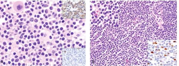
strated an aberrant T-cell population. Histological sec- tions of the bone marrow biopsy showed diffuse inter- stitial proliferation of CD3+ small lymphocytes (Figure 3A). Individual lymphocytes had irregular nuclear con- tours with occasional convolutions and small nucleoli, with CD4+ and CD8- phenotypes. They were positive for CD2, CD3, CD4, CD5, and CD7, but negative for CD8, CD10, CD13, CD19, CD20, CD23, CD33 and TdT.
The excised inguinal node showed effacement of
nodal architecture with marked expansion of interfollic- ular areas infiltrated by monomorphic populations of small mature T-cells with irregular nuclear contours and occasional small nucleoli. Prominent high endothelial venules were frequently observed and often infiltrated by neoplastic cells (Fig. 3B). Mitotic or apoptotic figures were not conspicuous, and the Ki-67 labeling index was about 30∼40%. Essentially all infiltrating small lymphoid cells showed diffuse positive staining for CD4 and CD5. They were negative for CD8, CD30, cyclin D1, and ALK1. CD56 immunostaining was patchy and weak in tumor cells (about 5%).
EBER was negative on in situ hybridization per- formed in histological sections of the bone marrow and inguinal lymph node. PCR analysis of the inguinal lymph node revealed monoclonal rearrangement of T- cell receptor (TCR) gamma chain. PCR analyses for HTLV-1 using histological sections of the inguinal lymph node were negative.
DISCUSSION
The leukemic cells of T-PLL are slightly larger than normal lymphocytes, and are characterized by a central
Fig. 2. Giemsa-stained smear of ascitic fluid: Individual lym- phocyte shows eccentric cytoplasmic protrusion or blebs giv- ing the appearance of so called hand-mirror morphology (Giemsa).
Fig. 3. Bone marrow and inguinal lymph node biopsy: (A) The bone marrow biopsy demonstrates lymphoid cells showing nuclear atypical and irregular nuclear contour with occasional convolution and small nucleolus. They are positive for CD3 and negative for CD20 (upper inset, CD3; lower inset, CD20). (B) The inguinal lymph node is diffusely infiltrated by small-sized monomorphic lym- phocytes with small amount of pale cytoplasm with 30~40% of Ki-67 labeling index (right lower inset, Ki67). (H&E, inset; immuno- histochemical stain).