ISSN 2234-3806 • eISSN 2234-3814
https://doi.org/10.3343/alm.2017.37.5.443 www.annlabmed.org 443
Ann Lab Med 2017;37:443-445
https://doi.org/10.3343/alm.2017.37.5.443
Letter to the Editor
Diagnostic Hematology
Hidden “Preleukemic Phase” of Chronic Myeloid Leukemia Presenting Without Leukocytosis in the Peripheral Blood Unrelated to Chemotherapy in a Patient Diagnosed With Diffuse Large B Cell
Lymphoma
Dong-Won Yoo, M.D.1, Sang Hyuk Park, M.D.2, Jongyoun Yi, M.D.3, In-Suk Kim, M.D.1, Hyung-Hoi Kim, M.D.3, Chulhun L. Chang, M.D.1, and Eun Yup Lee, M.D.3
Department of Laboratory Medicine1, Pusan National University School of Medicine, Pusan National University Yangsan Hospital, Yangsan; Department of Laboratory Medicine2, Ulsan University Hospital, University of Ulsan College of Medicine, Ulsan; Department of Laboratory Medicine3, Pusan National University School of Medicine, Pusan National University Hospital, Busan, Korea
Dear Editor,
Almost all cases of CML show typical clinical features such as marked leukocytosis in the peripheral blood (PB), splenomeg- aly, and granulocytic hyperplasia in the PB and bone marrow (BM). CML without leukocytosis in the PB is rare but could oc- cur if “undiagnosed” CML patients received cytoreductive ther- apy due to the patient’s predisposing hematologic malignancies.
In contrast, the presence of the BCR-ABL1 mutation without the clinical features of CML and unrelated to cytoreductive therapy (so-called “preleukemic phase” or “smoldering” CML) is very rare [1-3]. Here, we report a rare case of hidden preleukemic phase of CML presenting without leukocytosis in the PB unre- lated to prior chemotherapy in a diffuse large B cell lymphoma (DLBCL) patient. To our knowledge, this is the first case of pre- leukemic phase of CML in the Korean population.
A 67-yr-old man was admitted owing to abdominal pain in April 2016. At admission, he exhibited multiple lymphadenopa-
thy and was diagnosed as having DLBCL after right supraclavic- ular lymph node biopsy. His hemogram results at admission were as follows: white blood cell count, 8.98×10⁹/L; hemoglobin, 15.2
×10 g/L; platelets, 2.04×10¹²/L; segmented neutrophils, 71.7%;
lymphocytes, 10.5%; monocytes, 12.6%; eosinophils, 1.0%; and basophils, 4.2%. PB smear showed normal granulocytic matu- ration pattern without atypical lymphocytes or immature granu- locytes (Fig. 1A). The patient’s BM aspirates and touch print re- sults showed slightly hypercellular marrow with mild granulocytic hyperplasia (myeloid:erythroid ratio=6.93:1, and differential co- unts as follows: myeloblasts, 2.0%; promyelocytes, 2.4%; my- elocytes, 14.8%; metamyelocytes, 14.8%; band neutrophils, 15.9%; segmented neutrophils, 19.3%; eosinophils, 2.4%; and basophils, 0.0%) without atypical lymphocytes (Fig. 1B and 1C).
The BM biopsy section showed 60% cellularity with mild granu- locytic hyperplasia, eosinophilia, and megakaryocytic hyperpla- sia (4.5/high power field) without evidence of neoplastic lymphoid
Received: December 19, 2016 Revision received: March 1, 2017 Accepted: May 23, 2017
Corresponding author: Sang Hyuk Park
Department of Laboratory Medicine, Ulsan University Hospital, University of Ulsan College of Medicine, 877 Bangeojin Sunhwan-doro, Dong-gu, Ulsan 44033, Korea
Tel: +82-10-4422-4308, Fax: +82-52-250-8269 E-mail: [email protected]
© Korean Society for Laboratory Medicine.
This is an Open Access article distributed under the terms of the Creative Commons Attribution Non-Commercial License (http://creativecommons.org/licenses/by-nc/4.0) which permits unrestricted non-commercial use, distribution, and reproduction in any medium, provided the original work is properly cited.
1 / 1 CROSSMARK_logo_3_Test
2017-03-16 https://crossmark-cdn.crossref.org/widget/v2.0/logos/CROSSMARK_Color_square.svg
Yoo D-W, et al.
Preleukemic phase of CML in a DLBCL patient
444 www.annlabmed.org https://doi.org/10.3343/alm.2017.37.5.443 Fig. 1. Peripheral blood smear, bone marrow aspiration, touch print, biopsy, and immunohistochemical staining results of the patient at ini- tial diagnosis. (A) Peripheral blood smear results of the patient demonstrating the absence of leukocytosis, atypical lymphocytes or imma- ture granulocytes (400×, Wright stain). (B, C) Bone marrow aspiration smear and touch print indicating slightly hypercellular marrow with mild granulocytic and megakaryocytic hyperplasia without atypical lymphocytes (B, 200×; C, 400×; Wright stain). (D) Bone marrow biopsy section demonstrating approximately 60% cellularity with mild granulocytic hyperplasia, eosinophilia, and megakaryocytic hyperplasia with- out evidence of neoplastic lymphoid cell infiltration (40×, hematoxylin & eosin stain). (E, F) Immunohistochemical stain results of CD45RO (200×) and CD20 (200×) demonstrating the infiltration of a few reactive T and B lymphocytes, respectively.
A
C
E
B
D
F
Yoo D-W, et al.
Preleukemic phase of CML in a DLBCL patient
https://doi.org/10.3343/alm.2017.37.5.443 www.annlabmed.org 445
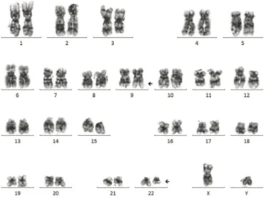
cell infiltration (Fig. 1D), demonstrated by the CD45RO and CD20 immunohistochemical stain results (Fig. 1E and 1F). Based on these findings, the initial diagnosis was given as (1) no evidence of BM involvement of DLBCL, and (2) mild granulocytic and mega- karyocytic hyperplasia, suggestive of reactive changes. However, karyotyping results were 46,XY,t(9;22)(q34;q11.2)[19]/46,XY[1]
(Fig. 2) and major BCR/ABL1 reverse transcriptase PCR dem- onstrated the presence of major BCR/ABL1 fusion transcript.
From these results, the final diagnosis was revised as preleuke- mic phase of CML without leukocytosis in the PB, unrelated to prior chemotherapy associated with initial diagnosis of DLBCL.
After the final diagnosis, the patient received R-CHOP chemo- therapy plus imatinib to manage both DLBCL and preleukemic phase of CML. In the follow-up examinations performed six months after diagnosis, the patient’s karyotype returned to normal and at present, the patient is scheduled to undergo 12 months fol- low-up examinations after diagnosis.
Although previous studies have reported a latency period of several years between the detection of the t(9;22)(q34;q11.2) and the development of CML clinical features [1-3], hidden pre- leukemic phase of CML in patients without a history of cytore- ductive therapy is still considered rare. CML without leukocyto- sis in the PB can occur if true CML patients received cytoreduc- tive therapy related to other hematologic malignancies, but these
patients should not be regarded as preleukemic phase of CML because they would have typical CML symptoms. A recent study tried to identify clues that may be helpful in detecting preleuke- mic phase of CML in asymptomatic patients and reported the presence of peripheral basophilia (57% of cases), and increased number of small, hypolobulated megakaryocytes in BM, which may distinguish preleukemic CML from other leukemoid reac- tions [3]. Our present case also showed peripheral basophilia and mild megakaryocytic hyperplasia with small-sized, hypolob- ulated forms in BM biopsy, which would support results from the previous study [3]. A likely explanation for the preleukemic phase of CML is that BCR-ABL1 occurs in multipotent stem cells and is considered an oncogenic driver mutation. BCR-ABL1 con- fers a growth advantage over normal cells, and massive expan- sion of BCR-ABL1-positive cells facilitates the acquisition of ad- ditional mutations leading to the development of overt CML [3].
Currently, there are no guidelines for the treatment of patients with preleukemic phase of CML. Since the development of pre- leukemic CML cells into overt leukemia would be associated with an increase in BCR-ABL1 levels, tyrosine kinase inhibitor (TKI) therapy may delay the transformation to overt CML in these pa- tients, and this hypothesis is supported by a previous study [3].
Our patient was also treated with TKI and the effect was success- ful. More follow-up results would add some clues about the effi- cacy of TKI treatment in cases of preleukemic CML.
Authors’ Disclosures of Potential Conflicts of Interest
No potential conflicts of interest relevant to this article were re- ported.
REFERENCES
1. Hudnall SD, Northup J, Panova N, Suleman K, Velagaleti G. Prolonged preleukemic phase of chronic myelogenous leukemia. Exp Mol Pathol 2007;83:484-9.
2. Bennett JM, Dsouza KG, Patel M, O’Dwyer K. “Preleukemic or smolder- ing” chronic myelogenous leukemia (CML): BCR-ABL1 positive: A brief case report. Leuk Res Rep 2014;4:12-4.
3. Aye le L, Loghavi S, Young KH, Siddiqi I, Yin CC, Routbort MJ, et al. Pre- leukemic phase of chronic myelogenous leukemia: morphologic and immunohistochemical characterization of 7 cases. Ann Diagn Pathol 2016;21:53-8.
Fig. 2. Karyotype analysis results of the patient after initial evaluation of the peripheral blood smear and bone marrow examinations. Karyo- type analysis results indicate the presence of the t(9;22)(q34;q11.2) in 19 of 20 cells evaluated.