ISSN 2234-3806 • eISSN 2234-3814
196 www.annlabmed.org http://dx.doi.org/10.3343/alm.2013.33.3.196 Ann Lab Med 2013;33:196-199
http://dx.doi.org/10.3343/alm.2013.33.3.196
Case Report
Diagnostic Hematology
A Case of CD4 + T-Cell Large Granular Lymphocytic Leukemia
Jaewook Kim, M.D.1, Chan Jeoung Park, M.D.1, Seongsoo Jang, M.D.1, Young-Uk Cho, M.D.1, Sang Hyuk Park, M.D.1, Eul-Ju Seo, M.D.1, Hyun-Sook Chi, M.D.1, and Cheolwon Suh, M.D.2
Departments of Laboratory Medicine1 and Internal Medicine2, University of Ulsan College of Medicine, Asan Medical Center, Seoul, Korea
We report here a case of a 59-yr-old man with CD4+ T-cell large granular lymphocytic leu- kemia (T-LGL). Peripheral blood examination indicated leukocytosis (45×109 cells/L) that consisted of 34% neoplastic lymphoid cells. Other laboratory results indicated no specific abnormalities except for serum antinuclear antibody titer (1:640), glucose (1.39 g/L), and hemoglobin A1c (7.7%) levels. Computed tomography indicated multiple small enlarged lymph nodes (<1 cm in diameter) in both the axillary and inguinal areas, a cutaneous nodule (1.5 cm in diameter) in the left suboccipital area, and mild hepatosplenomegaly.
Bone marrow examination revealed hypercellular marrow that consisted of 2.4% neoplas- tic lymphoid cells. The neoplastic lymphoid cells exhibited a medium size, irregularly shaped nuclei, a moderate amount of cytoplasm, and large granules in the cytoplasm. Im- munohistochemical analysis indicated CD3+, CD4+, T-cell receptor βF1+, granzyme B+, and TIA1+. Flow cytometric analysis of the neoplastic lymphoid cells revealed CD3+, cytoplas- mic CD3+, CD4+, and CD7+. Cytogenetic analysis indicated an abnormal karyotype of 46,XY,inv(3)(p21q27),t(12;17)(q24.1;q21),del(13)(q14q22)[2]/46,XY[28]. The patient was diagnosed with CD4+ T-LGL and received chemotherapy (10.0 mg methotrexate). This is the second case of CD4+ T-LGL that has been reported in Korea.
Key Words: CD4+ T-LGL skin lesion, Leukocytosis
Received: June 27, 2012 Revision received: August 7, 2012 Accepted: November 20, 2012
Corresponding author: Chan Jeoung Park Department of Laboratory Medicine, Asan Medical Center, University of Ulsan College of Medicine, 86 Asanbyeongwon-gil, Songpa-gu, Seoul 138-736, Korea Tel: +82-2-3010-4508
Fax: +82-2-478-0884 E-mail: [email protected]
© The Korean Society for Laboratory Medicine.
This is an Open Access article distributed under the terms of the Creative Commons Attribution Non-Commercial License (http://creativecom- mons.org/licenses/by-nc/3.0) which permits unrestricted non-commercial use, distribution, and reproduction in any medium, provided the original work is properly cited.
INTRODUCTION
T-cell large granular lymphocytic leukemia (T-LGL) is a hetero- geneous disorder that is characterized by the expansion of a discrete or monoclonal population of large granular lymphocytes in the peripheral blood (PB) [1].
T-LGL usually expresses CD3, CD8, and T-cell receptor (TCR) α/β. CD5 and/or CD7 are variably expressed and are often aber- rantly diminished on malignant circulating LGL cells [2, 3]. T-LGL typically expresses cytotoxic granular proteins such as TIA1, gran- zyme B, and granzyme M [4, 5]. Immunohistochemical analysis of bone marrow (BM) biopsies with antibodies to these antigens
and CD8 can be used to confirm a diagnosis of T-LGL [4-6].
The clinical course of T-LGL is indolent in most cases [7].
CD8+ T-LGL is associated with mild to moderately stable lym- phocytosis, neutropenia, splenomegaly, and occasionally ane- mia [8]. Lymphadenopathy is very rare [9]. In addition, T-LGL demonstrates a strong association with autoimmune diseases, especially rheumatoid arthritis [8].
In contrast, the monoclonal expansion of CD4+ T-LGL has been reported only sporadically in the literature [7]. It is marked by its association with malignant diseases and characteristically shows the absence of cytopenia, splenomegaly, and autoim- mune disease [7]. Here, we report a case of CD4+ T-LGL.
Kim J, et al.
CD4+ T-LGL
197
http://dx.doi.org/10.3343/alm.2013.33.3.196 www.annlabmed.org
CASE REPORT
A 59-yr-old man with a skin rash that had been present for 6 months was admitted to the hospital for an evaluation. He was diagnosed with hypertension and diabetes mellitus. PB exami- nation revealed the following: white blood cell count, 45×109
cells/L (consisting of 34% neoplastic lymphoid cells, 10% seg- mented neutrophils, 47% lymphocytes, 6% monocytes, and 3% eosinophils); hemoglobin, 131 g/L; mean corpuscular volume, 90.9 fL; and 419×109 platelets/L. Neoplastic lymphoid cells dis- played large granules (Fig. 1). PB neoplastic lymphoid cells were surface CD3+, cytoplasmic CD3+, CD4+, CD7+, CD8-, CD16-,
Fig. 1. Neoplastic lymphoid cells. (A) The neoplastic lymphoid cells with large cytoplasmic granules in the peripheral blood (Wright-Giemsa stain, ×1,000). (B) The neoplastic lymphoid cells in bone marrow aspirates with a medium, irregularly shaped nuclei, a moderate amount of cytoplasm, and large cytoplasmic granules (Wright-Giemsa stain, ×1,000).
A B
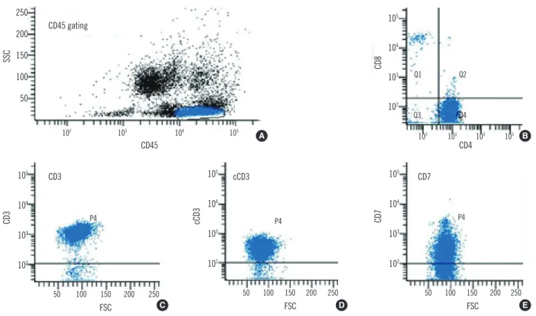
Fig. 2. Immunophenotyping of neoplastic lymphoid cells in peripheral blood by flow cytometry. (A) Gating of neoplastic lymphoid cells with bright CD45 expression and low SSC, (B) CD4 positivity (96% among gated cells) and CD8 negativity, (C) surface CD3 positivity (95%), (D) cytoplasmic CD3 positivity (93%), and (E) CD7 positivity (73%).
Abbreviations: SSC, side scatter characteristics; FSC, forward scatter characteristics.
102 103 104
CD45 CD45 gating
SSC
250 200 150 100 50
105 102 103 104
CD4
CD8
105
104
103
102
105 Q2
Q1
Q4 Q3
A B
50 50 50
P4 P4 P4
100
100 150 200 250 100 150 200 250 150 200 250
FSC FSC FSC
CD7
cCD3
CD3
105
104
103
102 105
104
103
102 105
104
103
102
C D E
CD3 cCD3 CD7
Kim J, et al.
CD4+ T-LGL
198 www.annlabmed.org http://dx.doi.org/10.3343/alm.2013.33.3.196 CD19-, CD20-, and CD56- (Fig. 2). Other laboratory results in-
cluded the following: serum antinuclear antibody titer, 1:640; glucose, 1.39 g/L; hemoglobin A1c, 7.7%; total protein/albumin, 7.1/3.9 g/dL; AST/ ALT, 12/12 IU/L; and total bilirubin, 5 g/L.
Multiple small enlarged lymph nodes ( <1 cm in diameter) in both the inguinal and axillary areas, and mild hepatospleno- megaly were noted on the abdominal and pelvic computed to- mography (CT) scans. A cutaneous nodule (1.5 cm in diameter) was also observed in the left suboccipital area, but this seemed to be a reactive enlargement of the lymph nodes. BM study re- vealed hypercellular marrow that consisted of 2.4% neoplastic lymphoid cells. The neoplastic lymphoid cells exhibited a me- dium size, irregularly shaped nuclei, a moderate amount of cy- toplasm, and large granules in the cytoplasm (Fig. 1). Immuno- histochemical analysis of the BM biopsy showed CD3+, CD4+, TCR βF1+, granzyme B+, and TIA1+ (Fig. 3). TCR γ gene rear- rangement by BIOMED-2 PCR assays (InVivoScribe, San Diego, CA, USA) was negative. Cytogenetic analysis indicated an ab- normal karyotype: 46,XY,inv(3)(p21q27),t(12;17)(q24.1;q21), del(13)(q14q22)[2]/46,XY[28]. The patient was diagnosed with CD4+ T-LGL and received chemotherapy (10.0 mg methotrexate/
week for 4 months). After the treatment, PB examination indi- cated the following values: white blood cell count, 24×109 cells/
L with 25% neoplastic lymphoid cells; hemoglobin, 137 g/L; and
395×109 platelets/L. The patient tolerated the treatment well, and his skin lesions improved.
DISCUSSION
T-LGL represents 2% of the cases of monoclonal proliferation of B cell, T cell, and natural killer cell mature lymphocytic leuke- mia in Western countries [7, 9]. The male:female ratio of the re- ported cases is approximately one, and the majority of cases occur in the 38-72 yr age group [9, 10]. The patient described here was a 59-yr-old man.
Despite their indolent clinical behavior, clear clinical differ- ences exist between CD4+ T-LGL and the classical CD8+ T-LGL, particularly with regard to the absence of neutropenia, anemia, splenomegaly, rheumatoid arthritis, or other autoimmune dis- eases, and the higher incidence of association with malignant diseases in the former group [7, 11]. In our case, neutropenia, anemia, rheumatoid arthritis, and other autoimmune diseases were absent. Although the patient demonstrated mild spleno- megaly and was not diagnosed with any associated malignant diseases, our results support the notion that CD4+ T-LGL is a clonal disorder with clinicopathological characteristics that are distinct from the more common CD8+ T-LGL.
Only one case of CD4+ T-LGL displaying skin lesions has been
Fig. 3. Immunohistochemical findings in the bone marrow biopsy. (A) CD3+, (B) CD4+, (C) CD8-, (D) T-cell receptor βF1+, and (E) gran- zyme B+ (immunohistochemical stain, ×400).
A B
E D
C
Kim J, et al.
CD4+ T-LGL
199
http://dx.doi.org/10.3343/alm.2013.33.3.196 www.annlabmed.org
reported so far [7]. In our case, the patient presented with a skin lesion, and this skin lesion demonstrated atypical T-cell in- filtration on skin biopsy. Generally, T-LGL involves the PB, BM, liver, and spleen, but our case supports the notion that CD4+ T- LGL can also involve the skin.
In our case, clonal T-cell proliferation was diagnosed after an- tinuclear antibodies were discovered, which suggests a role for the immune system in preferentially developing and expanding cytotoxic CD4+ T-cell clones. There are data that support the hy- pothesis that T-LGL arises from sustained immune stimulation and associated tumors [7, 9, 12, 13], but further studies are re- quired to establish both the relationship between CD4+ T-LGL and classical CD8+ T-LGL, and the clinicopathological character- istics that distinguish these leukemias.
In conclusion, in this report we described a 59-yr-old man with CD4+ T-LGL. To the best of our knowledge, only one case of CD4+ T-LGL has been reported in Korea so far [14], and ours is the second.
Authors’ Disclosures of Potential Conflicts of Interest
No potential conflicts of interest relevant to this article were re- ported.
REFERENCES
1. Semenzato G, Zambello R, Starkebaum G, Oshimi K, Loughran TP Jr.
The lymphoproliferative disease of granular lymphocytes: updated crite- ria for diagnosis. Blood 1997;89:256-60.
2. Morice WG, Kurtin PJ, Leibson PJ, Tefferi A, Hanson CA. Demonstra- tion of aberrant T-cell and natural killer-cell antigen expression in all cases of granular lymphocytic leukaemia. Br J Haematol 2003;120: 1026-36.
3. Lundell R, Hartung L, Hill S, Perkins SL, Bahler DW. T-cell large granu-
lar lymphocyte leukemias have multiple phenotypic abnormalities in- volving pan-T-cell antigens and receptors for MHC molecules. Am J Clin Pathol 2005;124:937-46.
4. Morice WG, Jevremovic D, Hanson CA. The expression of the novel cy- totoxic protein granzyme M by large granular lymphocytic leukaemias of both T-cell and NK-cell lineage: an unexpected finding with implications regarding the pathobiology of these disorders. Br J Haematol 2007;137: 237-9.
5. Morice WG, Kurtin PJ, Tefferi A, Hanson CA. Distinct bone marrow find- ings in T-cell granular lymphocytic leukemia revealed by paraffin sec- tion immunoperoxidase stains for CD8, TIA-1, and granzyme B. Blood 2002;99:268-74.
6. Osuji N, Beiske K, Randen U, Matutes E, Tjonnfjord G, Catovsky D, et al. Characteristic appearances of the bone marrow in T-cell large granu- lar lymphocyte leukaemia. Histopathology 2007;50:547-54.
7. Lima M, Almeida J, Dos Anjos Teixeira M, Alguero Md Mdel C, Santos AH, Balanzategui A, et al. TCRαβ+/CD4+ large granular lymphocytosis: a new clonal T-cell lymphoproliferative disorder. Am J Pathol 2003;163: 763-71.
8. Melenhorst JJ, Sorbara L, Kirby M, Hensel NF, Barrett AJ. Large granu- lar lymphocyte leukaemia is characterized by a clonal T-cell receptor re- arrangement in both memory and effector CD8(+) lymphocyte popula- tions. Br J Haematol 2001;112:189-94.
9. Swerdlow SH, Campo E, et al. eds. WHO classification of tumors of hae- matopoietic and lymphoid tissues. 4th ed. Lyon: IARC, 2008:272-3. 10. Olteanu H, Karandikar NJ, Eshoa C, Kroft SH. Laboratory findings in
CD4(+) large granular lymphocytoses. Int J Lab Hematol 2010;32:e9-16. 11. Garrido P, Ruiz-Cabello F, Bárcena P, Sandberg Y, Cantón J, Lima M, et
al. Monoclonal TCR-Vβ13.1+/CD4+/NKa+/CD8-/+dim T-LGL lymphocytosis:
evidence for an antigen-driven chronic T-cell stimulation origin. Blood 2007;109:4890-8.
12. Bigouret V, Hoffmann T, Arlettaz L, Villard J, Colonna M, Ticheli A, et al.
Monoclonal T-cell expansions in asymptomatic individuals and in pa- tients with large granular leukemia consist of cytotoxic effector T cells expressing the activating CD94:NKG2C/E and NKD2D killer cell recep- tors. Blood 2003;101:3198-204.
13. Wlodarski MW, O’Keefe C, Howe EC, Risitano AM, Rodriguez A, War- shawsky I, et al. Pathologic clonal cytotoxic T-cell responses: nonran- dom nature of the T-cell-receptor restriction in large granular lympho- cyte leukemia. Blood 2005;106:2769-80.
14. Song SH, Chung SM, Hwang DW, Choi JY, Yoo KD, Hong HS, et al. T- cell large granular lymphocytic leukemia: A case report. Korean J He- matol 2009;44:139-43.