INTRODUCTION
Primary non-Hodgkin’s lymphoma of bone (PLB) is a rare disease that accounts for less than 2 percent of all lym- phomas in adults (1). The histologies of the vast majority of cases present as diffuse large B-cell lymphoma, while T-cell lymphomas are extremely rare (2-10). Although there are a few publications regarding the characteristics, treatment options, and prognosis of PLB, the optimal management remains unclear. Recently, several reports have supported the combined modality of chemotherapy and radiotherapy (11-13). Multiple myeloma is a B-cell neoplasia that is usu- ally associated with osteolytic lesions and paraprotein in the plasma and/or in the urine (14-16). Because of the uniquely different cell lines of the two lymphoid malignancies, con- current disease has rarely been reported. To our knowledge, the simultaneous development of primary T-cell lymphoma of bone and multiple myeloma has never been reported. In a view of the same bone lesion in the case of PLB and multi- ple myeloma, the clinical discrimination of the two diseases is difficult. Through this case, we experienced the importance of reevaluation of disease regarding the possibility of other underlying diseases when it was deteriorated despite ade- quate treatment.
CASE REPORT
A 48-yr-old male presented with right ankle pain, mass, and general weakness over 2 months. On physical examina- tion, he appeared acutely ill but no other specific findings were noted.
The complete blood cell counts showed leucocytes 8.7×
109/L, hemoglobin 15.5 g/dL, and platelets 245×109/L.
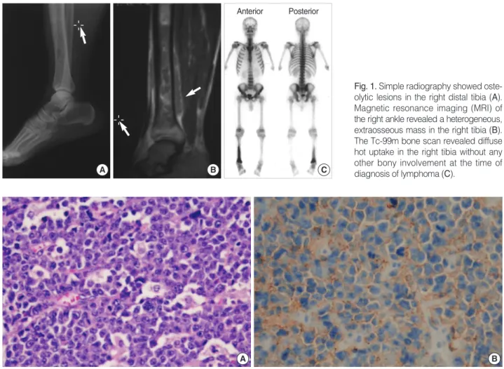
Blood chemistry showed total protein 6.6 g/dL, albumin 4.6 g/dL, lactate dehydrogenase 236 U/L, blood urea nitro- gen 14.2 mg/dL, and creatine 0.9 mg/dL. Urinalysis was normal. Simple radiography showed an osteolytic lesion in the right distal tibia. Magnetic resonance imaging (MRI) of the right ankle revealed a heterogeneous osteolytic and ext- raosseous mass in the right tibia (Fig. 1A, B). The bone biopsy showed diffuse infiltration of large tumor cells with vesicu- lar prominent nuclei, abundant cytoplasm, and numerous mitotic figures. Immunohistochemical staining showed an LCA, CD3, CD45RO, and CD15 phenotype (Fig. 2). These findings led to the diagnosis of a T-cell lymphoma. We then performed the staging work-up. Chest and abdomen com- puted tomography (CT) showed no abnormal findings such as lympadenopathy or hepatosplenomegaly. The Tc99m-methy- lene diphosphonate (MDP) bone scan revealed hot uptake in the right distal tibia without any other bony involvement
544
Jun-Eul Hwang, Sang-Hee Cho, Ok-Ki Kim, Hyun-Jeong Shim, Se-Ryeon Lee, Jae-Sook Ahn, Duk-Hwan Yang, Yeo-Kyeoung Kim, Je-Jung Lee, Hyeoung-Joon Kim, and Ik-Joo Chung
Department of Internal Medicine, Division of Hemato-Oncology, Chonnam National University Medical School, Gwangju, Korea
Address for correspondence Ik-Joo Chung, M.D.
Department of Internal Medicine, Chonnam National University Hwasun Hospital, 160 Ilsim-ri, Hwasun-eup, Hwasun, Jeollanam-do 519-809, Korea
Tel : +82.61-379-7626, Fax : +82.61-379-7628 E-mail : [email protected]
J Korean Med Sci 2008; 23: 544-7 ISSN 1011-8934
DOI: 10.3346/jkms.2008.23.3.544
Copyright � The Korean Academy of Medical Sciences
Newly Developed Multiple Myeloma in a Patient with Primary T-Cell Lymphoma of Bone
Primary non-Hodgkin’s lymphoma of bone (PLB) is rare, and generally presents as a single extensive and destructive bone lesion. Histopathologically, most cases present as diffuse large B-cell lymphoma, and T-cell lymphoma is rare. By contrast, multiple myeloma is a disease defined as the neoplastic proliferation of a single clone of plasma cells producing a monoclonal immunoglobulin. We report a case of multiple myeloma that developed during treatment of PLB in a type of T-cell. A 48-yr-old man was diagnosed as T-cell PLB, stage IE, 18 months ago. The patient received the chemoradiotherapy and salvage chemotherapy for PLB. However, the lymphoma progressed with generalized bone pain, and laboratory findings showed bicytopenia and acute renal failure. On bone marrow biopsy, the patient was diagnosed as having multiple myeloma newly developed with primary T-cell lymphoma of bone. In spite of chemotherapy, the patient died of renal failure.
Key Words : Lymphoma, Non-Hodgkin; Multiple Myeloma; T-Cell Lymphoma of Bone
Received : 20 April 2007 Accepted : 15 November 2007
Multiple Myeloma in a Primary Lymphoma of Bone 545
(Fig. 1C). The peripheral blood smear was normal, and the bone marrow biopsy showed no lymphoma involvement.
The patient was diagnosed as having primary T-cell lym- phoma of bone, stage IE. Based on the International Prog- nostic Index, the patient belonged to a low risk group (17).
The patient received 6 cycles of CHOP chemotherapy (cyclophosphamide 750 mg/m2 i.v. day 1, doxorubicin 50 mg/m2i.v. day 1, vincristine 1.4 mg/m2i.v. day 1, and pred- nisolone 100 mg p.o. days 1-5). Follow-up MRI showed a partial response. After addition of the involved-field radia- tion therapy up to 5,700 cGy as once daily fraction and two more cycles of chemotherapy, the tumor mass was further decreased, but still remained on MRI. F-18 fluorodeoxyglu- cose PET-CT showed the remaining hypermetabolic lesion on tibia, and also revealed hot uptake at the inferior ramus of the right pubis (Fig. 3A). We thought that the disease was progressive and treated the patient with salvage chemother- apy including ICE regimen (ifosfamide 5 g/m2i.v. day 2, and carboplatin AUC of 5 i.v. day 2, and etoposide 100 mg/
m2 i.v. days 1-3) and the DHAP regimen (dexamethasone 40 mg i.v. days 1-4, cytarabine 2 g/m2i.v. day 2, and cisplatin 100 mg/m2i.v. D1), but the disease further progressed. The patient received the MINE regimen (ifosfamide 1,333 mg/
Fig. 1. Simple radiography showed oste- olytic lesions in the right distal tibia (A).
Magnetic resonance imaging (MRI) of the right ankle revealed a heterogeneous, extraosseous mass in the right tibia (B).
The Tc-99m bone scan revealed diffuse hot uptake in the right tibia without any other bony involvement at the time of diagnosis of lymphoma (C).
A B C
Fig. 2. Diffuse infiltration of large tumor cells with vesicular prominent nucleoli, abundant cytoplasm, and numerous mitotic cells (A, H&E,
×200). Immunohistochemical staining for CD45RO showed positive (B, ×200).
A B
Fig. 3. After chemoradiotherapy, F-18 FDG PET-CT showed the residual hypermetabolic lesion on tibia, but also revealed hot up- takes at the inferior ramus of the right pubis (A). Despite salvage chemotherapy, F-18 FDG PET-CT revealed diffuse hot uptake in a skeletal area (B).
A B
Anterior Posterior
S19 S7
546 J.-E. Hwang, S.-H. Cho, O.-K. Kim, et al.
m2i.v. days 1-3, mitoxanthrone 8 mg/m2i.v. day 1, and etopo- side 65 mg/m2i.v. days 1-3) as the fourth line chemothera- py. After three cycles of MINE chemotherapy, restaging work-up showed a partial response. However, at the time of the fourth chemotherapy cycle, he complained of generalized bone pain. Complete blood cell counts revealed bicytopenia (leucocytes 4.5×10⁹/L, hemoglobin 8.8 g/dL, platelets 85×
109/L), and the peripheral blood smear showed atypical lym- phocytes (2%). Serum creatine was 2.0 mg/dL. Whole body PET revealed multiple hot uptake in the skeletal area (Fig.
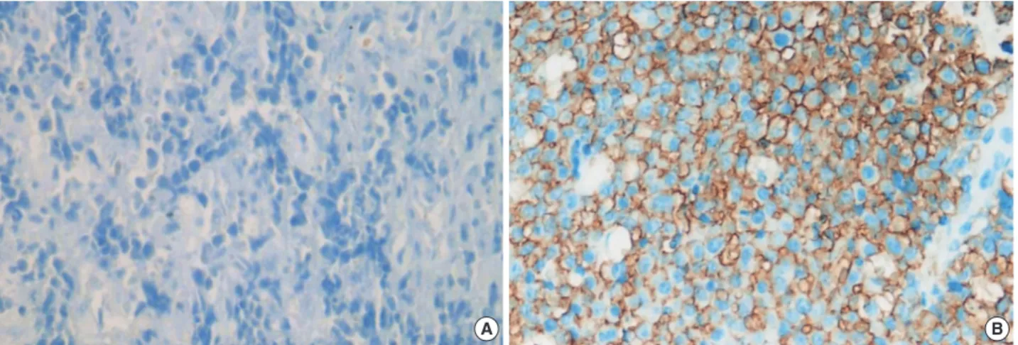
3B). Unexpectedly, the bone marrow biopsy showed diffuse infiltration of plasma cells with about 80% of bone marrow cells. Monoclonal gammopathy was detected in urine pro- tein electrophoresis. Urine immunoelectrophoresis identi- fied the monoclonal band as light chain lambda, and the free serum light chain lambda level in urine was 2,810 mg/L (range, 5.71-26.3 mg/L). Bone marrow biopsy showed dif- fuse uptake of CD138, which was not expressed in the lym- phoma mass (Fig. 4). We concluded that the multiple myelo- ma was newly developed in the patient with primary T-cell lymphoma of bone. The general condition of the patient was rapidly deteriorated with thrombocytopenia and acute renal failure. The patient received a combination chemother- apy with cyclophosphamide and prednisolone, and plasma- pheresis was performed to correct paraproteinemia and hyper- calcemia. Despite aggressive supportive care, the patient died of renal failure.
DISCUSSION
PLB generally presents as a solitary, extensive, destructive bone lesion (1). It is a very rare disease that accounts for less than 2 percent of all lymphomas in adults and accounts for approximately 3-5% of all malignant bone tumors and 4- 7% of all extranodal non-Hodgkin’s lymphomas. Patholog- ic fracture and systemic ‘B’ symptoms (fever, weight loss,
and night sweats) also develop at the time of diagnosis (4, 5). Most patients suffer from bone pain, which is not relieved by rest. A palpable mass due to soft tissue extension is seen in about one-half of cases (10). It is usually characterized by lytic bones lesions located in the metaphysis of long bones or in the axial skeleton (2-7). Histologic diagnosis may be obtained by percutaneous or open biopsy. Histopathologi- cally, the majority of PLB are diffuse large B-cell type, while T-cell lymphomas are extremely rare (4-7). Management strate- gies include a combined modality of chemotherapy and radio- therapy (11-13). Although prognosis, survival, and complete remission rates of B-cell PLB were well-described and high complete remission rates have been reported, the treatment response and prognosis of T-cell PLB remain obscure and data are lacking (7-9).
Multiple myeloma is a lymphoproliferative disease as is lymphoma (14). It is characterized by the neoplastic prolifer- ation of a single clone of plasma cells producing a monoclon- al immunoglobulin. This clone of plasma cells proliferates in the bone marrow and often results in skeletal osteolytic lesions. Common clinical features include recurrent bacteri- al infection, anemia, hypercalcemia, and renal insufficiency, and treatments include chemotherapy and radiotherapy (16).
An interesting feature of this case is the development of multiple myeloma over approximately 1.5 yr following the appearance of a primary T-cell lymphoma of bone. He suf- fered from two lymphoid neoplasms of distinctly different cell lines: the first originating from T-cells and, 1.5 yr later, the second of B-cell origin. Bryant et al. reported plasma cell myeloma in a patient with a cutaneous T-cell lymphoma (17). The investigators assumed that the malignant plasma cells evolved under the sustained inducer stimulus of the neoplastic T-cells. Wickenhauser et al. also reported a newly- detected multiple myeloma in a patient with a long-stand- ing anaplastic cutaneous T-cell lymphoma (20). In the pre- sent case, through immunohistochemical CD138 staining, we confirmed that the multiple myeloma originated from
Fig. 4. CD138 expression in the lymphoma mass (A) and bone marrow biopsy at the diagnosis of multiple myeloma (B) (×200).
A B
Multiple Myeloma in a Primary Lymphoma of Bone 547
cells that differed from previously detected lymphoma cells.
Neoplastic T-lymphocytes are known to retain immunoreg- ulatory capabilities similar to those of their normal counter- part (18, 19). We also speculated that the proliferation of T- helper cells might have resulted in a sustained stimulation of B-cells, followed by proliferation of certain B-cell clones.
It is also possible, although with a low order of possibility, that the two infrequent malignancies have arisen indepen- dently of each other. In this case, it is difficult to determine whether the bone lesion originated from multiple myeloma or lymphoma, based only on imaging studies.
In conclusion, it is important to think about lymphoid malignancies of different pathology when an underlying lymphoid neoplasm shows rapid progression. In addition, considering the scarcity of the association between T-cell lymphoma and multiple myeloma, detection and reporting of new cases are of great value in the study of the potential relationships of immunoregulatory derangements caused by primary lymphoid tumors.
REFERENCES
1. Dubey P, Ha CS, Besa PC, Fuller L, Cabanillas F, Murray J, Hess MA, Cox JD. Localized primary malignant lymphoma of bone. Int J Radiat Oncol Phys 1997; 37: 1087-93.
2. Gianelli U, Patriarca C, Moro A, Ponzoni M, Giardini R, Massimi- no M, Alfano RM, Armiraglio E, Nuciforo P, Bosari S, Coggi G, Parafioriti A. Lymphomas of the bone: a pathological and clinical study of 54 cases. Int J Surg pathol 2002; 10: 257-66.
3. Limb D, Dreghorn C, Murphy JK, Mannion R. Primary lymphoma of bone. Int Orthop 1994; 18: 180-3.
4. Ostrowski ML, Unni KK, Banks PM, Shives TC, Evans RG, O’Co- nnell MJ, Taylor WF. Malignant lymphoma of bone. Cancer 1986;
58: 2646-55.
5. Winkler RE, Ruchlemer R, Heyd J. Multifocal T-cell lymphoma of bone. Am J Hematol 1999; 61: 154.
6. Zinzani PL, Carrillo G, Ascani S, Barbieri E, Tani M, Paulli M, Ste- foni V, Sabattini E, Alinari L, Binazzi R, Tura S, Baccarani M, Pileri SA. Primary bone lymphoma: experience with 52 patients. Haema- tologica 2003; 88: 280-5.
7. Baar J, Burkes RL, Gospodarowicz M. Primary non-Hodgkin’s lymphoma of bone. Semin Oncol 1999; 26: 270-5.
8. Durr HR, Muller PE, Hiller E, Maier M, Baur A, Jansson V, Refior HJ. Malignant lymphoma of bone. Arch Orthop Trauma Surg 2002;
122: 10-6.
9. Heyning FH, Hogendoorn PC, Kramer MH, Hermans J, Kluin-Nele- mans JC, Noordijk EM, Kluin PM. Primary non-Hodgkin’s lym- phoma of bone: a clinicopathological investigation of 60 cases.
Leukemia 1999; 13: 2094-8.
10. Mulligan ME, McRae GA, Murphey MD. Imaging features of pri- mary lymphoma of bone. AJR Am J Roentgenol 1999; 173: 1691-7.
11. Mendenhall NP, Jones JJ, Kramer BS, Hudson TM, Carter RL, Enneking WF, Marcus RB Jr, Million RR. The management of pri- mary lymphoma of bone. Radiother Oncol 1987; 9: 137-45.
12. Fairbanks RK, Bonner JA, Inwards CY, Strickler JG, Habermann TM, Unni KK, Su J. Treatment of stage IE primary lymphoma of bone. Int J Radiat Oncol Biol Phys 1994; 28: 363-72.
13. Fidias P, Spiro I, Sobczak ML, Nielsen GP, Ruffolo EF, Mankin H, Suit HD, Harmon DC. Long term results of combined modality thera- py in primary bone lymphomas. Int J Radiat Oncol Biol phys 1999;
45: 1213-8.
14. Kyle RA, Gertz MA, Witzig TE, Lust JA, Lacy MQ, Dispenzieri A, Fonseca R, Rajkumar SV, Offord JR, Larson DR, Plevak ME, Ther- neau TM, Greipp PR. Review of 1027 patients with newly diagnosed multiple myeloma. Mayo Clin Proc 2003; 78: 21-33.
15. Lahtinen R, Laakso M, Palva I, Virkkunen P, Elomaa I. Randomis- ed, placebo controlled multicentre trial of clodronate in multiple myeloma. Finnish Leukaemia Group. Lancet 1992; 340: 1049-52.
16. Blade J, Kyle RA. Multiple myeloma in young patients: Clinical presentation and treatment approach. Leuk Lymphoma 1998; 30:
493-501.
17. A predictive model for aggressive non-Hodgkin’s lymphoma. The International Non-Hodgkin’s Lymphoma Prognostic Factors Pro- ject. N Engl J Med 1993; 329: 987-94.
18. Bryant E, Ronan SG, Iossifides IA. Plasma cell myeloma in a patient with a cutaneous T-cell lymphoma. Cancer 1982; 50: 2122-5.
19. Broder S, Poplack D, Whang-Peng J, Durm M, Goldman C, Muul L, Waldmann TA. Characterization of a suppressor cell leukemia:
evidence for the requirement of an interaction of two T cells in the development of human suppressor effector cells. N Engl J Med 1978;
298: 66-72.
20. Uchiyama T, Sagawa K, Takatsuki K, Uchino H. Effect of adult T cell leukemia cells on pokeweed mitogen-induced normal B-cell dif- ferentiation. Clin Immunol Immunopathol 1978; 10: 24-34.
21. Wickenhauser C, Borchmann P, Diehl V, Scharffetter-Kochanek K.
Development of IgG lambda multiple myeloma in a patient with cutaneous CD30+ anaplastic T-cell lymphoma. Leuk Lymphoma 1999; 35: 201-6.