INTRODUCTION
Kikuchi’s disease is a benign, self-limited disease with well- defined clinical entity that typically involves the cervical lymph nodes of adolescents and young adults and occurs commonly in Asian (1). Hemophagocytic syndrome is a histiocytic reac- tive process of strong immunologic activation, such as severe infection, malignancy, autoimmune diseases, and metabolic diseases (2). It is characterized by histiocytic proliferation, hemophagocytosis, fever, hepatosplenomegaly, generalized lymphadenopathy, hypertriglyceridemia, and hypofibrino- genemia.
There are eight cases of hemophagocytic syndrome simul- taneously associated with Kikuchi’s disease in the literature (3-9). The prognosis and treatment for hemophagocytic syn- drome in Kikuchi’s disease is still unknown. We report a case of a 13-yr old girl with simultaneous Kikuchi’s disease and hemophagocytic syndrome.
CASE REPORT
A 13-yr-old girl was admitted to our hospital because of a 4-week history of fever, generalized maculopapular rash, pro- gressive cervical lymph node swelling, and an attack of gen- eralized tonic-clonic seizure 2 days before admission. On exam- ination, she had a temperature of 38℃and submandibular lymphadenopathy (3 to 4 cm in diameter). The liver and spleen were not palpable. She developed progressive pancytopenia
(white blood cell count, 1,500/ L; hemoglobin 10.3 g/dL;
platelets 39,000/ L). Hepatic dysfunction (AST 1156 IU/L and ALT 657 IU/L), cholestasis (total bilirubin 2.47 mg/dL and direct bilirubin 1.88 mg/dL) and acute renal failure (blood urea nitrogen 18.15 mg/dL, creatinine 1.96 mg/dL) were pre- sent. Prothrombin time and international normalized ratio were normal and activated partial thromboplastin time was mildly prolonged (64.1 sec). Her triglycerides and ferritin lev- els were increased to 144 mg/dL (normal range, 37-134 mg/
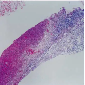
dL) and 14,955.2 ng/mL (normal range, 6-282 ng/dL), respec- tively. Antinuclear antibody, anti-double stranded DNA anti- body, cytomegalovirus immunoglobulin (Ig) M, herpes sim- plex virus IgM, and viral serology for hepatitis A, B, and C were negative. Epstein-Barr virus IgM was under 1:10 (nor- mal range under 1:10) and Epstein-Barr virus IgG was 1:160 (normal range under 1:10). Bacterial and viral cultures of blood, urine, and stool were negative. On the following day, she developed an acute respiratory distress syndrome that required mechanical ventilation. A bone marrow aspiration and biopsy showed many histiocytes and obvious hemophago- cytic histiocytes (20% of total histiocytes) (Fig. 1). Fine-nee- dle aspiration biopsy of lymph nodes revealed paracortical necrotizing lesions with typical features of Kikuchi’s disease (Fig. 2, 3).
Intravenous immunoglobulin (0.4 g/kg/day for 5 days) and methylprednisolone (1 g/d for 3 days) were administered. There were no further seizure attack any more and we did not pre- scribed any antiepileptic drug. The clinical symptoms and signs improved gradually. On day 3, she was administered
Young Mi Kim, Yoon Jin Lee, Sang Ook Nam, Su Eun Park, Ji Yoen Kim*, Eun Yup Lee�
Department of Pediatrics, Surgical Pathology*, and Clinical Pathology�, College of Medicine, Pusan National University, Busan, Korea
Received : 4 June 2002 Accepted : 9 October 2002
Address for correspondence Su Eun Park, M.D.
Department of Pediatrics, College of Medicine, Pusan National University, 10, 1-Ga, Ami-dong, Suh-gu, Busan 602-739, Korea
Tel : +82.51-240-7298 Fax : +82.51-248-6205 E-mail : [email protected]
592 J Korean Med Sci 2003; 18: 592-4
ISSN 1011-8934
Copyright � The Korean Academy of Medical Sciences
Hemophagocytic Syndrome Associated with Kikuchi's Disease
A 13-yr-old female was admitted to our hospital with fever, seizure, and cervical lym- phadenopathy. Laboratory data showed pancytopenia, elevation of serum transam- inase, lactate dehydrogenase, triglyceride, and ferritin levels. Lymph node biopsy revealed features of Kikuchi’s disease and there were signs of histiocytosis and hemophagocytic phenomenon in bone marrow. She recovered after treatment with intravenous immunoglobulin and corticosteroids therapy. Hemophagocytic syndrome can be associated with Kikuchi’s disease especially in childhood and seems to have a less aggressive clinical course and better prognosis.
Key Words : Histiocytic Necrotizing Lymphadenitis; Kikuchi’s Disease; Histiocytosis
Hemophagocytic Syndrome Associated with Kikuchi’s Disease 593
with etoposide (150 mg/m2), which was discontinued due to subsequent pancytopenia (white blood cell count 410/ L;
hemoglobin 9.2 g/dL; platelets 19,000/ L on day 12). We continued supportive care and prescribed oral dexamethasone (10 mg/m2/day every 8 hr for 2 weeks, followed by 7.5 mg/m2/ day every 8 hr for 2 weeks, 5 mg/m2/day every 8 hr for 2 weeks, and 2.5 mg/m2/day every 8 hr for 1 weeks) and bactrim (5 mg/
kg in 2 divided doses, three times per week) with dramatic re-
sponse. She required mechanical ventilation for 7 days. There was complete resolution of the hemophagocytic syndrome with- in 1.5 months and no evidence of disease recurrence during the following 8 months.
DISCUSSION
Diagnosis of hemophagocytic syndrome can be made with fever of unknown etiology for 7 or more days, unexplained cytopenia affecting at least 2 cell lines, abnormal liver func- tion tests, disseminated intravascular coagulopathy, and bone marrow that contains greater than 3% histiocytes undergo- ing hemophagocytosis. Clinical severity ranges from complete recovery to rapid deterioration. The mortality rate is 20- 42% (10).
Our patient had necrotizing lymphadenitis followed by ful- minant hemophagocytic syndrome. Eight cases of hemophago- cytic syndrome combined with necrotizing lymphadenitis have been reported in the literature thus far (3-9). Four patients aged from 14 to 17 yr and three were Asians. Two patients had underlying systemic lupus erythematosus and one patients was pregnant. Six patients recovered after treatment with corticos- teroids with or without intravenous immunoglobulin.
Kikuchi’s disease is usually self-limited and supportive treat- ment alone is sufficient for the disease. The treatment and prog- nosis of childhood Kikuchi’s disease associated with hemoph- gocytic syndrome remain unclear. According to our review of the literature, childhood Kikuchi’s disease is more frequently associated with hemophagocytic syndrome and seems to have a less aggressive course and better prognosis than the adult
Fig. 1.Bone marrow aspirate shows histiocytes engulfing erythro- cytes (Wright stain, ×1,000).
Fig. 2. Low power view of necrotizing lymphadenitis shows necro- sis with infiltration of numerous lymphocytes and histiocytes (H&E stain, ×40).
Fig. 3. High power view of necrotizing lymphadenitis shows numer- ous apoptotic bodies (H&E stain, ×400).
594 Y.M. Kim, Y.J. Lee, S.O. Nam, et al.
counterpart.
Our case shows that childhood hemophagocytic syndrome can be associated with Kikuchi’s disease and assume a relative- ly benign course. Administration of intravenous immunoglob- ulin and corticosteroids may be the treatment of choice of hemophagocytic syndrome associated with Kikuchi’s disease and may provide satisfactory results. Chemotherapy, such as etoposide-containing regimen, can be reserved for those who fail to respond to intravenous immunoglobulin and corticos- teroids.
REFERENCES
1. Stephan L, Elanine SJ. Histiocytoses. In: Philip AP, David GP, Prin- ciples and practice of pediatric oncology. Baltimore: Williams &
Wilkins 1997; 739-40.
2. Imashuku S. Advances in the management of hemophagocytic lym- phohistiocytosis. Int J Hematol 2000; 72: 1-11.
3. Chen JS, Chang KC, Cheng CN, Tsai WH, Su IJ. Childhood hemo- phagocytic syndrome associated with Kikuchi’s disease. Haemato- logica 2000; 85: 998-1000.
4. Wano Y, Ebata K, Masaki T, Takeshita S, Ogawa N, Kim CG, Okata
J, Saito H, Hirose Y, Tohyama T, Sugai S. Histiocytic necrotizing lymphadenitis (Kikuchi-Fujimoto’s disease) accompanied by hemo- phagocytosis and salivary gland swelling in a patient with systemic lupus erythematosus. Rinsho Ketsueki 2000; 41: 54-60.
5. Chmait RH, Meimin DL, Koo CH, Huffaker J. Hemophagocytic syn- drome in pregnancy. Obstet Gynecol 2000; 95: 1022-4.
6. Yabe H, Sinzato I, Hashimoto K. Necrotizing lymphadenitis present- ing as mesenteric lymphadenopathy. Rhinsho Ketsueki 1999; 40: 658- 62.
7. Ozaki Y, Kagawa H, Yasuzawa M, Yoshimura C, Shimizu T, Nomu- ra S, Kitajima H, Kishimoto Y, Komiyama Y, Okamura A, Fukuhara S. Anti-fibrinogen antibody detected in a patient with systemic lupus erythematosus and disseminated intravascular coagulation. Rhinsho Ketsueki 1998; 39: 436-41.
8. Okuda T, Yumoto Y. Subacute necrotizing lymphadenitis with a clin- ical course mimicking virus-associated hemophagocytic syndrome.
Rhinsho Ketsueki 1994; 35: 689-93.
9. Mahadeva U, Allport T, Bain B, Chan WK. Haemophagocytic syn- drome and histiocytic necrotising lymphadenitis (Kikuchi’s disease).
J Clin Pathol 2000 ; 53: 636-8.
10. Tsuda H. Hemophagocytic syndrome (HPS) in children and adults.
Int J Hematol 1997; 65: 215-6.